Stem Cells A cluster of nascent retinae generated


 of cells: All diploid (body) cells have the same chromosomes. So they")


 ● Prokaryotes have a simple")

 http: //en.")

")


- Slides: 35
Stem Cells A cluster of nascent retinae generated from 3 D embryonic stem cell cultures, by UCL News on Flickr (CC): http: //flic. kr/p/ff. PBPT
We will watch next class….
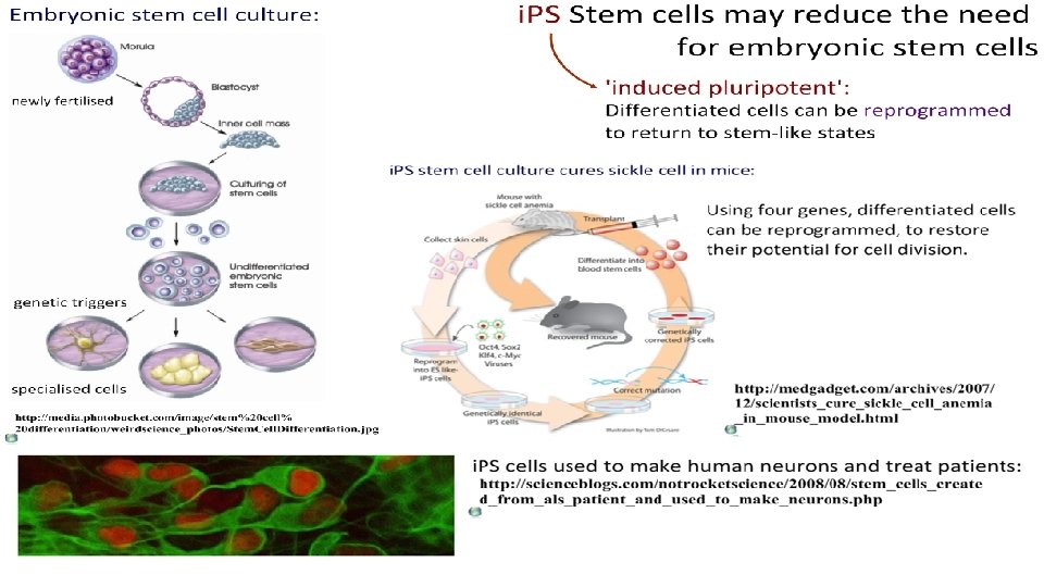
Stem Cells retain the capacity to divide and can differentiate along divergent pathways. Totipotent Can differentiate into any type of cell. Pluripotent Can differentiate into many types of cell. Multipotent Can differentiate into a few closely-related types of cell. Unipotent Can regenerate but can only differentiate into their associated cell type (e. g. liver stem cells can only make liver cells). Image from: http: //en. wikipedia. org/wiki/Stem_cell
Stem Cells retain the capacity to divide and can differentiate along divergent pathways. By Fwfu at en. wikibooks [Public domain], from Wikimedia Commons
Stem Cells retain the capacity to divide and can differentiate along divergent pathways. Screenshot from this excellent tutorial: http: //www. ns. umich. edu/stemcells/022706_Intro. html
Differentiation (specialization) of cells: All diploid (body) cells have the same chromosomes. So they carry all the same genes and alleles. BUT Not all genes are expressed (activated) in all cells. The cell receives a signal. This signal activates or deactivates genes. Genes are expressed accordingly and the cell is committed. Eventually the cell has become specialized to a function. Key Concept: Structure vs Function How do the structures of specialized cells reflect their functions? How does differentiation lead to this? Screenshot from this excellent tutorial: http: //www. ns. umich. edu/stemcells/022706_Intro. html
Therapeutic Uses of Stem Cells Treatment for Leukemia From: Problem Cancer of the blood or bone marrow, resulting in abnormally high levels of poorly-functioning white blood cells. Treatment Chemotherapy and radiotherapy can be used to destroy the white blood cells, but these need to be replaced with healthy cells. Bone marrow transplants are often used for this. Role of Stem Cells Hematopoetic Stem Cells (HSCs) can be harvested from bone marrow, peripheral blood or umbilical cord blood. As these can differentiate to form any type of white blood cell, they can be used to repopulate the bone marrow and produce new, healthy blood cells. The use of a patient’s own HSCs means there is far less risk of immune rejection than with a traditional bone marrow transplant. http: //en. wikipedia. org/wiki/Pluripotential_hemopoietic_stem_cell Animation of this process: Animated tutorials from: http: //outreach. mcb. harvard. edu/animations/thera 7 c. swf
Uses of Stem Cells • Stargardt’s disease: Early vision loss usually leading to blindness due to a gene mutation • Retinal pigment epithelial stem cells can be used to regenerate and support function of the eye’s light sensitive cells that have been damaged
Two Minute Essay What is a stem cell? How do stem cells differentiate into specialized cells? Outline one therapeutic use of stem cells.
Ethics of Stem Cells • Specially created embryos • is it right to create embryos just for medical use? • what about the ones that do not get used? • when do we say “life” begins? • Umbilical cord blood • Adult’s own tissues
Assignment for today STEM CELLS
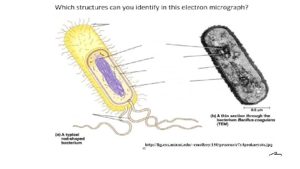
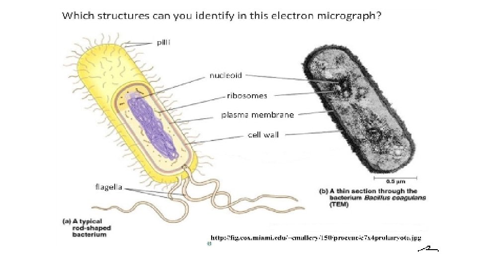
Objective 1. 2 Ultrastructure of cells Understanding (Statement objectives) ● Prokaryotes have a simple cell structure without compartments. ● Eukaryotes have a compartmentalized cell structure. ● Prokaryotes divide by binary fission. ● Electron microscopes have a much higher resolution than light microscopes. Applications ● Describe the structure and function of organelles within exocrine gland cells of the pancreas. ● Describe the structure and function of the organelles within palisade mesophyll of the leaf. Nature of science ● Developments in scientific research follow improvements in apparatus: Describe how the invention of electron microscopes led to greater understanding of cell structure. Skills ● Draw and label a diagram of the ultrastructure of prokaryotic cells based on electron micrographs. ○ Drawings of prokaryotic cells should show the cell wall, pili and flagella, and plasma membrane enclosing cytoplasm that contains 70 S ribosomes and a nucleoid with naked DNA. ● Draw and label a diagram of the ultrastructure of eukaryotic cells based on electron micrographs. ○ Drawings of eukaryotic cells should show a plasma membrane enclosing cytoplasm that contains 80 S ribosomes and a nucleus, mitochondria and other membranebound organelles are present in the cytoplasm. Some eukaryotic cells have a cell wall. ● Interpret of electron micrographs to identify organelles and deduce the function of specialized cells. ● Compare eukaryotic and prokaryotic cells. ○ Naked DNA versus DNA associated with proteins ○ DNA in cytoplasm versus DNA enclosed in a nuclear envelope ○ No mitochondria versus mitochondria ○ 70 S versus 80 S ribosomes ○ Eukaryotic cells have internal membranes that compartmentalize their functions. ● State three differences between plant and animal cells.
Prokaryotes Image: Hospital-associated MRSA by NIAID on Flickr http: //flic. kr/p/a 4 RLq 5
Prokaryotes “Before nucleus: ” evolutionary precursors to eukaryotes. Escherichia coli (E. coli) http: //en. wikipedia. org/wiki/Escherichia_coli
Prokaryotic Cell Parts mesosome cell wall plasma membrane pili cytoplasm nucleoid ribosomes flagella Cell structures animation: Prokaryotic cell parts are not generally membrane-bound, so we don’t refer to them as organelles.
Prokaryotic Cell Parts mesosome cell wall: protective protein-based coating (Gram + / Gram -) plasma membrane: selectively permeable, controls entry & exit of materials to and from the cell. pili: attach to other bacteria for DNA transfer cytoplasm: contains enzymes for metabolic reactions nucleoid: closed-loop of bacterial DNA in a condensed area ribosomes: protein synthesis (transcription & translation) flagella: whiplash-like motion causes movement Cell structures animation:
mesosomes Prokaryotic Cell Parts These don’t really exist naturally as bacterial cell parts, and could be an example of a paradigm shift in thinking. They were observed in some electron micrographs and thought to be in-folds of membrane used for division, respiration or making cell walls… … turns out they are an artifact of the preparation method for some electron microscope images. Cell structures animation: http: //en. wikipedia. org/wiki/Mesosome
Past-paper question: E. coli TEM image Identify these structures: I. III. IV. Calculate the magnification of the image. Image from IB Biology Question. Bank CDRom – get a copy here: https: //store. ibo. org/biology
Past-paper question: E. coli TEM image Identify these structures: I. Plasma membrane II. Cell wall / pili III. Nucleoid IV. Cytoplasm / ribosomes Calculate the magnification of the image. 1. Measure the scale bar in mm. 2. Multiply x 1000 to convert to μm. That is the magnification. How long is the Image from IB Biology Question. Bank CDRom – get a copy here: https: //store. ibo. org/biology bacterium?
PROKARYOTES E P R O D U C E through binary fission two-parts splitting
PROKARYOTES E through P R O D U C E binary fission The closed-loop DNA of the bacterium makes copies through semiconservative DNA replication. New plasmids are pulled to opposing poles by the spindle fibres. The bacterium divides in two.
http: //www. youtube. com/watch? v=g. Ewz. Dydci. Wc
https: //youtu. be/x 00 otj. SL 6 GQ
Prokaryotes divide by binary fission. Life cycle of E. coli from: http: //en. wikipedia. org/wiki/Escherichia_coli
What about conjugation?