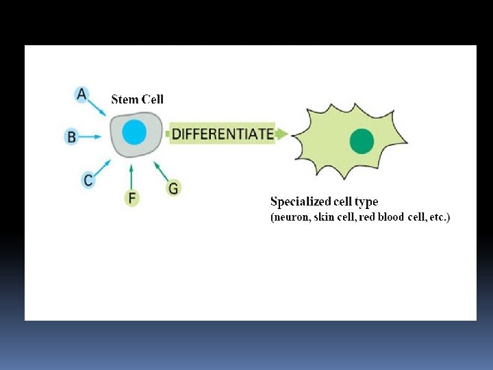
Stem Cell differentiation Differentiation is the process of


 A Blastocyst is a")


- Slides: 37
Stem Cell differentiation
Differentiation is the process of becoming specialized stem cells from unspecialized cells. The signals inside and outside cells that trigger stem cell differentiation. The internal signals are controlled by a cell's genes. The external signals include chemicals secreted by other cells, physical contact microenvironment. with neighboring cells in the
The process of cell differentiation starts with the fertilization of the female egg. As soon as the egg is fertilized, cell multiplication is initiated resulting in the formation of a sphere of cells known as the blastocyst. It is this sphere of cells that attach to the uterine wall and continues to differentiate. As the blastocyst differentiates, it divides and specializes to form a zygote that attaches to the womb for nutrients. As it continues to multiply and increase in size, the differentiation process results in the formation of different organs. During the final phase of cell differentiation, there is formation of several types of differentiated cells from one of the precursor or a progenitor cell.
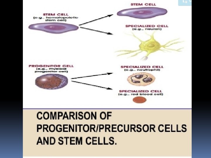
Progenitor cell A progenitor cell that has cell is a biological a tendency to differentiate into a specific type of cell and is pushed to differentiate into its "target" cell. The most important difference between stem cells and progenitor cells is that stem cells can replicate indefinitely, whereas progenitor cells can divide only a limited number of times.
blood stem cell differentiation only specialized types of blood cell: red blood cells, white blood cells, platelets
Distinguishing Features of Progenitor Cells and Stem Cells. Stem cells A progenitor cell A stem cell is an unspecialized cell that develops into a variety of specialized cell types. A progenitor cell is unspecialized that is capable of undergoing cell division and yielding two specialized cells. A stem cell divides and gives rise to one additional stem cell and a specialized cell. Example: a hematopoietic stem cell produce a second generation stem cell and a RBC. Example: a progenitor cell undergoing cell division to yield two specialized cells (a neutrophil and a red blood cell).
Adult stem cells may exhibit two types of differentiation pathways; Normal differentiation; to form specialized cell types of the tissue in which they reside. Transdifferentiation or plasticity; cells may also exhibit the ability to form specialized cell types of tissues other than the place where they reside.
Normal Differentiation The following are examples of differentiation pathways of adult stem cells. Hematopoietic stem cells give rise to all types of blood cells: red blood cells, B lymphocytes, T lymphocytes, natural killer cells, neutrophils, basophils, eosinophils, monocytes, macrophages, and platelets. Bone marrow stromal cells (mesenchymal stem cells) give rise to a variety of cell types: bone cells (osteocytes), cartilage cells (chondrocytes), fat cells (adipocytes), and other kinds of connective tissue cells such as those in tendons.
Neural stem cells in the brain give rise to its three major cell types: nerve cells (neurons) and two categories of non-neuronal cells- astrocytes and oligodendrocytes. Epithelial stem cells in the lining of the digestive tract occur in deep crypts and give rise to several cell types: absorptive cells, goblet cells and enteroendocrine cells. Skin stem cells occur in the basal layer of the epidermis and at the base of hair follicles. The epidermal stem cells give rise to keratinocytes, which migrate to the surface of the skin and form a protective layer. The follicular stem cells can give rise to both the hair follicle and to the epidermis.
Dr. Ziad W Jaradat ©
Trans-differentiation or plasticity of adult stem cells The ability of adult stem cell to differentiate into multiple cell types is called plasticity or transdifferentiation. The following list offers examples of adult stem cell plasticity that have been reported during the past few years. Hematopoietic stem cells may differentiate into: three major types of brain cells (neurons, oligodendrocytes, and astrocytes) skeletal muscle cells cardiac muscle cells liver cells. Bone marrow stromal cells may differentiate into: cardiac muscle cells and skeletal muscle cells. Brain stem cells may differentiate into: blood cells and skeletal muscle cells.
Plasticity of adult stem cells
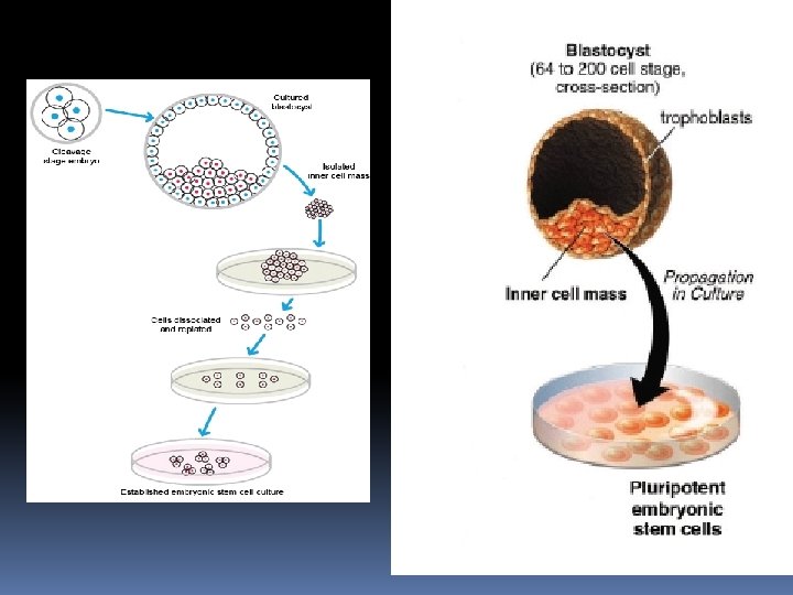
How are embryonic stem cells harvested? • Growing cells in the laboratory is called as cell culture. • Human ES cells are derived from 4 -5 day old blastocyst • Blastocyst structures include: – Trophoblast: outer layer of cells that surrounds the blastocyst & forms the placenta – Blastocoel: (“blastoseel”) the hollow cavity inside the blastocyst that will form body cavity – Inner cell mass: a group of approx. 30 cells at one end of the blastocoel: • Forms 3 germ layers that form all embryonic tissues (endoderm, mesoderm, ectoderm)
Stages of Embryogenesis Day 2 2 -cell embryo Day 1 Fertilized egg Day 11 -14 Tissue Differentiation Day 3 -4 Multi-cell embryo Day 5 -6 Blastocyst
Human Embryonic Stem Cells Blastocyst Inner Cell Mass (Stem Cells) A Blastocyst is a hollow ball of cells with a small clump of stem cells inside
Blastocyst from In Vitro Fertilization Clinic Human Embryonic Stem Cells Pipette Stem Cells A Blastocyst is a hollow ball of cells with a small clump of stem cells inside Stem Cells To remove the stem cells, the Blastocyst is opened and the stem cells removed with a pipette
Blastocyst from In Vitro Fertilization Clinic Human Embryonic Stem Cells Pipette Stem Cells The stem cells are placed in a dish and are fed and cared for (each blastocyst = 1 stem cell line) Stem Cells Pipette
Growth factors Stem Cells Pancreatic Islet Chemicals Petri Dish Muscle cell Neuron Different chemicals / molecules are added to the stem cells to make them become specific types of cells.
Isolation of adult stem cells From the body From amniotic fluid From umbilical chord
From the body itself Adult stem cells have been found in the brain, bone marrow, blood vessels, skeletal muscle, skin, teeth, heart, gut, liver, and other organs and tissues. Adult stem cells can be isolated from the body in different ways, depending on the tissue. Blood stem cells, for example, can be taken from a donor’s bone marrow, from blood in the umbilical cord when a baby is born, or from a person’s circulating blood. Mesenchymal stem cells, which can make bone, cartilage, fat, fibrous connective tissue. Neural stem cells (which form the brain’s three major cell types) have been isolated from the brain and spinal cord.
From amniotic fluid Amniotic fluid, which bathes the fetus in the womb, contains fetal cells including mesenchymal stem cells, which are able to make a variety of tissues. Many pregnant women elect to have amniotic fluid drawn to test for chromosome defects, the procedure known as amniocentesis.
Cord Blood • Umbilical cord blood is also known as placental blood. • It is the blood that flows in the circulation of the developing fetus in the womb. • After the baby’s birth, the left over blood in the umbilical cord and placenta is called cord blood. • This blood is a rich source of stem cells.
Collecting cord blood stem cells • This blood is collected by the physician after the baby is born and the cord is cut. • It takes less than 5 minutes and there is no pain, harm or risk to mother or newborn. • This cord blood containing the stem cells, is sent to a “Cord Blood Bank” either private or public where it is processed and the stem cells are preserved in liquid nitrogen.
Umbilical cord stem cells • Adult stem cells of infant origin • Less expensive
Culturing of Stem cells The culture conditions and types of media used for stem cell culture depend on the type of stem cell. There a wide range of protocols and products available for both maintaining stem cells in an undifferentiated state and for differentiating them into different lineages and cell types.
Cell Culture Techniques for ESC • Isolate & transfer of inner cell mass into plastic culture dish that contains culture medium • Cells divide and spread over the dish • Inner surface of culture dish is typically coated with mouse embryonic skin cells that have been treated so they will not divide.
• This coating is called a FEEDER LAYER – Feeder cells provide ES cells with a sticky surface for attachment – Feeder cells release nutrients • Recent discovery: methods for growing embryonic stem cells without mouse feeder cells – Significance – eliminate infection by viruses or other mouse molecules • ES cells are removed gently and plated into several different culture plates before crowding occurs
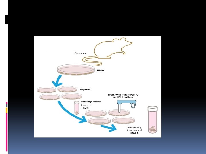
Feeder Cell Layers All the ESC cultures are maintained on feeder cell layers. Inactivated mouse embryonic fibroblasts (MEFs) were used as feeder layers that provide factors and a substrate that allow ESCs to grow and divide. There are several problems associated with MEFs, including the potential for the introduction of mousederived infectious agents, undesirable protein transfer.
Methodology MEFs can either be purchased or freshly generated in the lab. Fibroblasts from ~14– 15 -day-old fetal mice are cultured and expanded for 3– 4 days. To be used as a feeder layer, MEFs must be mitotically inactivated, either by irradiation with UV light or incubation with mitomycin C. Stocks of MEF cells can be frozen either before or after inactivation.
Feeder-free culture There are two types of feeder cell-free media: defined media and conditioned media. These media will support the growth of stem cells and contain factors that inhibit differentiation. The components of the media and the supplements vary depending on the type of stem cell.
Defined medium is a serum-free medium that has been supplemented with recombinant growth factors and other molecules that are required to support the growth and pluripotency of cells. Media formulated for stem cell culture often contain factors such as leukemia inhibitory factor and bone morphogenetic protein to prevent differentiation. Some defined media for human cell culture may contain bovine serum albumin (BSA).
Conditioned medium Cells in culture secrete factors into the media that support cell growth. After cells have grown and divided for a period of time, the media are removed. This conditioned media can then be used as a supplement along with fresh media. An advantage of conditioned media is that they contain more factors than defined media. The disadvantage is that the contamination with virus. Human skin fibroblasts are the most common cell type used to make conditioned media for human cells.
Stem cells not growing on a feeder layer need a matrix for attachment and growth. A culture matrix contains extracellular matrix (ECM) proteins and polysaccharides such as vitronectin and proteoglycans. There are many matrices available with varying combinations of proteins and carbohydrates. Different types and sources of stems cells require different matrices, and different matrix components can either maintain pluripotency or help drive differentiation.