Stefan Sivkov MD Ph D Development Onset 22


 - sclera (non-transparent, bluish in")


,")

 Avascular ii) Transparent iii) Limbus - connection with sclera: Highly vascular Contains sinus")


, central")
 inner layer. n N. opticus – axons of")
 Stratum neuroepitheliale")


")


- Slides: 39
Stefan Sivkov, MD, Ph. D
Development А. Onset 22 day of gestation; from neuroectoderm, ectoderm & mesoderm. B. Diencephalic vesicle gives origin to optic vesicle; after invagination develops double-layered optic cup. i) Inner layer develops into в pars nervosa of retina ii) Outer layer gives pars pigmentosa of retina, pigmentous epithelium of iris and ciliary body. C. Ectoderm gives origin of : i) Lens. ii) Corneal epithelium and conjunctiva. D. Mesoderm gives origin of: i) Stroma of sclera and cornea. ii) Stroma of choroid.
Development
Three parts n Optic apparatus – controls the light entering the eye n Detector system – for black / white and color vision n Nerve pathways – conveys signals to brain
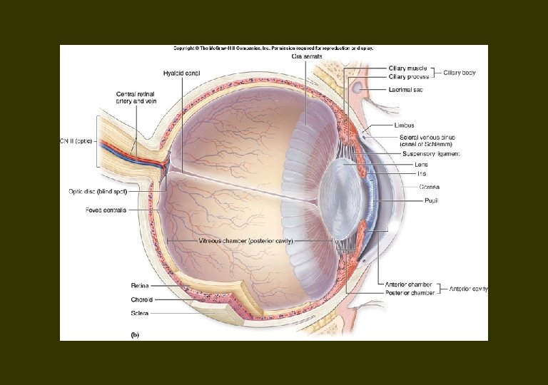
Eye ball 3 coats Tunica Fibrosa: cornea, sclera Tunica Vasculosa: choroidea, corpus ciliare, iris Tunica Nervosa: retina 3 chambers Anterior eye chamber Posterior eye chamber Corpus vitreum
Tunicae bulbi Outer, tunica fibrosa bulbi - cornea (transparent) - sclera (non-transparent, bluish in children, yellowish in adults) Middle, tunica vasculosa bulbi - iris and central opening, рupilla - corpus ciliare composed of: musculus ciliaris, processus ciliaris, zonula ciliaris with fibrae zonulares & spatia zonularia - choroidеа Inner, tunica interna bulbi (retina) - blind part, рars caeca retinae (from мargo pupillaris iridis to оra serrata): Pars iridica retinae (double-layered, pigmented cells) Pars ciliaris retinae (double-layered, pigmented inner layer) - рars optica retinae (stratified)
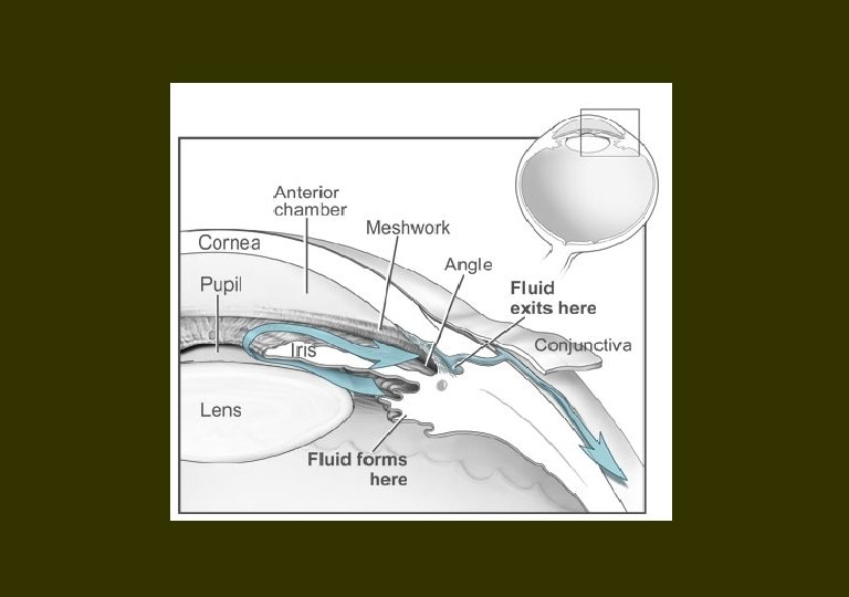
Eye chambers Three chambers filled with fluid: n Camera anterior bulbi (between cornea & iris), n Camera рosterior bulbi (between iris, fibrae zonulare & lens) n Corpus vitreum (between lens & retina). The nfirst two filled with humor aquosus, Corpus vitreum filled with humor vitreus.
Tunica fibrosa
Sclera Posterior 4/5 of tunica fibrosa; Collagen & elastic fibers, fibroblasts (substantia propria sclerae), 3 layers: § episclera: outer § substantia propria sclerae: middle § lamina fusca: inner, collagen & elastic fibers pigment cells; borders choroid
Cornea А. Anterior 1/5 of the coat Б. 5 – layered: - Epithelium anterius: squamous stratified nonceratizing, 5 - 6 layers: - Lamina limitans anterior (Bowman): 7 -12 mcm, collagen fibers, lacks cells, barrier agains infection, does not regenerates. - Substantia prоpria: 200 laminae of parallel collagen fibers. - Lamina limitans posterior (Descemet): 5 - 10 mcm, basal lamina of endothelium. - Endothelium posterius: simple squamous epithelium
i) Avascular ii) Transparent iii) Limbus - connection with sclera: Highly vascular Contains sinus venosus sclerae (Schlemm canal)
Tunica vasculosa Three parts: 1. Posterior - сhoroidea 2. Middle - сorpus ciliare 3. Anterior - iris
Choroidea n n 2/3 of the coat Attached posteriorly to the sclera – Spatium perichoroidale – Lamina suprachoroidea n Composed of: – – – n Connective tissue Pigment cells Blood vessels 3 layers: – Lamina vasculosa – Lamina choriocapillaris – Lamina basalis (Bruch)
Choroidea
Corpus ciliare n n Orbiculus ciliaris – posterior Corona ciliaris - anteromedial – Processus ciliares (70 -80) n n Dense capillary network Produce humor aquosus Fibrae zonulares Musculus ciliaris - lateral n n n Fibrae meridionales (Brucke) Fibrae radiales Fibrae circulares (Muller) – Takes part in (n. oculomotorius)
Coprpus ciliare Orbiculus ciliaris, corona ciliaris
Accomodation n Focusing for near vision n Contraction of the pupil n Convergence
Accomodation n M. ciliaris contracts and moves forward n That relaxes zonule ciliares and lens becomes more convex n The eye adapts to near vision
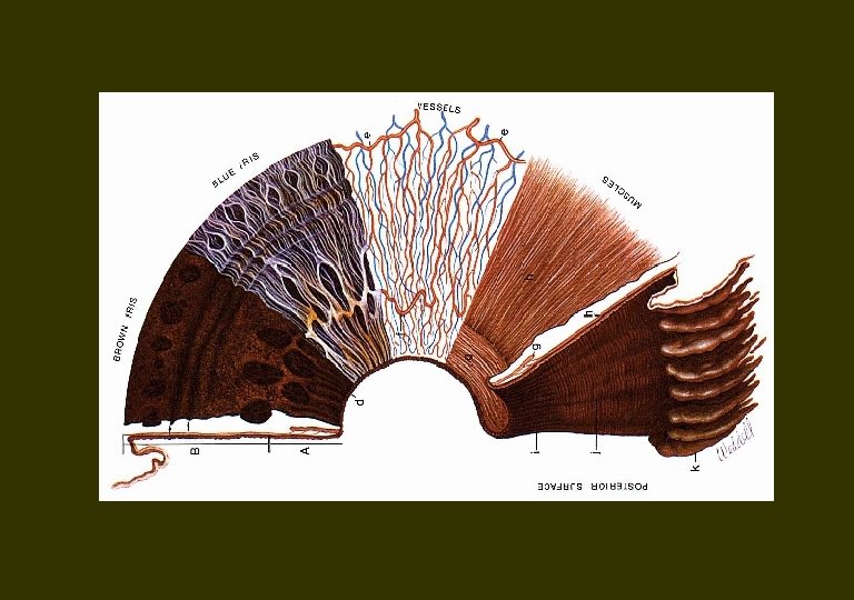
Iris n Color is genetically determined. n Pigment: а. melanin & b. lipochrome. n Melanin - gene of 15 chromosome. n Lypochrome - gene of 19 chromosome.
Iris n n n Anterior part Opening in the middle, pupilla. Margo pupillaris & margo ciliaris Annulus iridis minor & anulus iridis major Structure – Epithelium anterius – Stroma iridis – M. sphincter & dilatator pupillae – Epithelium pigmentosum Attached to the sclera by lig. pectinatum
Iris. Ligamentum pectinatum
Retina n n Eye fundus discus nervi optici (2 x 1. 5 mm), central - excavation. 4 mm lateral is an oval spot, fovea, in the center of macula lutea. Centarl part of retina – d=21 mm from the center of discus n. optici. Diameter of retina - 42 mm.
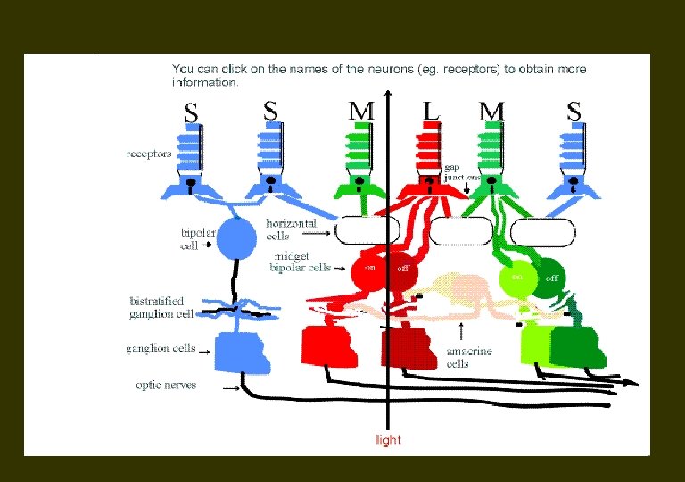
Structure n Retina (0. 5 mm) inner layer. n N. opticus – axons of the ganlion cells. n Ganglion cells, efferent, the innermost. n Photoreceptive cells, rod and cones, outer.
RETINA 1. Pars pigmentosa - detachment of retina 2. Pars nervosa (1) Stratum neuroepitheliale Bacillus, Conus (2) Stratum nucleare externum Bipolar cell Horizontal cell Amacrine cell (3) Stratum nucleare internum Ganglion cell Nervus Opticus (II)
Structure of retina n Three cellular layers Outer nuclear – bodies of rods & cones Inner nuclear – bodies of bipolar, horizontal & amacrine cells Ganglion – bodiesa of ganglion cells. n Two synaptic layers Outer plexiform Inner plexiform
Synaptic layers Stratum plexiforme externum Stratum plexiforme internum
Ora serrata. Pars optica & pars ceca retinae
Fovea centralis
Rods and cones
Optic nerve
Visual Pathway
Visual Pathway Modality: Vision Receptor: Photoreceptor Cell of Retina Cranial Nerve: II (Optic nerve) 1 st Neuron: Bipolar Cell 2 nd Neuron: Ganglion Cell optic nerve optic chiasm optic tract 3 rd Neuron: Lateral Geniculate Nucleus optic radiation Termination: Primary Visual Area (V I) Brodmann area 17
Visual Pathway 1. Optic nerve 2. optic chiasm 3. optic tract 4. lateral geniculate body 5. optic radiation 6. visual cortex (striate cortex) 7. Meyer’s loop 8. lateral ventricle
Visual Pathway 1. Optic nerve 2. Optic chiasm 3. Optic tract 4. Lateral geniculate body 5. Optic radiation 6. Visual cortex (Striate cortex)
Clinical Features of Visual Pathway Lesion 1. optic nerve 2. optic chiasm 3. optic tract 4. 5. optic radiation A. unilateral blindness B. bitemporal hemianopsia C. left homonymous hemianopsia D. left inferior homonymous quadranopsia E. left superior homonymous quadranopsia