
Staphylococci Fatin Naeem Abbas Staphylococus Morphological characteristic Cultural

Staphylococci Fatin Naeem Abbas
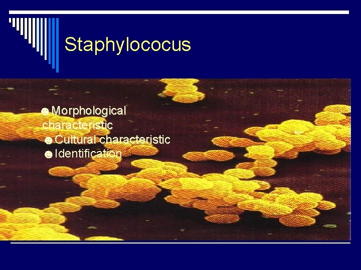
Staphylococus ☻Morphological characteristic ☻Cultural characteristic ☻Identification
Specimen: o Pus o wounds o sputum o urine o Faeces o C. S. F. o nasopharyngeal swabs o
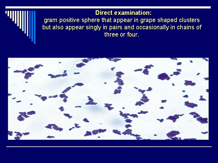
. Direct examination: gram positive sphere that appear in grape shaped clusters but also appear singly in pairs and occasionally in chains of three or four.

Culture: The primary isolation medium is blood agar after 12 -18 hours incubation at 37ºC large opaque slightly convex colonies appear that have a smooth and may be white creamy or yellow they may be surrounded by zone of hemolysis.
Mannitol salt and DNase test agars are used in the clinical laboratory to differentiate the pathogenic Staphylococcus spp. which ferment mannitol from the nonpathogenic Micrococcus spp. The pathogen most often encountered is Staphylococcus aureus that causes common food poisoning, osteomyelitis, bacterial pneumonia, and other diseases. The coagulase test is used to distinguish between pathogenic and nonpathogenic members of the genus Staphylococcus. All pathogenic strains of S. aureus are coagulase positive whereas the nonpathogenic species (S. epidermidis) are coagulase negative. The coagulase increases virulence by surrounding those bacteria that produce it with a clot that protects the bacteria from phagocytosis and antibodies.
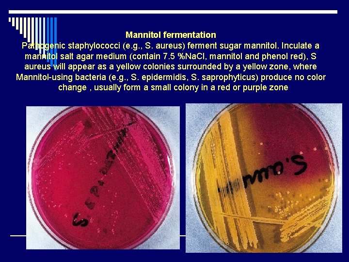
Mannitol fermentation Pathogenic staphylococci (e. g. , S. aureus) ferment sugar mannitol. Inculate a mannitol salt agar medium (contain 7. 5 %Na. Cl, mannitol and phenol red), S aureus will appear as a yellow colonies surrounded by a yellow zone, where Mannitol-using bacteria (e. g. , S. epidermidis, S. saprophyticus) produce no color change , usually form a small colony in a red or purple zone

DNase test flood the DNase test agar plate with 1 N HCl. A zone of clearing around the colony indicates a positive DNase test (i. e. , the DNA in the medium has been degraded)
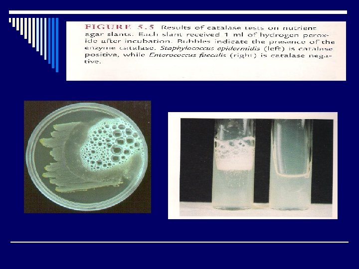
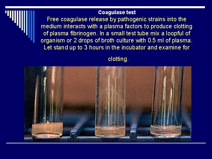
Coagulase test Free coagulase release by pathogenic strains into the medium interacts with a plasma factors to produce clotting of plasma fibrinogen. In a small test tube mix a loopful of organism or 2 drops of broth culture with 0. 5 ml of plasma. Let stand up to 3 hours in the incubator and examine for clotting.
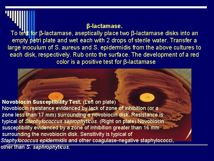
β-lactamase. To test for β-lactamase, aseptically place two β-lactamase disks into an empty petri plate and wet each with 2 drops of sterile water. Transfer a large inoculum of S. aureus and S. epidermidis from the above cultures to each disk, respectively. Rub onto the surface. The development of a red color is a positive test for β-lactamase Novobiocin Susceptibility Test. (Left on plate) Novobiocin resistance evidenced by lack of zone of inhibition (or a zone less than 17 mm) surrounding a novobiocin disk. Resistance is typical of Staphylococcus saprophyticus. (Right on plate) Novobiocin susceptibility evidenced by a zone of inhibition greater than 16 mm surrounding the novobiocin disk. Sensitivity is typical of Staphylococcus epidermidis and other coagulase-negative staphylococci, other than S. saphrophyticus.
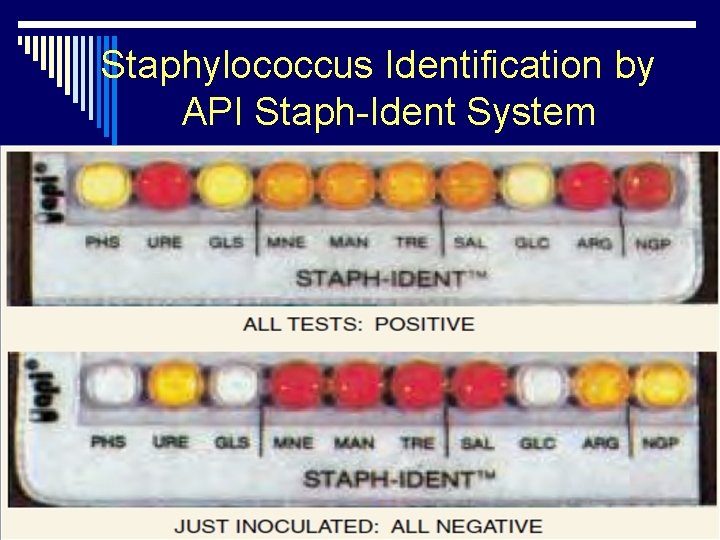
Staphylococcus Identification by API Staph-Ident System
- Slides: 12