STANLEY ROBBINS MD in his office Boston University

STANLEY ROBBINS, MD, in his office, Boston University School of Medicine in 1978

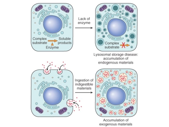
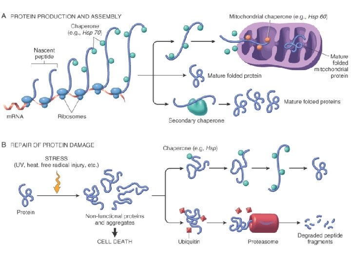
Intracellular accumulations of endogenous or exogenous substances - Lipids: fat may accumulate in the liver as fatty change - Proteins: abnormal protein accumulation is often irreversible. - Glycogen: glycogen storage diseases - Complex Lipids: sphinglolipidoses and other lipid accumulations

Intracellular accumulations of endogenous or exogenous substances - Complex carbohydrates: mucopolysaccharidoses and other complex carbohydrate diseases. - Minerals: iron, as hemosiderin, or carbon, as anthracotic pigment - Pigments: lipofuscin is a benign brown pigment of lipid origin that may accumulate with age, or melanin from melanomas, or bilirubin as in jaundice
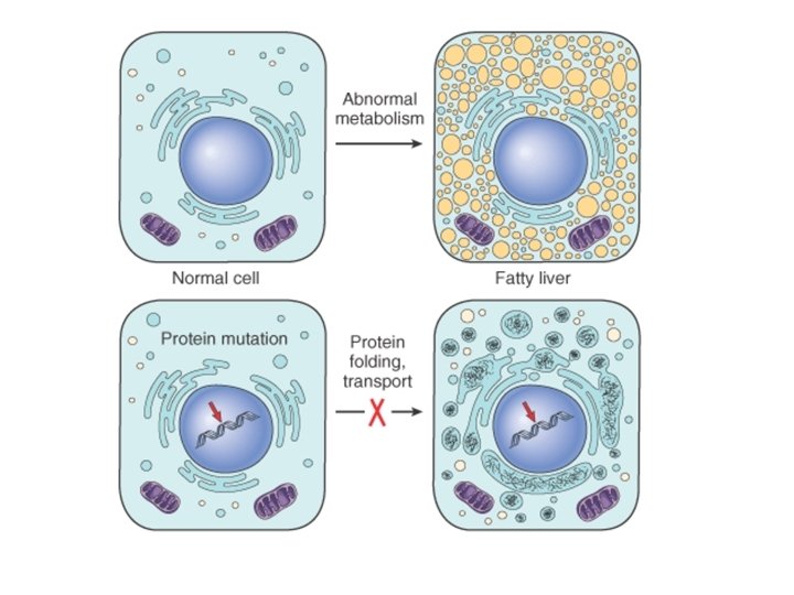

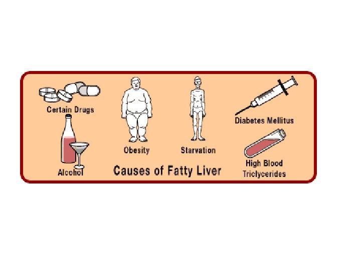
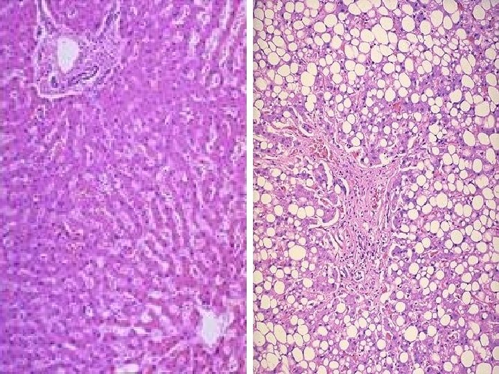
Fatty Change • Hepatic lipid accumulation is characterized by intracellular accumulation of triglycerides, and due to the failure of metabolic removal. • Defects in fat metabolism are often induced by alcohol consumption, and also associated with diabetes, obesity, and toxins. • Fatty change is most often seen in the liver (and heart), and is generally reversible.
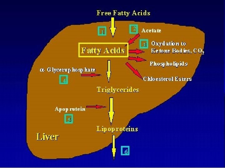

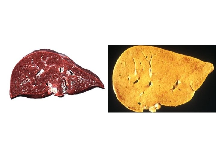
FATTY CHANGE – Gross features • hepatomegaly • pale, yellow color • greasy appearance

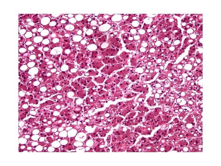
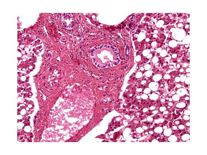
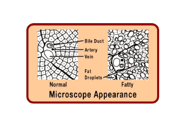
FATTY CHANGE – Microscopic features • Fat vacuoles coalesce and displace the nucleus to the periphery of the cell • vacuoles appear clear, with well-defined edges • lipid accumulations must be distinguished from accumulations of water or glycogen, using special preparation and stain – Oil Red. O.

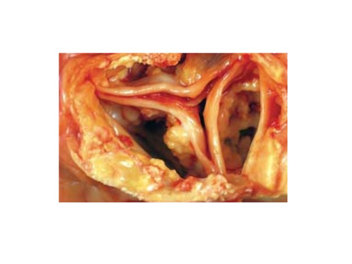
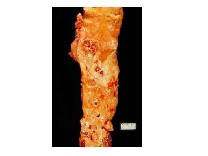
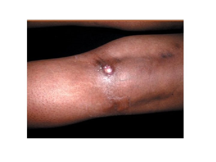
Calcification • Dystrophic calcification – Abnormal calcium deposition in dead or degenerating tissues • In areas of necrosis • Atheromas of advanced atherosclerosis

Calcification Metastatic calcification – Abnormal calcium deposition in “normal” tissues secondary to hypercalcemia (in soft tissues that are not the site of previous damage )

Metastatic Calcification – Hyperparathyroidism – Destruction of bone tissue – Vitamin D related disorders – Renal failure

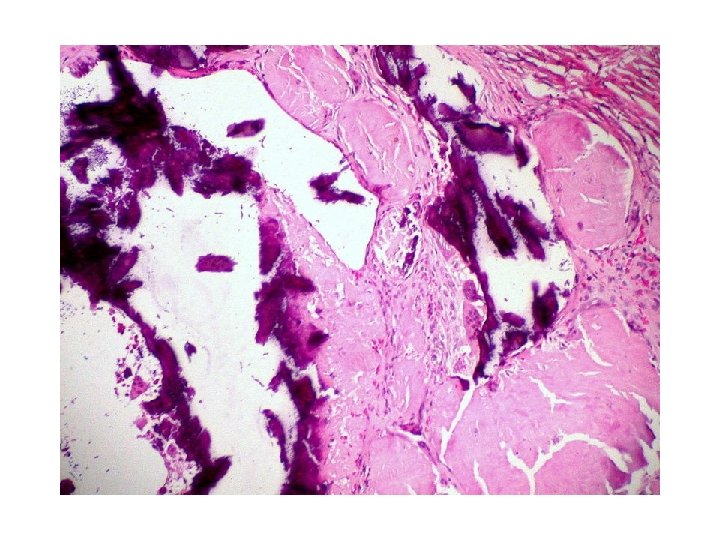
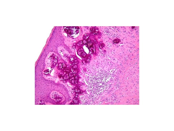
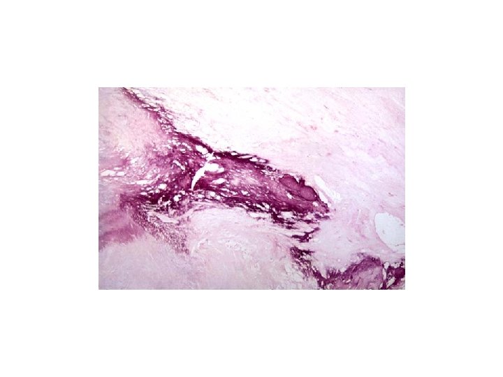
Morphology With H&E stain : basophilic amorphous granular appearance Von Kossa stain – black deposits
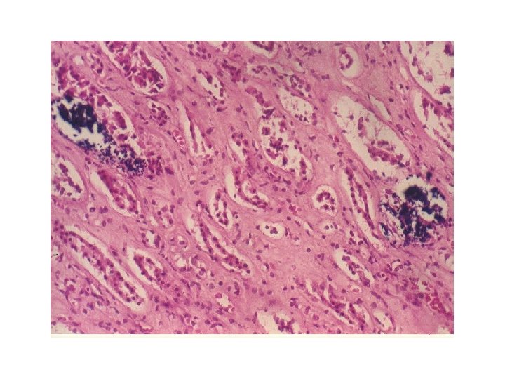
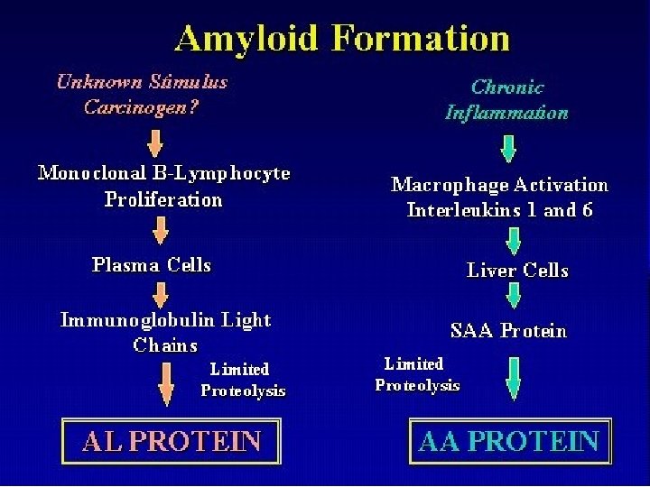
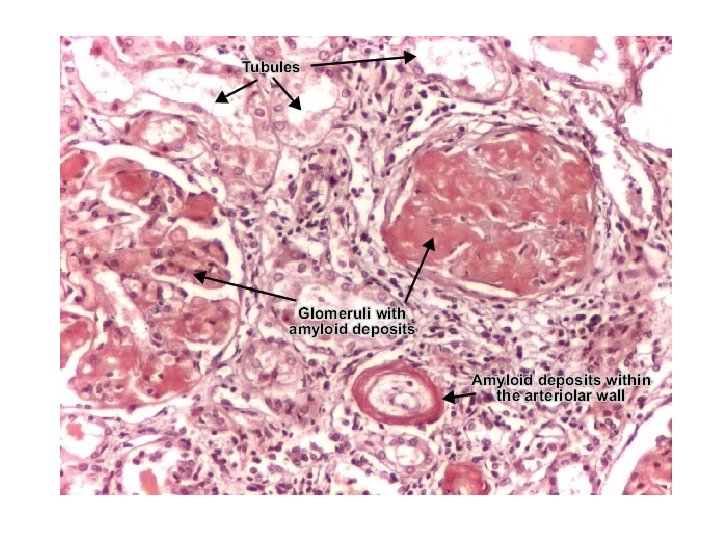
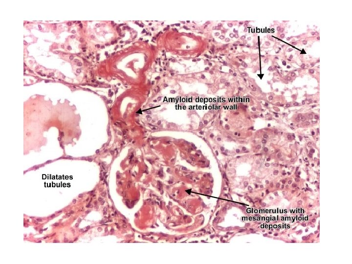

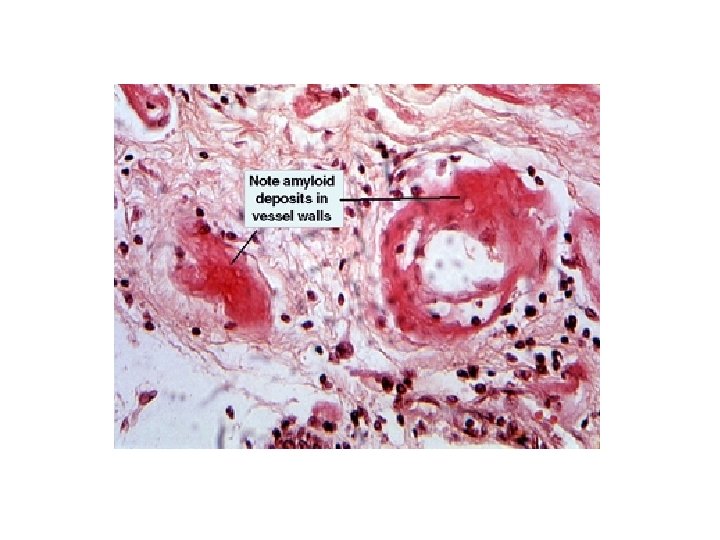
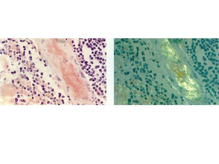
Amyloid Congo red-stained amyloid shows light green birefringence when subjected to polarized light.

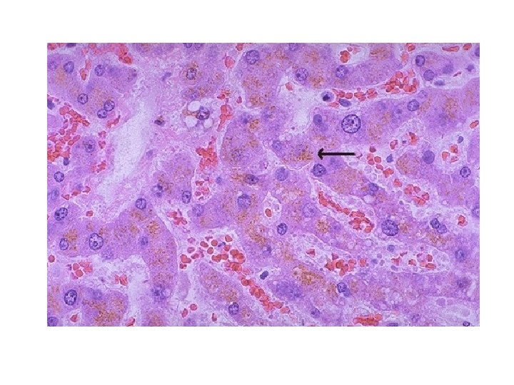
Lipofuscin • “Wear & tear” pigment • derived through lipid peroxidation of polyunsaturated lipids of subcellular membranes • accumulates in tissues undergoing slow, regressive changes – common in liver and heart of aging patients or patients with severe malnutrition and cancer cachexia • appears as a yellow-brown, finely granular, intracytoplasmic (or perinuclear) pigment
 Brown discoloration of intestinal muscle due to vitamin E")
Lipofuscin/ceroid (Latin: fuscus=brown, “brown lipid”) Brown discoloration of intestinal muscle due to vitamin E deficiency and smooth muscle lipofuscinosis


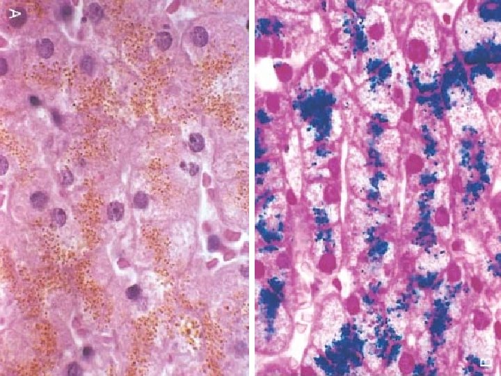
Haemosiderin • • derived from hemoglobin golden yellow, granular or crystalline pigment storage form of iron forms in response to local or systemic excess of iron ferritin forms hemosiderin granules • local excess: from gross or minute hemorrhage (eg. Bruise) • systemic excess: from increased absorption of dietary iron, impaired use of iron, hemolytic anemia, transfusions

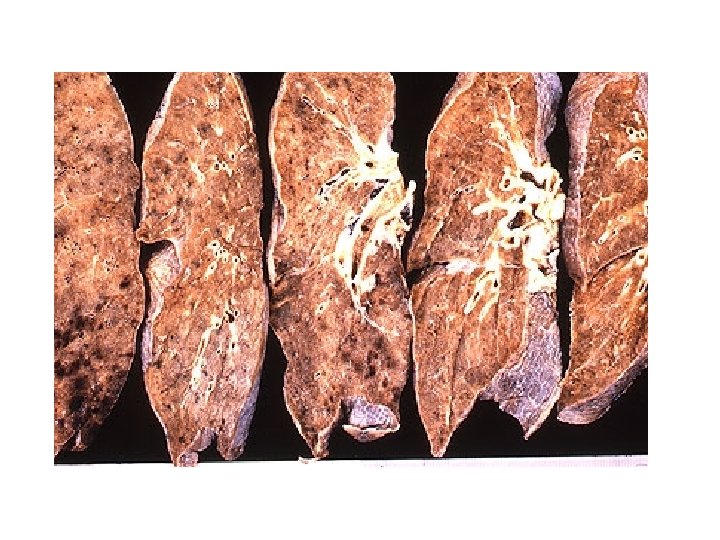
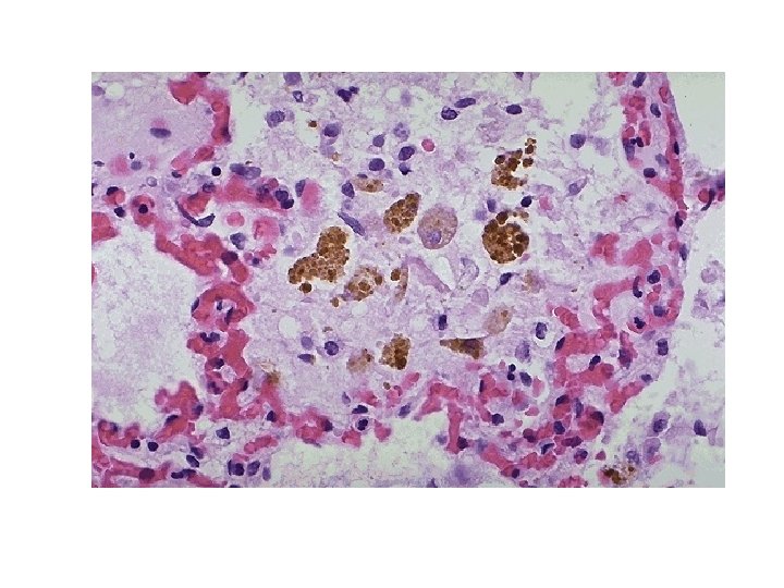
Hemosiderin Chronic passive congestion with hemosiderosis and edema in a lung

Hemosiderin Chronic passive congestion with hemosiderin in alveolar macrophages

PRUSSIAN BLUE

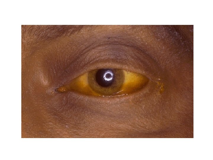
Bilirubin
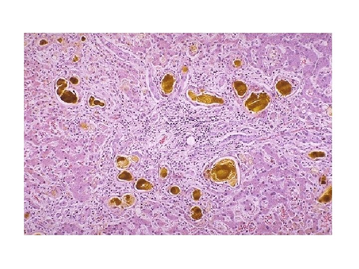
 casts in hepatic canaliculi in chronic liver disease")
Bilirubin (bile) casts in hepatic canaliculi in chronic liver disease
")
Melanin (Greek, melas=black)
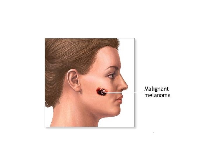
 Melanin in melanoma tumor cells")
Melanin (Greek, melas=black) Melanin in melanoma tumor cells
- Slides: 52