Spore stain Acid fast stain Spore staining SchaefferFulton

 Principle Ø Bacteria in genera such as Bacillus and Clostridium")


 Principle Ø A few species of bacteria in the")


- Slides: 13
Spore stain & Acid fast stain
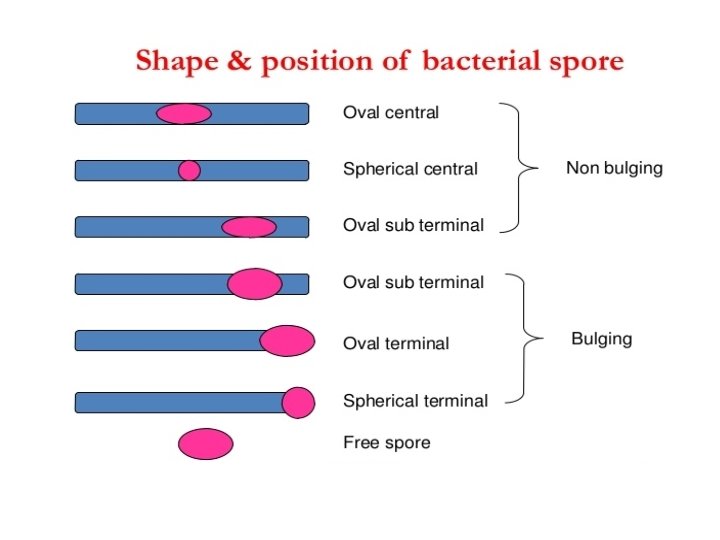
Spore staining (Schaeffer-Fulton method) Principle Ø Bacteria in genera such as Bacillus and Clostridium produce a quite resistant structure capable of surviving for long periods in an unfavorable environment and then giving rise to new bacterial cell. Ø This structure is called an endospore since it develops within the bacterial cell. Ø The location and size of endospore vary with the species
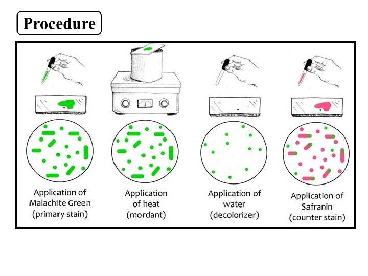
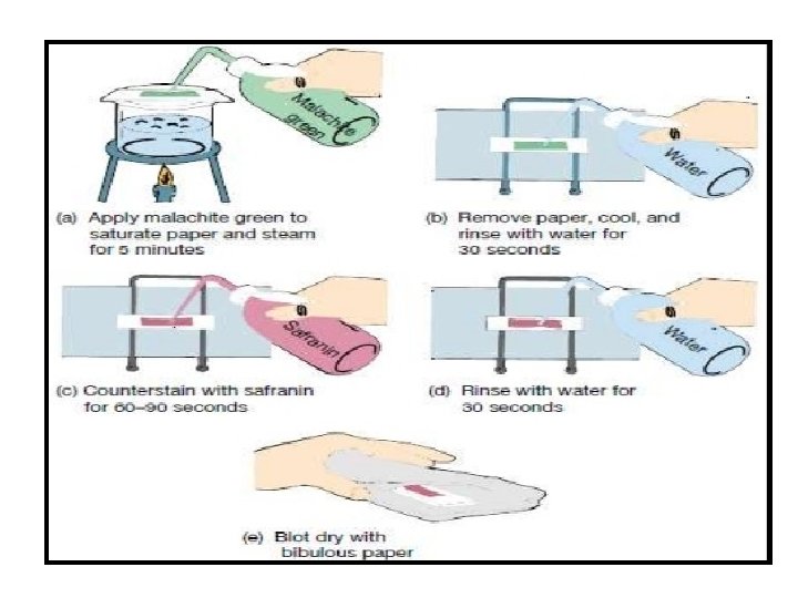
Ø Endospore do not stain easily but , once stained they strongly resist decolorization. Ø The endospore are stained with malachite green , heat is used to provide stain penetration. Ø The rest of the cell is then decolorized and counter stained a light red safranin.
Bacillus subtilus
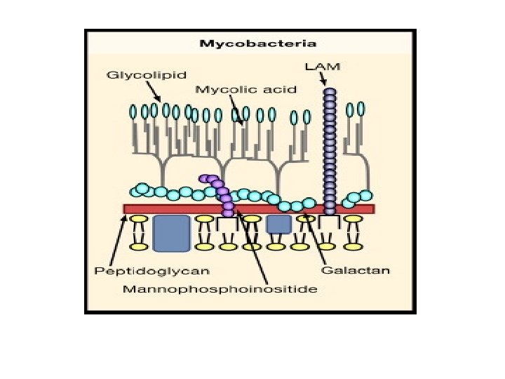
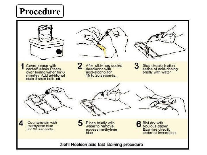
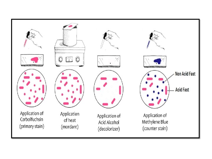
Acid fast staining (Ziehl-Neelson stain) Principle Ø A few species of bacteria in the genera Mycobacteria and Nocardia do not readily stain with simple stain , however these microorganisms can be stained by heating them with carbolfuchsin. Ø The heat drives the stain in to the cell once the microorganisms have taken up the carbolfuchsin , they are not easily decolorized by acid-alcohol and hence are termed acid fast. Ø This acid fasten is due to the high lipid content (mycolic acid) in the cell wall these microorganisms.
Ø Microorganisms that are not acid fast , termed non acid fast appear (Blue due) staining with methylene blue after they have been decolorized by the acid alcohol.
Mycobacterium tuberculosis Escherichia coli