
Spontaneous intracranial haemorrhage A rare presentation of Arachnoid

- Slides: 9
Spontaneous intracranial haemorrhage : A rare presentation of Arachnoid cyst - Dr. Chandra Raychaudhari
Clinical Presentation • A 14 -year-old boy presented with acute onset headache and three episodes of vomiting followed by brief loss of consciousness. • There was no history of trauma or head injury. No other co-morbid conditions. • Patient was advised plain and contrast CT scan and MRI.
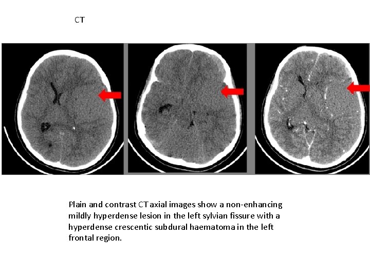
CT Plain and contrast CT axial images show a non-enhancing mildly hyperdense lesion in the left sylvian fissure with a hyperdense crescentic subdural haematoma in the left frontal region.
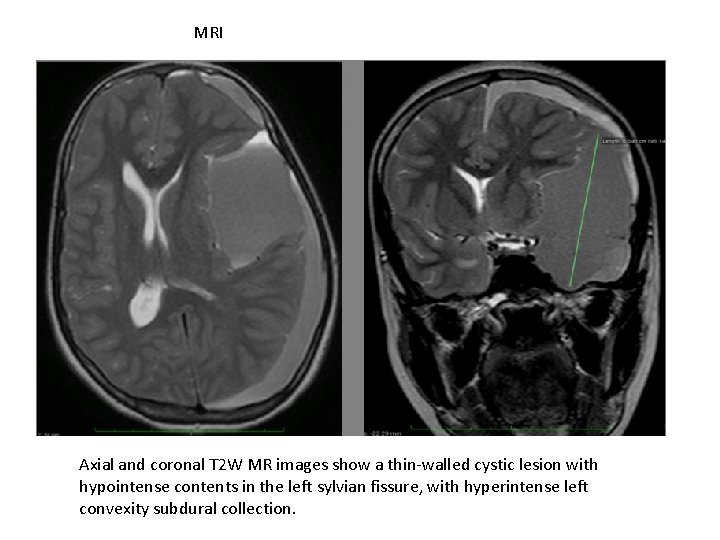
MRI Axial and coronal T 2 W MR images show a thin-walled cystic lesion with hypointense contents in the left sylvian fissure, with hyperintense left convexity subdural collection.
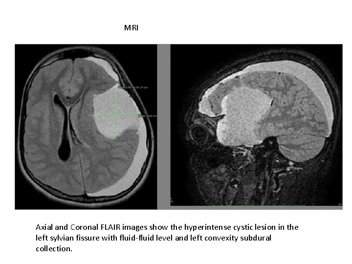
MRI Axial and Coronal FLAIR images show the hyperintense cystic lesion in the left sylvian fissure with fluid-fluid level and left convexity subdural collection.
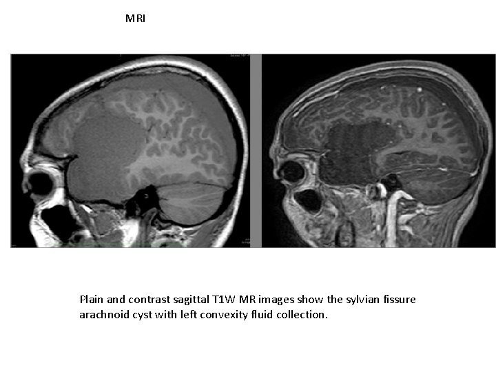
MRI Plain and contrast sagittal T 1 W MR images show the sylvian fissure arachnoid cyst with left convexity fluid collection.
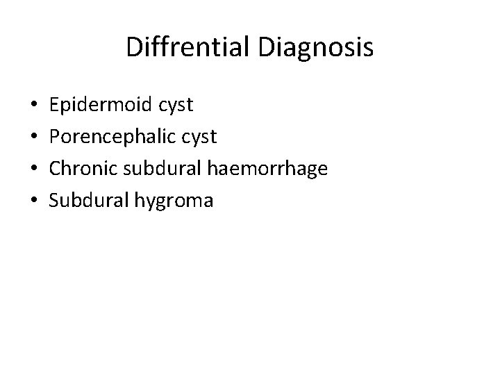
Diffrential Diagnosis • • Epidermoid cyst Porencephalic cyst Chronic subdural haemorrhage Subdural hygroma
Conclusion • Arachnoid cysts demonstrate non-enhancing, well circumscribed, non-calcified, and extraaxial cystic mass lesions on CT or MR. • Arachnoid cysts constitute 1 % of intracranial space occupying lesions, and their detection has increased with the use of CT and MRI.
THANK YOU