Spleen Largest lymphoid organ located on the left


 at high power (40 x) sinus cord sinus U-M Histology Collection")


- Slides: 16
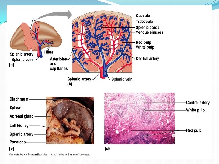
Spleen Largest lymphoid organ located on the left side of the abdominal cavity beneath diaphragm, it covered by the visceral peritoneum, below it thick capsule of dense irregular c. t contains some smooth muscle fibers, these capsule has trabeculae that extend inward and contains ophages. . It is served by spleenic artery and vein, which enter and the hilus.
Spleen
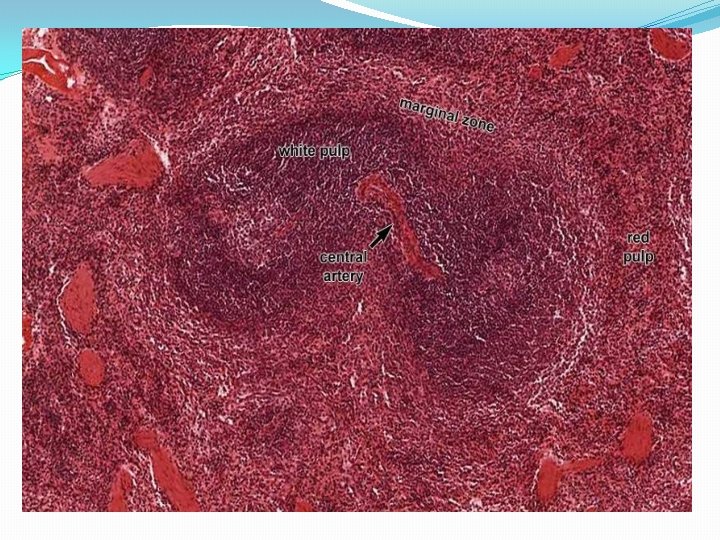
Histological Structure 1 -White pulp with lymphoid tissue. � 2 -Red pulp found between white pulp and trabeculae. � 3 -Marginal zone forms border between red and white � pulp.
White pulp _Is covered of branches of arteries central artery and diffused lymphatic tissue, form irregular masses around central artery this area is called (periarterial lymphatic sheath) (PALS), in PALS- T lymphocytes. ymphoid follicles comprise mostly B lymphocyte cells. _Reticular fibers. _W. p is also composed of dense aggregation of lymphocytes as nodules some with germinal center. marginal zone.
Marginal zone The periphery of lymphatic sheath and nodules, is � surrounded by a marginal zone that separates the white and red pulp. The marginal zone contains plasma cells, mainly B- � lymphocytes, macrophages and interdigitating, dendritic cells and marginal blood sinuses. This area play role in (immune response and filtering the � blood).
Red Pulp There are two components in the red pulp: _Spleenic cords of billroth. _Tortuous blood sinuses(venous sinuses). Spleenic cords of billroth: These vary in thickness and consist of a spongy cellular mass supported by reticular fibers. the collage nous trabeculae are continuous with the reticular fibers of the pulp. the lymphatic tissue is organized as cords or strands, contains large no. of RBC, lymphocytes, granular leukocytes, , embedded in meshwork of reticular t.
Venous Sinuses: Are large with wide irregular lumen, 12 -40 Mm wide. . sinuses occupy more space than the splenic cords. The walls of sinuses lack a muscular coat and display a rangement of endothelium and basal lamina. they have elongated fusiform flattened endothelial cells called stave cells. Discontinuous basal lamina, supported by (thick reticular fiber), there are space between endothelial cells permit exchange between sinusoid and adjacent tissue.
Spleen (red pulp) at high power (40 x) sinus cord sinus U-M Histology Collection
Splenic blood circulation Spleen is inserted in blood circulation, has special vascular channels in order to filter the blood and special types of blood circulation: -Closed blood circulation _Opened blood circulation (in human). The splenic arteries enter spleen through hilum, follow the trabeculae as trabecular artery, then in filtered by a sheath of lymphocytes in white pulp as central artery it terminate into penicillar arteries in to 3 -segments.
Pulp arterioles, sheathed arterioles, terminal arterial capillary. Then the blood is carried to red pulp directly then to s opened circulation. When terminal arterial capillaries opened in venous sinuses then to vein (trabecular vein) and leave spleen by splenic vein through hilum, this circulation is closed circulation.
Functions of spleen: _ Filtration of blood –removal of antigenic material and cellular debris by macrophages and dendritic cells, concentrated and presented to lymphocytes in the white pulp. _ Lymphocyte activation-both T and B lymphocytes are activated in the spleen, plasma cells migrate from the white pulp in to the red where they secrete IG in to the venous blood. _Destruction of olddamaged RBCs- phagocytosed by s broken down.
Thymus Central lymphoid organ, located in the mediastinum, thin capsule, lobular organization. Each lobule has outer cortex and inner medulla, cortex contains densely packed lymphocytes and scattered macrophages, medulla contain fewer lymphocytes, thymic (Hassall's) corpuscles and epithelial reticular cells.
Thymus Gland
Cortex Is the outer layer of thymus darkly stained due to dense aggregation of T lymphocytes (thymocytes). They don’t aggregates as nodules, in cortex macrophages ; epithelial reticular cells. _Epithelial reticular cells: Have acidophilic cytoplasm with processes that attached to the processed of other cells by desmosomes, they secret peptide hormone (thymosin, thymopoietin, serum thymus factor. They control T-cells production, regulate development and maturation of T-cells.