SPLEEN Dr Tabrez It is a haemolymph organ

SPLEEN Dr Tabrez

• It is a haemo-lymph organ • In fetal life- produces RBC In adult life- replaces worn out RBC & produces lymphocytes. Major repository of phagocytic macrophages

• Situation; – Left hypochondrium – Part of epigatrium • Axis; Lies oblique Axis coincides with Lt. 10 th rib
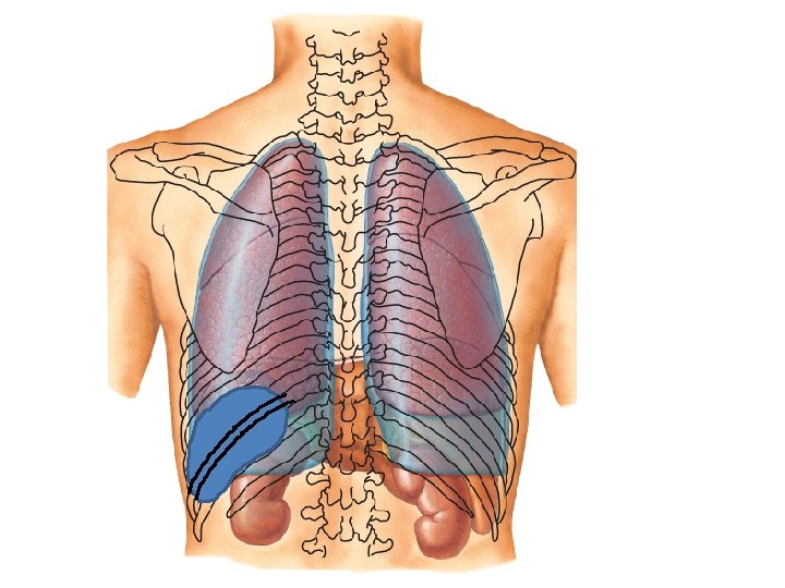

Lt 9 th rib Lt 10 th rib

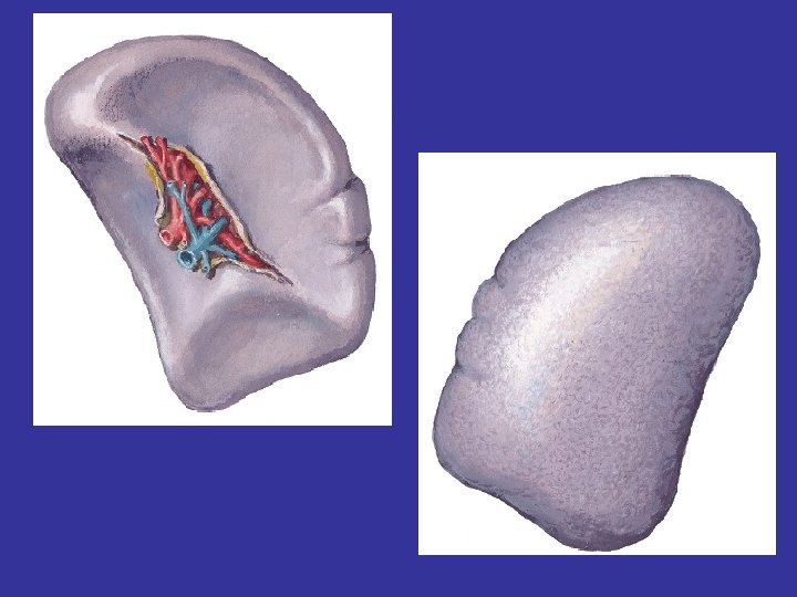
Shape: Oblong when colic impression is prominent Tetrahedral When colic imp. is absent Dimensions: Harris’s Dictum of odd numbers (1, 3, 5, 7, 9 to 11)

• 1” thickness • 3” breadth • 5” length • 7 oz weight • 9 to 11 ribs of Lt side- position of spleen • Left 10 th rib- coincides with axis of spleen

Characteristics • Highly vascular • Friable, elastic & pink in color • Moves with respiration • Not essential for life

Presenting parts • 2 ends- Medial Lateral • 2 surfaces- Diaphragmatic visceral • 2 Borders- superior inferior (intermediate border) • 2 angles- Anterior basal Posterior basal

External features of the spleen.

Types of spleen according to its shape.

Medial end Posterior extremity • Blunt & rounded • Situated – towards vertebral column • Lies opposite to T 10 spine, 3. 5 to 4 cm to the left of mid dorsal line

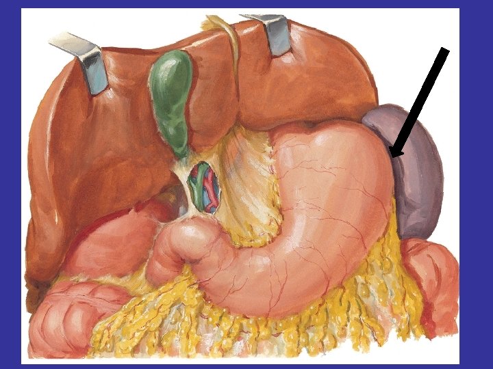
Lateral End Anterior Extremity • Broad – represented by a border • Extend from superior border to inferior border • Related with Lt colic flexure & phrenicocolic lig.

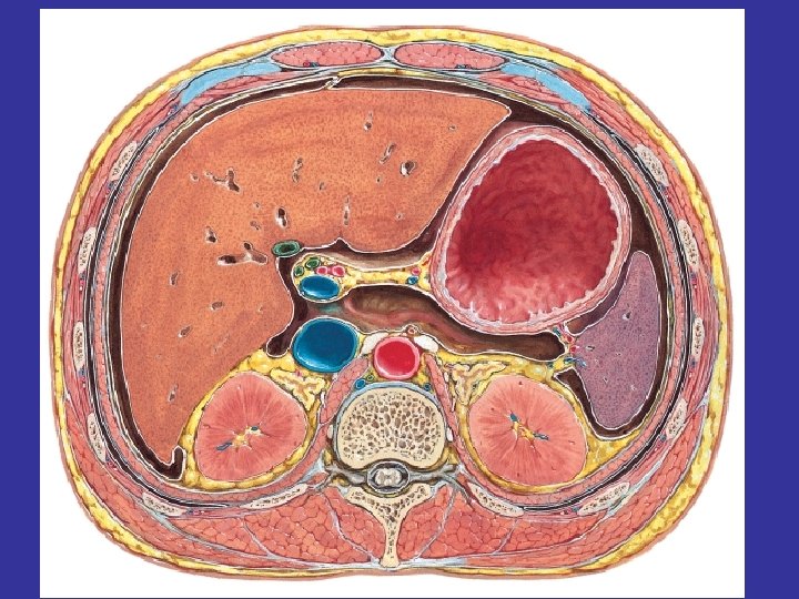
Diaphragmatic surface • Smooth & convex • Relations • Diaphragm- separated by recess of greater sac of peritoneum • Lower border of Lt lung & Lt pleural sac- in upper 1/3 • Costo-diaphragmatic recess- in lower 1/3 • 9 th to 11 th ribs of Lt side with IC spaces
 Intercostal space")
Diaphragm 9 to 11 ribs (left) Intercostal space

Visceral Surfaces • It has 4 impressions- gastric, renal, colic & pancreatic • Gastric Impression: Shallow, concave fossa Directed upward, forward & medially Hilum of spleen – lies in its lower part

GASTRIC IMPRESSION PANCREATI CIMPRESSIO N RENAL IMPRESSION COLIC IMPRESSION

Gastric Impression • Relations • Fundus of stomach- separated by recess of greater sac • Hilum- gives attachment to gastrosplenic lig. & lieno-renal lig.

Renal Impression • Anterior surface of left kidney • Sometimes- Left supra renal gland • Above structures are separated from spleen by recess of greater sac.

Colic Impression • Situated infront of lateral end • Left colic flexure Pancreatic Impression • Lies b/w colic impression & lateral end of hilum • Tail of pancreas b/w layers of lieno-renal ligament

Superior Border • Separates gastric impression from diaphragmatic surface • Close to lateral end – has 2 or 3 notches – Which indicates lobulated development of spleen Inferior Border • Separates renal impression from diaphragmatic surface

Anterior Basal angle Posterior Basal angle

Anterior Basal angle • Junction b/w superior border & lateral end • Lies in 9 th intercostal space or slightly behind left mid-axillary line • When spleen is enlarged – anterior basal angle can be palpated under left costal margin- hence it is clinical angle of spleen.

Posterior Basal angle • Junction of inferior border and lateral end of spleen

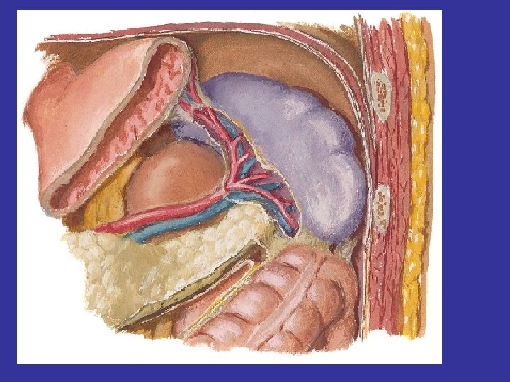
Ligaments of spleen • Gastrosplenic ligament • Lieno-renal ligament • Lieno-phrenic ligament • Phrenico-splenic ligament

Gastrosplenic Ligament • Bi-layered peritoneal fold • Extent – fundus of stomach to anterior lip of hilum of the spleen • Anterior layer- from greater sac- continuous with posterior layer of lieno-renal ligament • Posterior layer- from lesser sac- continuous with anterior layer of lieno-renal ligament. • Contains- short gastric vessels and left gastroepiploic vessels

Gastro splenic ligament Lieno renal ligament Phrenic colic ligament

Gastrosplenic Lig Spleen Lienorenal Lig

Lieno-renal ligament • Bi-layered peritoneal fold • Extent- posterior lip of hilum of the spleen to anterior surface of left kidney • Anterior layer- derived from lesser sac • Posterior layer- derived from greater sac • Contains- Splenic vessels, nerves & lymphatics & sometimes tail of pancreas

Lieno-phrenic Ligament • Upward continuation of lieno-renal ligament • Connects hilum of spleen to diaphragm • Suspends the spleen from above. Suspensory ligament of spleen

Phrenico-colic Ligament • Triangular peritoneal fold • Extends- left colic flexure to diaphragm opposite left 10 th or 11 th rib • It suspends the spleen from belowsustentaculum lienis

SEGMENTS OF THE SPLEEN • The splenic artery within the spleen usually gives two branches—superior and inferior. • These branches usually do not anastomose and each branch supplies its own territory(segment). • Thus, an avascular zone exists between these two territories.

• Thus, the spleen presents two segments, superior and inferior, separated by an avascular plane passing perpendicular to the long axis of the spleen. • The knowledge of these vascular segments is essential for segmental resection of the spleen to preserve the splenic tissue if required. .

DEVELOPMENT OF SPLEEN • The spleen develops between the two layers of the upper part of the dorsal mesogastrium from a number of condensations of mesenchymal cells. • These separate masses of mesenchymal cells (called splenunculi) fuse together to form the lobulated spleen. • The notches on the superior border of the adult spleen indicate the lobulated development of the organ.

ACCESSORY SPLEENS • The failure of fusion of splenunculi results in the formation of accessory spleens. • These are usually found in the derivatives of the dorsal mesogastrium, viz. : (a) in the gastrosplenic ligament, (b) in the lienorenal ligament, and (c) in the greater omentum.

• Arterial Supply. Splenic artery branch of celiac artery • Venous drainage- Splenic vein • Lymphatic drainage- pancreatico-splenic nodes • Nerve supply- Celiac plexus (symphathetic)

Functions of spleen • Filtration of unwanted elements from the blood by phagocytosis • Activation of T and B lymphocytes • Haemopoiesis and Lymphopoiesis

Clinical Correlates • While doing splenectomy care should be taken to avoid injury to the tail of the pancreas – that lies close to hilum of the spleen • Phrenico colic ligament prevents the direct downward displacement incase of enlargement of spleen • Kehr’s sign- obstruction of smaller branches (end arteries) of splenic artery – splenic infarction- causes referred pain to left shoulder.
 occurs in number of diseases. •")
Splenomegaly: • The enlargement of the spleen (splenomegaly) occurs in number of diseases. • The spleen may increase in size by as much as tenfold (massive splenomegaly). • Common causes of massive splenomegaly are: (a) malaria, (b) cirrhosis of liver, (c) chronic myeloid leukemia, and (d) kala-azar. • The very large spleen projects downward and medially toward the right iliac fossa in direction of the axis of the 10 th rib.
 is sometimes performed when the")
Splenectomy: • The splenectomy (surgical removal of the spleen) is sometimes performed when the spleen is ruptured or inadvertently nicked at operation. • It is also performed in the treatment of certain blood diseases. • The removal of spleen does not impair the immune response seriously. .

- Slides: 45