splanchnocranium Consists of part of skull that is


- Slides: 22
splanchnocranium - Consists of part of skull that is derived from branchial arches - The facial bones are the bones of the anterior and lower human skull
Bones n n n n n Ethmoid bone Inferior nasal concha Lacrimal bone Maxilla Nasal bone Palatine bone Vomer Zygomatic bone Mandible
Ethmoid bone n n n n The ethmoid is a single bone, which makes a significant contribution to the middle third of the face. It is located between the lateral wall of the nose and the medial wall of the orbit and forms parts of the nasal septum, roof and lateral wall of the nose, and a considerable part of the medial wall of the orbital cavity. In addition, the ethmoid makes a small contribution to the floor of the anterior cranial fossa. The ethmoid bone can be divided into four parts, the perpendicular plate, the cribriform plate and two ethmoidal labyrinths. Important landmarks include: • Perpendicular plate • Cribriform plate • Crista galli. • Ala. • Ethmoid labyrinths • Medial (nasal) surface. • Orbital plate. • Superior nasal concha. • Middle nasal concha. • Anterior ethmoidal air cells. • Middle ethmoidal air cells. • Posterior ethmoidal air cells. Attachments The falx cerebri (slide) attaches to the posterior border of the crista galli.
n n n lamina cribrosa 1 crista galli 2 lamina perpendicularis 3 labyrinthi ethmoidales 4 cellulae ethmoidales anteriores et posteriores 5 lamina orbitalis 6 n n concha nasalis media 7 processus uncinatus 8
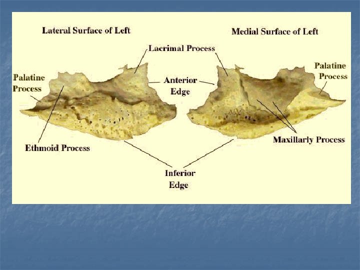
n n n Inferior nasal concha Each inferior nasal concha consists of a curved plate of bone attached to the lateral wall of the nasal cavity. Each consists of inferior and superior borders, medial and lateral surfaces, and anterior and posterior ends. The superior border serves to attach the bone to the lateral wall of the nose, articulating with four different bones. The anterior third of the superior border articulates with the conchal crest of the maxilla and the posterior third with the conchal crest of the perpendicular plate of the palatine bone. The middle third of the upper border shows three small processes: lacrimal, maxillary and ethmoidal processes. The concha’s longer inferior border lies free within the nasal cavity. It is thickened and often curves inwards. The inferior nasal concha exhibits the following landmarks: • Anterior end. • Posterior end. • Lateral surface. • Medial surface. • Ethmoidal process. • Lacrimal process. • Maxillary process.
Lacrimal bone n n n The two bones are small, thin and rectangular. They each lie in the anterior part of the medial wall of the orbit. Anteriorly, the lacrimal bone articulates with the frontal process of the maxilla. Posteriorly, the bone meets the orbital plate of the ethmoid. Superiorly, it joins the frontal bone and inferiorly, the maxilla. The lacrimal bone exhibits the following landmarks: • Nasal surface. • Orbital surface. • Posterior lacrimal crests. • Lacrimal grooves. • Fossa for lacrimal sac. • Lacrimal hamulus. • Descending process. Attachments The lacrimal part of the orbicularis oculi muscle arises from the lateral surface of the lacrimal bone.
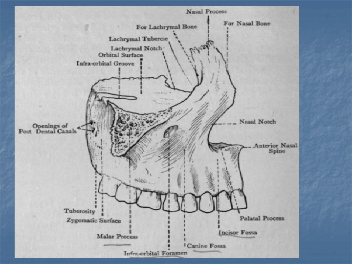
Maxilla n n n The maxillary bones occupy the central part of the face and support the teeth of the upper jaw. They contribute to much of the skeleton of the upper face including the nasal aperture, the bridge of the nose, the floor of the orbital cavities and the bones of the cheeks. Each bone consists of a body and four processes: the frontal, zygomatic, alveolar and palatine processes. The body forms the main bulk of the bone and possesses anterior, orbital, nasal and infratemporal (posterior) surfaces. The maxillary bones show many articulations. On the face, the maxillary bones articulate with each other, the nasal bones, the nasal cartilages and the frontal bone. Laterally, they articulate with the zygomatic bones. Each maxillary bone also joins with the vomer, the septal cartilage, the lacrimal bone, the ethmoid bone and the inferior nasal concha to contribute to the skeleton of the nasal fossa and the orbit. Attachments The maxilla provides attachment for buccinator, depressor septi, incisivus labii superioris, levator anguli oris, levator labii superioris, orbicularis oculi, and nasalis.
• The maxillary bone exhibits the following features: • • • • • Body • Infra-orbital foramen. • Infra-orbital groove. • Canine fossa. • Nasal notch. • Anterior nasal spine. • Alveolar canals. • Greater palatine groove. • Inferior meatus. • Maxillary hiatus. • Maxillary sinus. • Frontal process • Anterior lacrimal crest. • Agger nasi. • Ethmoidal crest. • Fossa for lacrimal sac. • • • • Lacrimal groove. • Conchal crest. • Middle meatus. • Zygomatic process • Alveolar process • Tuberosity. • Canine eminence. • Palatine process • Floor of nasal cavity. • Incisive canal. • Incisive fossa. • Nasal crest. • Alveolar ridges. • Depressions for palatine gland.
Nasal bone n n n n The two nasal bones form the upper part of the bridge of the nose. Each nasal bone is quadrilateral, being longer than it is wide. The nasal bone exhibits the following features: • Superior border. • Inferior border. • Medial border. • Lateral border. • Internal surface. • External surface. • Grooves for anterior ethmoidal nerve. • Vascular foramina. The superior border articulates with the nasal part of the frontal bone. The inferior border forms the superior boundary of the anterior nasal aperture. The lateral border meets the frontal process of the maxilla and the medial border meets its fellow in the midline. Attachments The procerus muscle is attached to the external surface of the nasal bone and the lateral nasal cartilage to the inferior border.
Palatine bone n n n The palatine bone is found at the back part of the nasal cavity between the maxilla and the pterygoid process of the sphenoid bone. It contributes to the floor and lateral wall of the nasal cavity, the roof of the mouth, and the floor of the orbit; it contributes to the formation of the pterygopalatine and pterygoid fossae; and one fissure, the inferior orbital fissure. The palatine bone is L-shaped, and consists of a horizontal plate and a vertical perpendicular plate and four processes: the pyramidal, orbital, maxillary and sphenoidal processes. The horizontal plate is a quadrilateral plate of bone that forms the posterior quarter of the hard palate when articulated with its fellow; it has a nasal surface and four borders: anterior, posterior, medial and lateral. The perpendicular plate is rectangular, the vertical dimension being approximately twice that of the antero-posterior dimension. It has two surfaces, the maxillary and nasal, and four borders: anterior, posterior, superior and inferior.
• The palatine bone exhibits the following features: • • • • Horizontal plate • Palatine surface. • Nasal surface. • Anterior border. • Posterior border. • Medial border. • Lateral border. • Palatine crest. • Posterior nasal spine. • Pyramidal process. • Lesser palatine foramina. • Perpendicular plate • Maxillary surface. • • • • Nasal surface. • Anterior border. • Posterior border. • Superior border. • Inferior border. • Pterygopalatine canal. • Palatovaginal canal. • Conchal crest. • Ethmoid crest. • Greater palatine foramina. • Greater palatine groove. • Maxillary process. • Sphenoidal process. • Orbital process. • Sphenopalatine notch.
Vomer n n The vomer is a single thin plate of bone, shaped like a ‘ploughshare’ and forms the postero-inferior portion of the nasal septum (slide). It possesses two lateral surfaces and four borders, anterior, inferior, superior and posterior. All borders accept the posterior border articulate with the adjacent bones. The posterior border is free and does not articulate with any other structures. It is slightly concave and slopes antero-inferiorly to form a prominent midline ridge between the two posterior nasal apertures.
n n n n n The vomer exhibits the following landmarks: • Ala. • Site of articulation with septal cartilage. • Site of articulation with ethmoid bone. • Site of articulation with maxilla and palatine bone. • Site of articulation with sphenoid bone. • Groove for nasopalatine nerve and vessels. • Groove for ethmoidal nerve. The anterior border is the longest border on the bone and slopes antero-inferiorly. It articulates with the nasal septal cartilage and with the perpendicular plate of the ethmoid bone. The inferior border lies anterior to the superior border and articulates with the median maxillary and palatine nasal crests. Finally, the superior border articulates with the vaginal process and the body of the sphenoid bone.
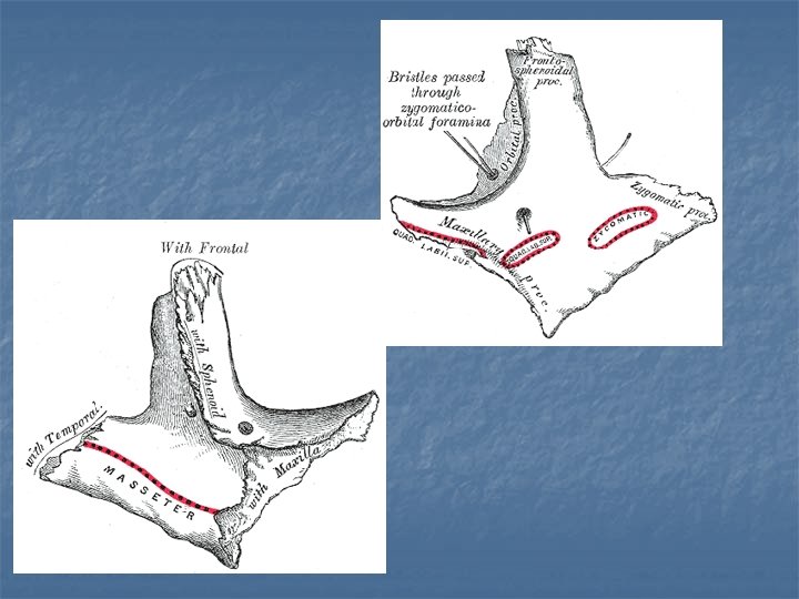
n n Zygomatic bone The two zygomatic bones form the skeleton of the cheeks. Each bone has: three surfaces; lateral (anterolateral), temporal (postero-medial) and orbital, five borders; orbital (antero-superior), maxillary (anteroinferior), temporal (postero-superior), postero-inferior and postero-medial and two processes, the frontal and temporal. The lateral surface is smooth and slightly convex. The entire surface of the zygomatic bone viewed medially is called the ‘temporal surface’. It can be subdivided into two regions. Anteriorly, it shows a roughened area for articulation with the zygomatic process of the maxilla. Posteriorly, the temporal surface includes the lower surface of the orbital plate and the temporal surface of the temporal process. This posterior region is smooth and forms the anterior boundary of the temporal fossa.
• The zygomatic bone exhibits the following landmarks: • • Body. • • Temporal process. • • Frontal process. • • Orbital surface (plate). • • Orbital border. • • Maxillary border. • • Temporal border. • • Postero-inferior border. • • Postero-medial border. • • Marginal tubercle. • • Zygomatico-orbital foramen. • • Zygomaticofacial foramen. • • Zygomaticotemporal foramen. • Attachments • The zygomatic bone provides attachment for the: • • Levator labii superioris. • • Masseter. • • Temporalis. • • Temporal fascia. • • Zygomaticus major. • • Zygomaticus minor.
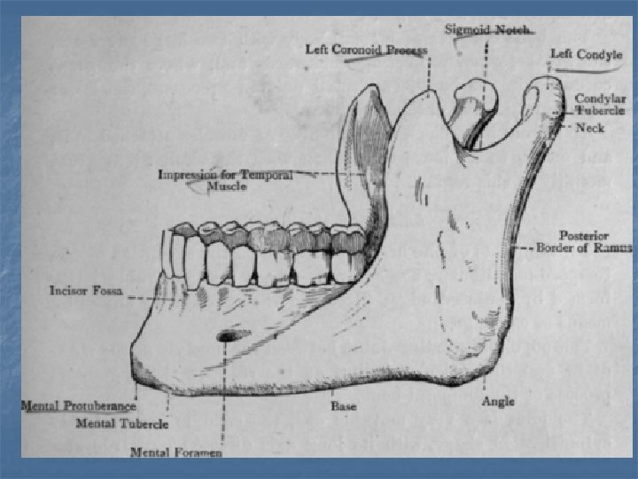
Mandible n The mandible consists of a horizontal, horseshoe-shaped body and two vertical rami. The body of the mandible supports the mandibular teeth within the alveolar processes. The rami of the mandible articulate with the temporal bones at the temporomandibular joints.
• The mandible exhibits • the following prominent • landmarks: • • • Body • • • Alveolar ridge (process). • • • Menti symphysis. • • • Mental protuberance. • • Mental tubercle. • • • Incisive fossa. • • Digastric fossa. • • • Inferior mental spine • (genial tubercle). • • • Superior mental spine • (genial tubercle). • • • Mylohyoid line. • • • Sublingular fossa. • • • Submandibular fossa. • Mental foramen. • Oblique line. • Rami • Coronoid process. • Condylar process. • Head of condylar process. • Neck of condylar process. • Pterygoid fovea. • Mandibular notch. • Anterior border. • Posterior border. • Lingula. • Mylohyoid groove. • Obtuse angle.
n n Attachments It gives attachments to anterior bellies of the digastric muscle, geniohyoid (slide), genioglossus, mylohyoid (slide), masseter, buccinator, medial and lateral pterygoid, mentalis, temporalis, incisivus labii inferioris, depressor anguli oris, platysma muscles and part of the orbicularis oris. It also gives attachment to the sphenomandibular and stylomandibular ligaments and the pterygomandibular raphe.