Spirochaetales Treponema Borrelia Leptospira General Overview of Spirochaetales

Spirochaetales Treponema Borrelia Leptospira

General Overview of Spirochaetales Ø Gram-negative Spirochete from Greek for “coiled hair” ØExtremely thin and can be very long Ø Tightly coiled helical cells with tapered ends Ø Motile by periplasmic flagella (a. k. a. , axial fibrils or endoflagella) Ø Outer sheath encloses axial fibrils wrapped around protoplasmic cylinder • Axial fibrils originate from insertion pores at both poles of cell • May overlap at center of cell in Treponema and Borrelia, but not in Leptospira • Differering numbers of endoflagella according to genus & species

Periplasmic Flagella Diagram

Tightly Coiled Spirochete OS = outer sheath AF = axial fibrils AF Leptospira interrogans
")
Cross section of Borrelia burgdorferi Cross-Section of Spirochete with Periplasmic Flagella (Outer sheath)

Genus Treponema T. pallidum Brachyspira paraluis- cuniculi Syphilis in Man Syphilis in Rabbit

Darkfield Microscopy of Treponema pallidum

General Characteristics of Genus Treponema Anaerobic or microaerophilic Corkscrew rods 5 -20 µm long Stains well with silver impregnation. Commensal of oral cavity, intestinal tract, and urinary tract. Don’t grow on artificial culture m

Rabbit syphilis Definition: It is a benign venereal disease of rabbits characterized by small vesicles, scabs or ulcers which heal in 10 - 14 days. Etiology: Brachyspira paraluis- cuniculi formely known as Treponema cuniculi


Diagnosis: Clinical picture. Sample: Skin scraping from the lesions. Discharges oozing from ulcers formed on genital organs. Direct film examined with dark ground microscopy short regular coils showing rotating undulating motility

Genus Borrelia Gram-negative, wide irregular helical coils approximately 10 - 30 µm in length. Stain well with Giemsa stain Microaerophilic & require complex growth medium Transmitted by arthropods (Tick and louse). Many are nonpathogenic and are commensals but may causes systemic infection in many species. Culture of borreliae from infected animal is confirmatory.

Avian spirochaetosis Definition: It is an infectious disease of many fowel species characterized by fever, cyanosis of the head & greenish diarrhea. Etiology: Borrelia anserine or Borrelia gallinarum

Clinical signs: Fever, emaciation & weight loss. Cyanosis of the head & greenish diarrhea. Marked anaemia & paralysis No relapses occur, the bird either die (mortality 100%) or recover completely. Postmortem lesions: Liver enlargement with necrotic foci. Splenomegaly with widespread hemorrhages

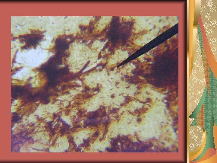
Diagnosis: Sampling: blood or infected tissues Brownish B. anserina. Yellowish background Film from blood & infected tissues stained with Giemsa, Gram or simple stains shows B. anserina Gram-negative, wide irregular spiral coils approximately 10 - 30 µm in length. With Fontana stain. anserina appear as brown wide irregular spiral coils against yellowish background.

Isolation and identification: Blood or suspected infected tissues as liver & Spleen is inoculated into: Yolk sac of 5 -6 days embryonated eggs B. anserina could be seen in blood or tissue under microaerophilic condition at 30 -35 ºC growth of B. anserina is monitored by dark field microscopy for 4 -6 weeks. Identification of B. anserina in infected tissue smears or cultures using immunofluorescence. Serologic detection of Ig. M or rising Ig. G by ELISA.

Genus leptospira General characteristic: Widely distributed in nature particularly in aquatic environment and animals. Tightly coiled spirals 5 - 15 μm in length with hooks at one or both ends. Shows Spiny undulating motility in liquid media. Leptospira differs from other spirochetes in lacking glycolipids and having diaminopimelic acid rather than ornithine in its eptidoglycan Requires vitamins B 1 & B 12, serum & long chain fatty acids for growth.

Leptospirosis Definition: It is a bacterial zoonotic disease caused by genus Leptospira that affects humans and a wide range of animals. Etiology: Leptospira species

Diagnosis: Leptospires can be demonstrated in tissue using dark field microscopy or fluorescent antibodies. Serology can be used to type serum for the presence of antibodies against different serovars. Microscopic agglutination test- most common test; serum dilutions are titered against live antigens. ELISA to detect anti-leptospira antibodies in serum. PCR
- Slides: 20