
Spinal Vascular Neurointervention Dr Adam Rennie Lysholm Department
- Slides: 62
Spinal Vascular Neurointervention Dr Adam Rennie Lysholm Department of Neuroradiology The National Hospital of Neurology and Neurosurgery Great Ormond Street Hospital for Children
Spinal Vascular Neurointervention • Arteriovenous Shunts – Pial – Dural • Tumours – Paraspinal – Spinal Column – Cord
Spinal angiography - technique • General anaesthetic • Selective injections of – – – – Vertebral arteries Thyrocervical trunks Costocervical trunks External carotid arteries Supreme intercostal trunks (usually T 2 -T 4) (Rt) Intercosto-bronchial trunk, Intercostal and lumbar segmental arteries T 5 -L 4 Median and lateral sacral arteries • Must be meticulous / persistent • Can take hours • Complication rate 0. 5 – 2%
Rotational 3 D Catheter angiography
PIAL AVS SPINE
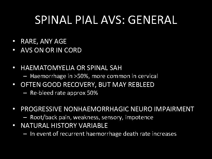
SPINAL PIAL AVS: GENERAL • RARE, ANY AGE • AVS ON OR IN CORD • HAEMATOMYELIA OR SPINAL SAH – Haemorrhage in >50%, more common in cervical • OFTEN GOOD RECOVERY, BUT MAY REBLEED – Re-bleed rate approx 50% • PROGRESSIVE NONHAEMORRHAGIC NEURO IMPAIRMENT – Root/back pain, weakness, sensory, impotence • NATURAL HISTORY VARIABLE – In event of recurrent haemorrhage death rate increases
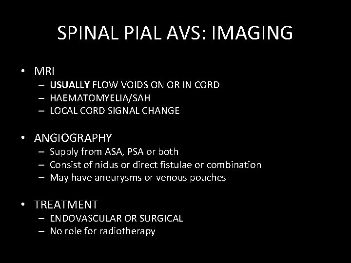
SPINAL PIAL AVS: IMAGING • MRI – USUALLY FLOW VOIDS ON OR IN CORD – HAEMATOMYELIA/SAH – LOCAL CORD SIGNAL CHANGE • ANGIOGRAPHY – Supply from ASA, PSA or both – Consist of nidus or direct fistulae or combination – May have aneurysms or venous pouches • TREATMENT – ENDOVASCULAR OR SURGICAL – No role for radiotherapy
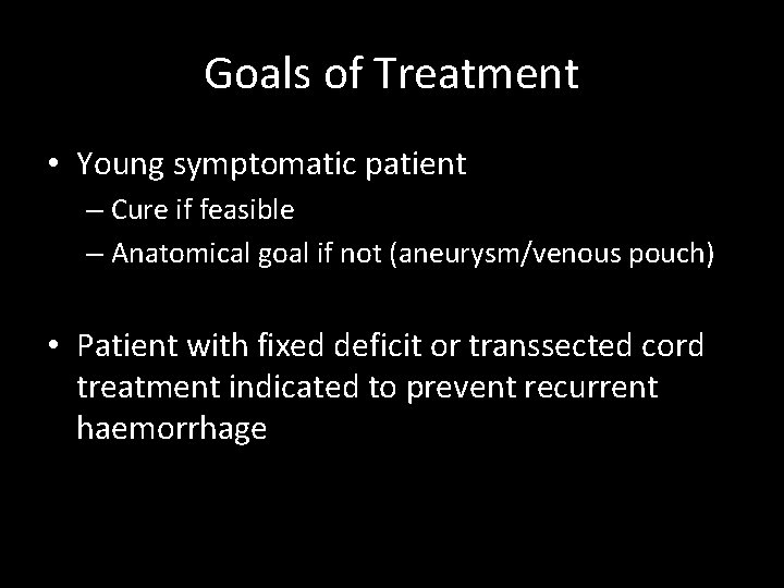
Goals of Treatment • Young symptomatic patient – Cure if feasible – Anatomical goal if not (aneurysm/venous pouch) • Patient with fixed deficit or transsected cord treatment indicated to prevent recurrent haemorrhage
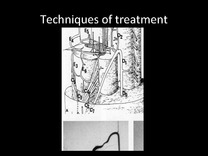
Techniques of treatment
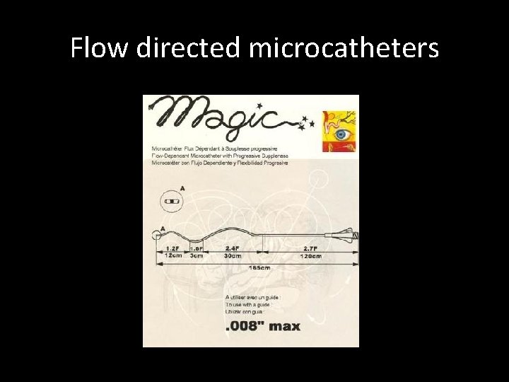
Flow directed microcatheters
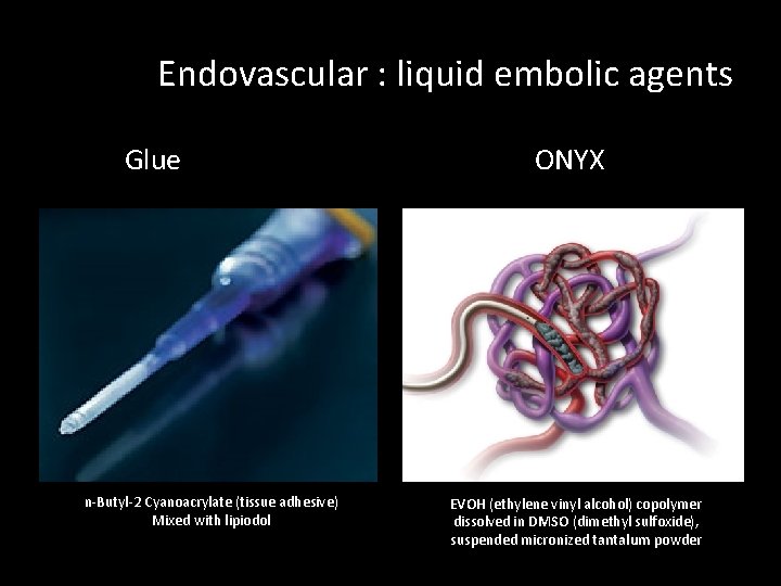
Endovascular : liquid embolic agents Glue n-Butyl-2 Cyanoacrylate (tissue adhesive) Mixed with lipiodol ONYX EVOH (ethylene vinyl alcohol) copolymer dissolved in DMSO (dimethyl sulfoxide), suspended micronized tantalum powder
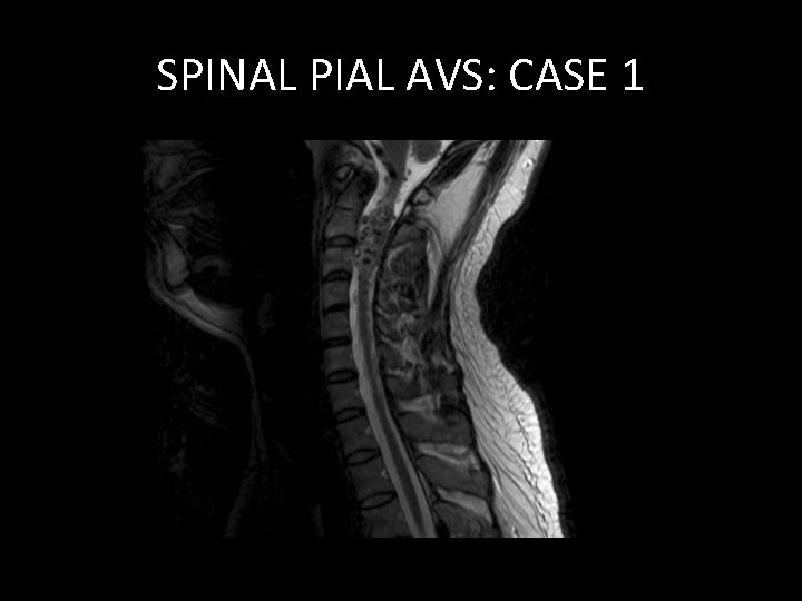
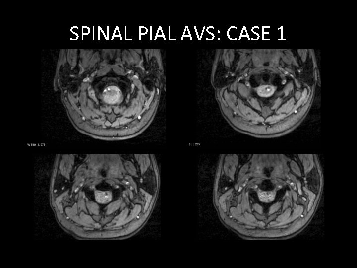
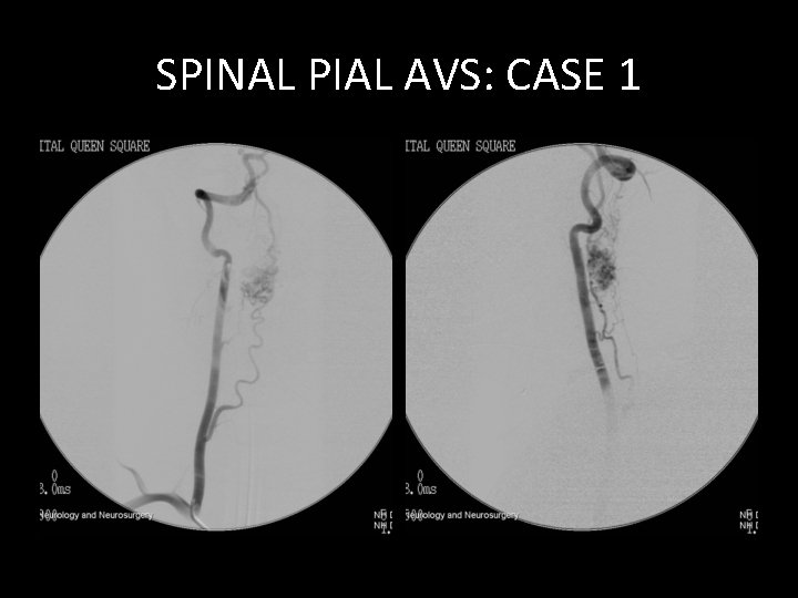
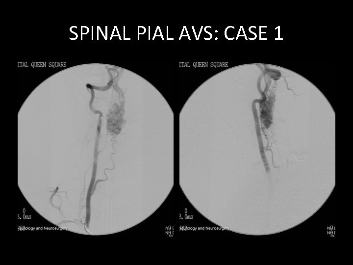
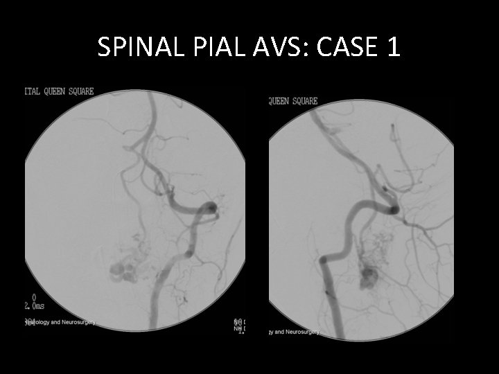
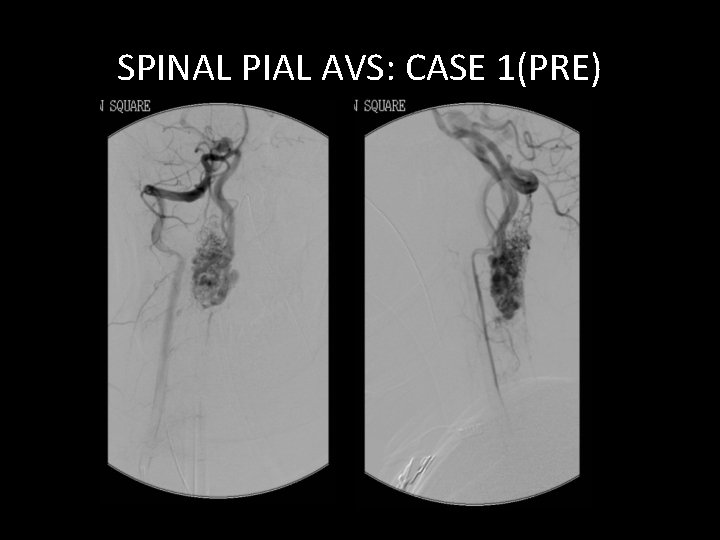
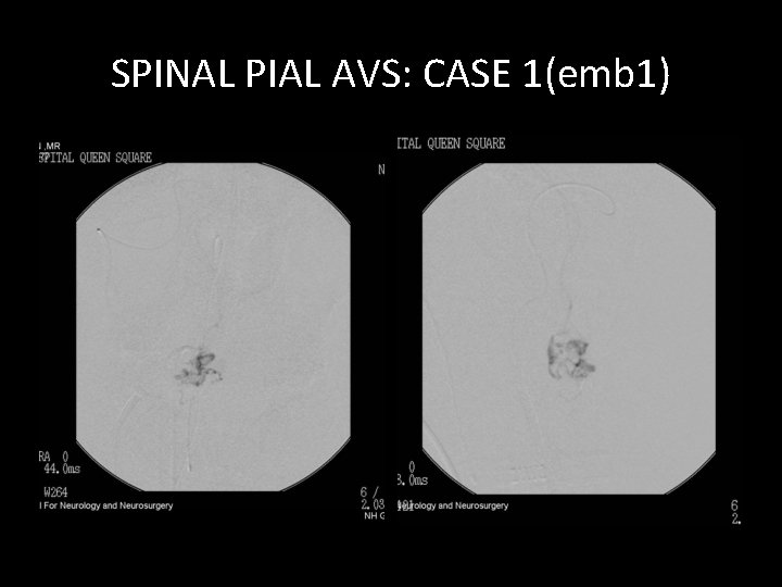
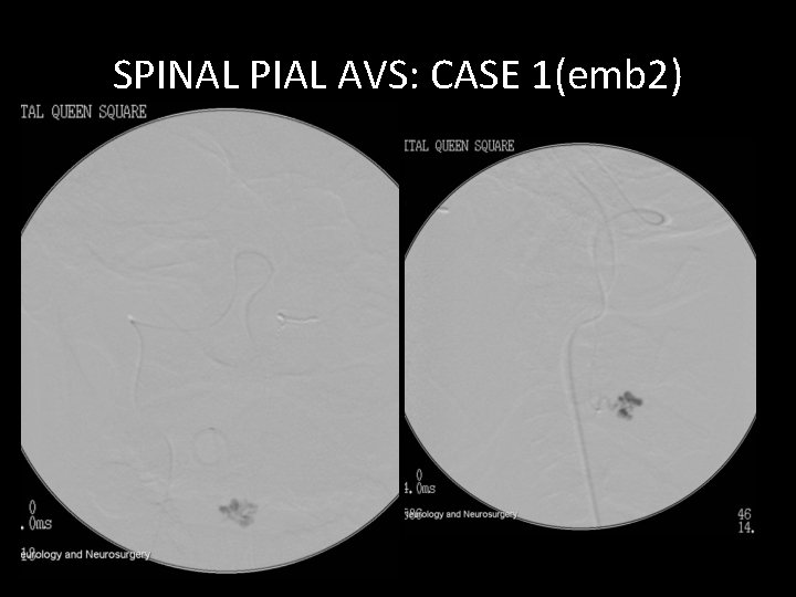
SPINAL PIAL AVS: CASE 1 • YOUNG MALE • PROGRESSIVE TETRAPARESIS • NO HISTORY OF BLEEDING
SPINAL PIAL AVS: CASE 1
SPINAL PIAL AVS: CASE 1
SPINAL PIAL AVS: CASE 1
SPINAL PIAL AVS: CASE 1
SPINAL PIAL AVS: CASE 1
SPINAL PIAL AVS: CASE 1(PRE)
SPINAL PIAL AVS: CASE 1(emb 1)
SPINAL PIAL AVS: CASE 1(emb 2)
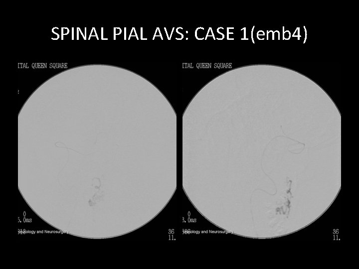
SPINAL PIAL AVS: CASE 1(emb 4)
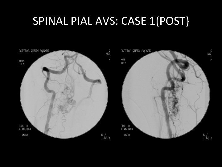
SPINAL PIAL AVS: CASE 1(POST)
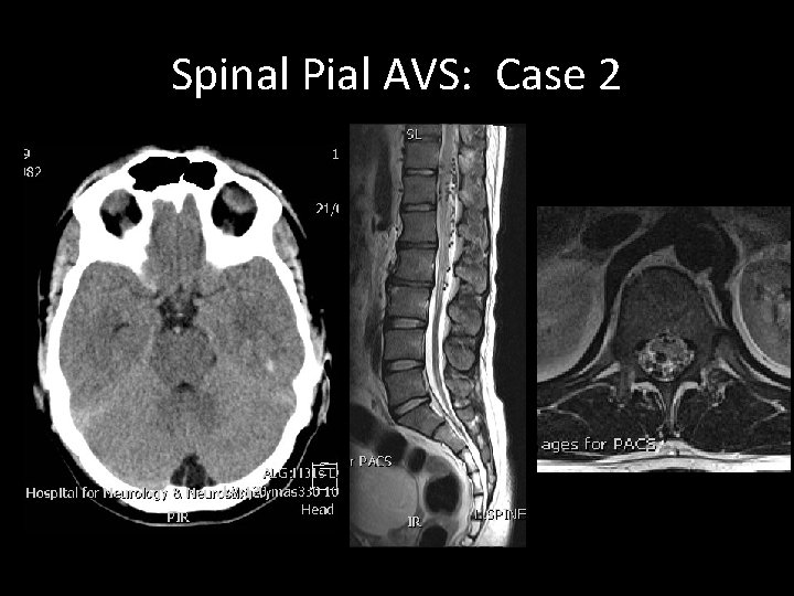
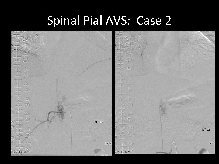
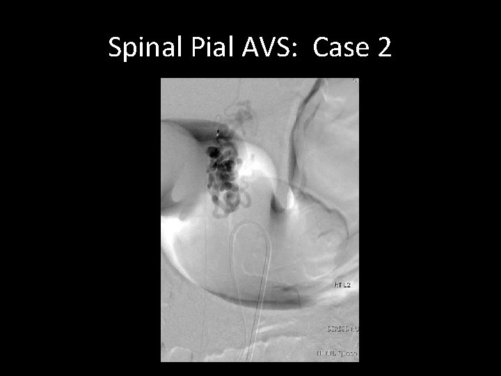
Spinal Pial AVS: Case 2 • Young male • Sudden onset of severe headache • CT Head, CTA and cerebral angiogram
Spinal Pial AVS: Case 2
Spinal Pial AVS: Case 2
Spinal Pial AVS: Case 2
DURAL AVS SPINE
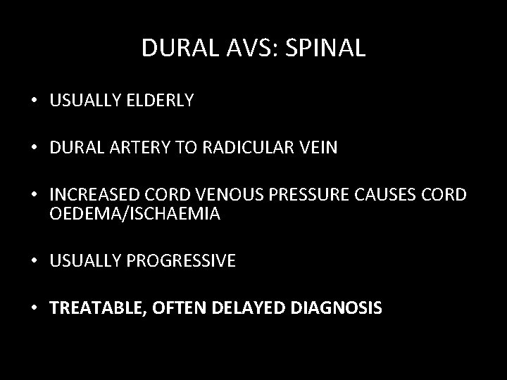
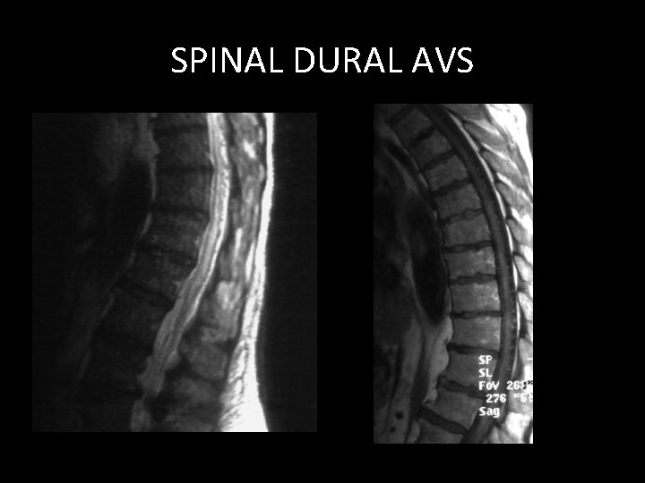
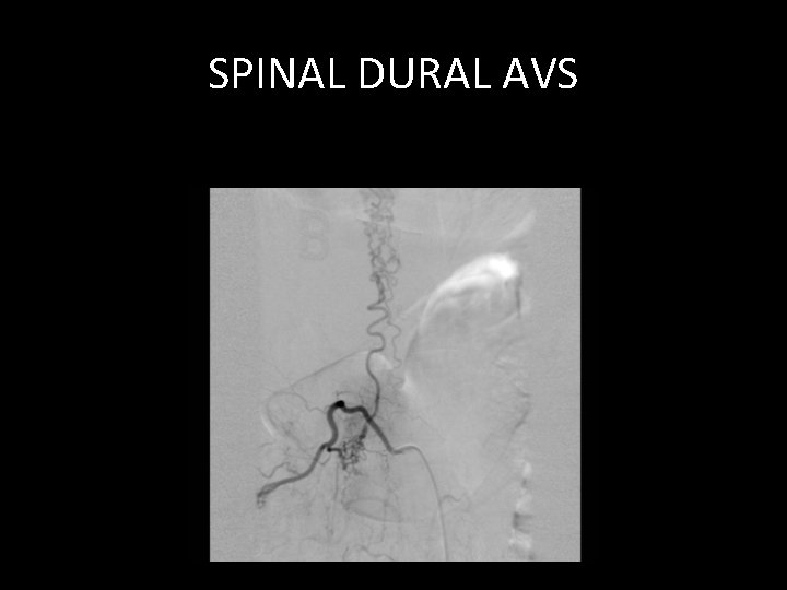
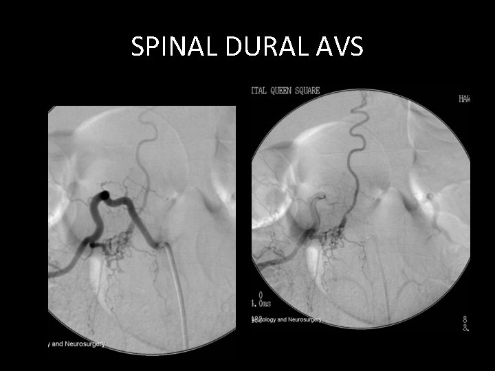
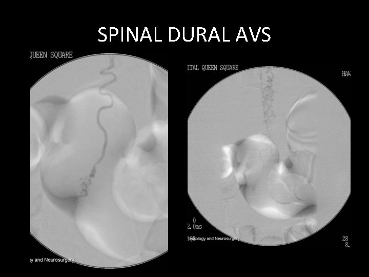
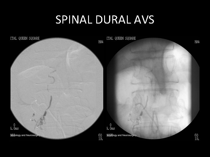
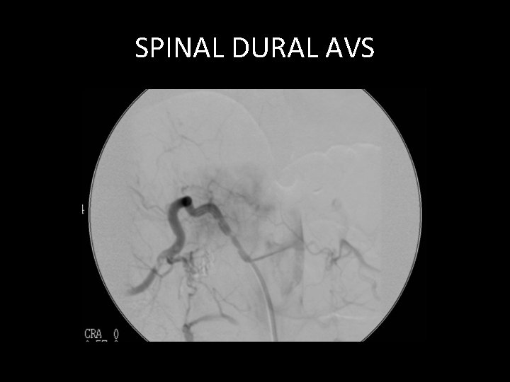
DURAL AVS: SPINAL • USUALLY ELDERLY • DURAL ARTERY TO RADICULAR VEIN • INCREASED CORD VENOUS PRESSURE CAUSES CORD OEDEMA/ISCHAEMIA • USUALLY PROGRESSIVE • TREATABLE, OFTEN DELAYED DIAGNOSIS
SPINAL DURAL AVS • 65 YEAR OLD MAN • 2 YEARS PROGRESSIVE PARAPARESIS • MORE RECENT INCONTINENCE • PREVIOUS NEGATIVE ANGIOGRAPHY/MRI
SPINAL DURAL AVS
SPINAL DURAL AVS
SPINAL DURAL AVS
SPINAL DURAL AVS
SPINAL DURAL AVS
SPINAL DURAL AVS
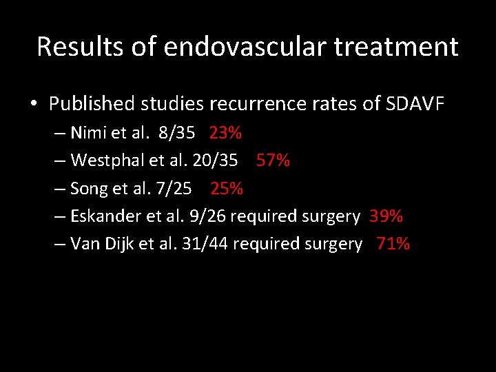
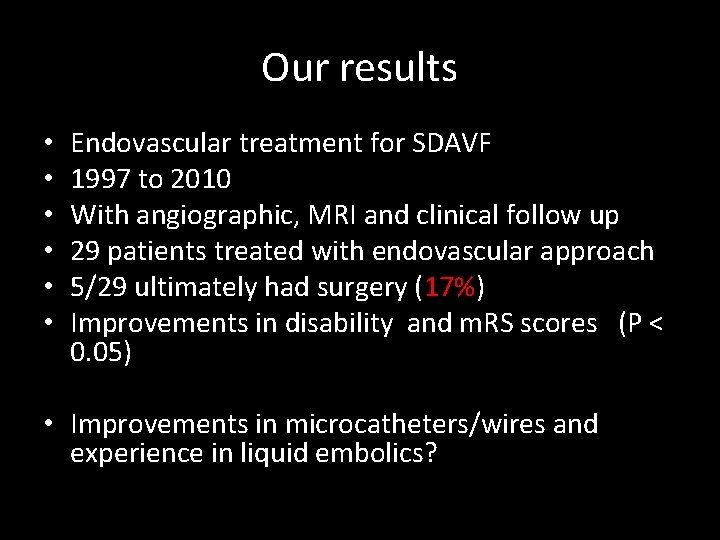
Results of endovascular treatment • Published studies recurrence rates of SDAVF – Nimi et al. 8/35 23% – Westphal et al. 20/35 57% – Song et al. 7/25 25% – Eskander et al. 9/26 required surgery 39% – Van Dijk et al. 31/44 required surgery 71%
Our results • • • Endovascular treatment for SDAVF 1997 to 2010 With angiographic, MRI and clinical follow up 29 patients treated with endovascular approach 5/29 ultimately had surgery (17%) Improvements in disability and m. RS scores (P < 0. 05) • Improvements in microcatheters/wires and experience in liquid embolics?
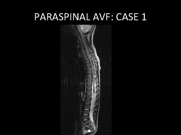
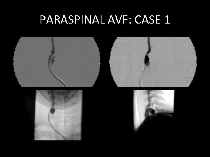
DIRECT LARGE A-VS PARAVERTEBRAL A-V FISTULA
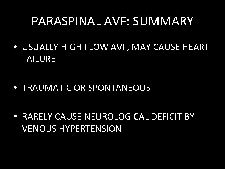
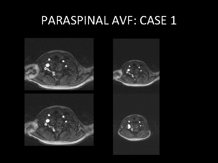
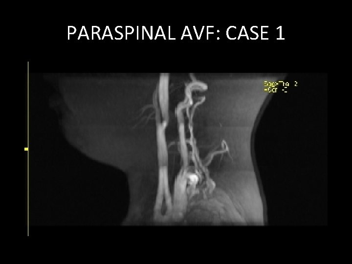
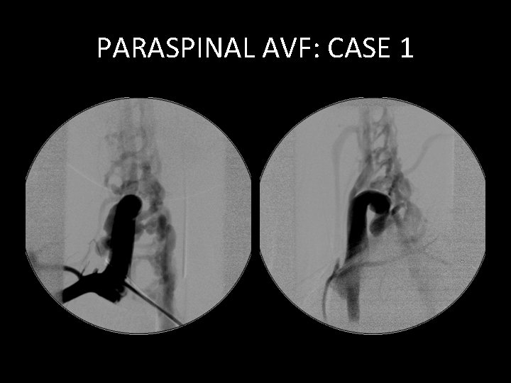
PARASPINAL AVF: SUMMARY • USUALLY HIGH FLOW AVF, MAY CAUSE HEART FAILURE • TRAUMATIC OR SPONTANEOUS • RARELY CAUSE NEUROLOGICAL DEFICIT BY VENOUS HYPERTENSION
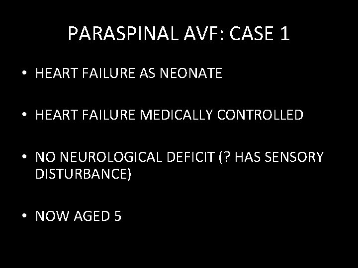
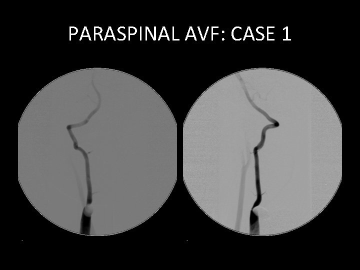
PARASPINAL AVF: CASE 1 • HEART FAILURE AS NEONATE • HEART FAILURE MEDICALLY CONTROLLED • NO NEUROLOGICAL DEFICIT (? HAS SENSORY DISTURBANCE) • NOW AGED 5
PARASPINAL AVF: CASE 1
PARASPINAL AVF: CASE 1
PARASPINAL AVF: CASE 1
PARASPINAL AVF: CASE 1
PARASPINAL AVF: CASE 1
PARASPINAL AVF: CASE 1
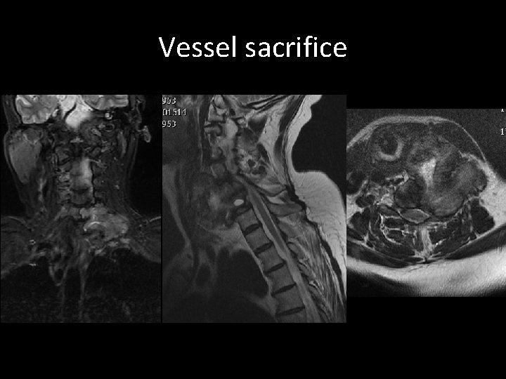
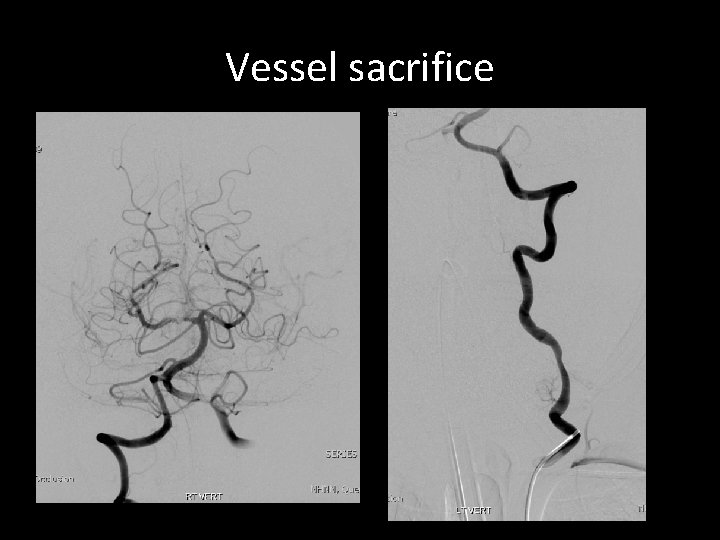
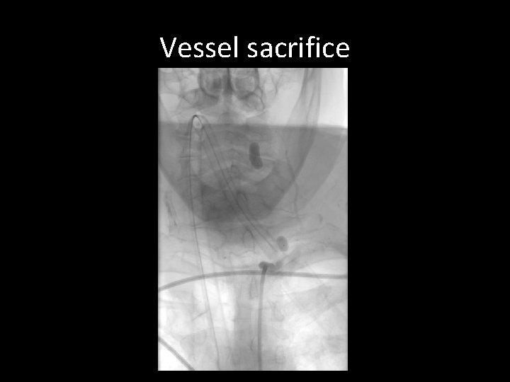
Other conditions treated • Vessel sacrifice pre resection • Tumour embolisation
Vessel sacrifice • 60 yrs female • Left side cervical nerve sheath tumour
Vessel sacrifice
Vessel sacrifice
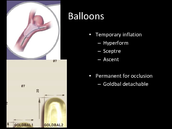
Balloons • Temporary inflation – Hyperform – Sceptre – Ascent • Permanent for occlusion – Goldbal detachable
Vessel sacrifice
TUMOURS SPINAL
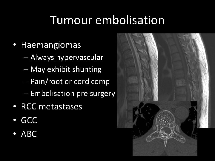
Tumour embolisation • Haemangiomas – Always hypervascular – May exhibit shunting – Pain/root or cord comp – Embolisation pre surgery • RCC metastases • GCC • ABC
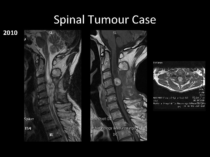
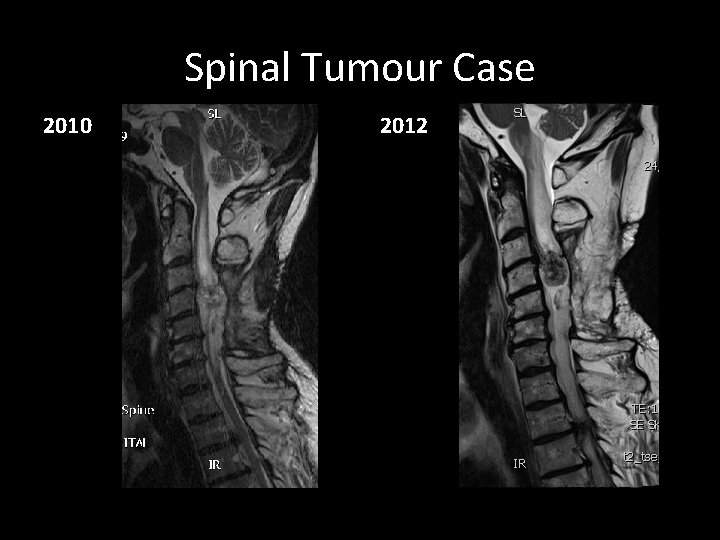
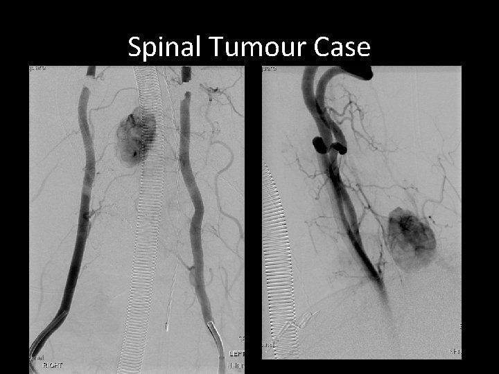
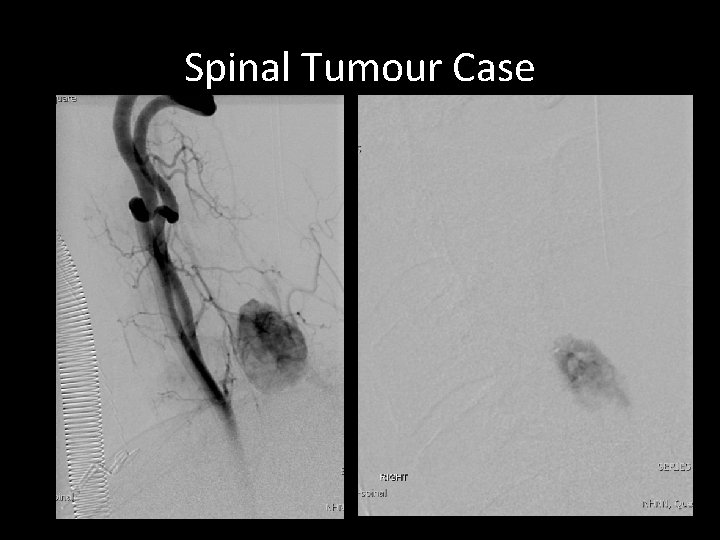
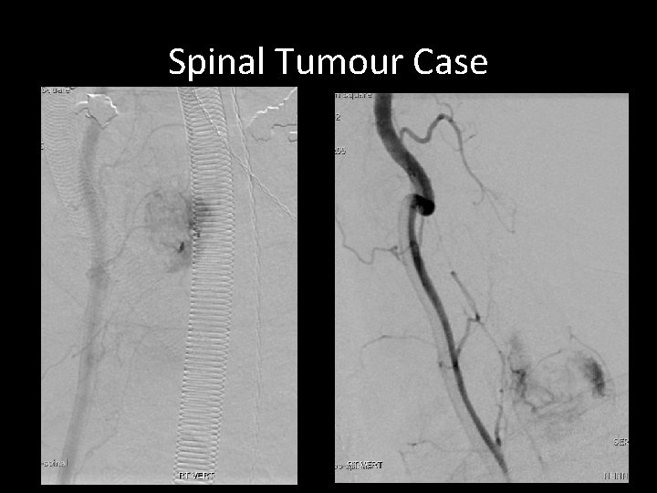
Spinal Cord Tumour Case • • • Cervical haemangioblastoma Attempted pre-operative embolisation 2010 Attempted surgical excision 2010 Cyberknife Increasing weakness 2012 Reattempted embolisation and excision
Spinal Tumour Case 2010
Spinal Tumour Case 2010 2012
Spinal Tumour Case
Spinal Tumour Case
Spinal Tumour Case
Summary • Numerous vascular spinal interventions • Risks and benefits should be carefully considered • Performed in experienced centre!
Contacts Neurointerventional Team: Dr Brew, Dr Robertson, Dr Solbach, Dr Rennie Dr Platts, Dr Rangi, Dr Cowley Lysholm Department of Neuroradiology The National Hospital of Neurology and Neurosurgery Queen Square neurorad@uclh. nhs. uk Tel. 020 34483446