Spinal cord injuries Overview Anatomy of the spinal


: – Dorsal (posterior) columns: deep touch,")


 and absent reflexes")


. Lippincott Williams and Williams:")
- Slides: 35
Spinal cord injuries
Overview: • Anatomy of the spinal cord • Case presentation • Spinal cord injuries – Classification – Complete and incomplete syndromes • Respiratory complications of spinal cord injuries • ICU management of spinal cord injuries • Pharmacological management
Gross anatomy • Begins at the foramen magnum of the skull, where it is continuous with the medulla oblongata • Cervical enlargement gives rise to the brachial plexus • Lumbar enlargement gives rise to the lumbosacral plexus • Tapers inferiorly to the conus medullaris – from here the filum terminale attaches to the coccyx • Lower end of the spinal cord lies at the lower border of L 1 • Vertebral column is much longer than the spinal column, so the cord segments do not correspond numerically to the vertebral bodies
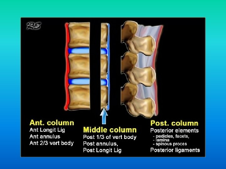
Columns of the spinal cord • Spinal column stabilised by three major ligaments; – Anterior longitudinal ligament – Posterior longitudinal ligament – Ligamentum flavum • Anterior column: Anterior 2/3 vertebral bodies and the anterior ligament • Middle column: Posterior 1/3 vertebral bodies and the posterior ligament • Posterior column: Ligamentum flavum and everything else • Injury involving > one column is considered unstable
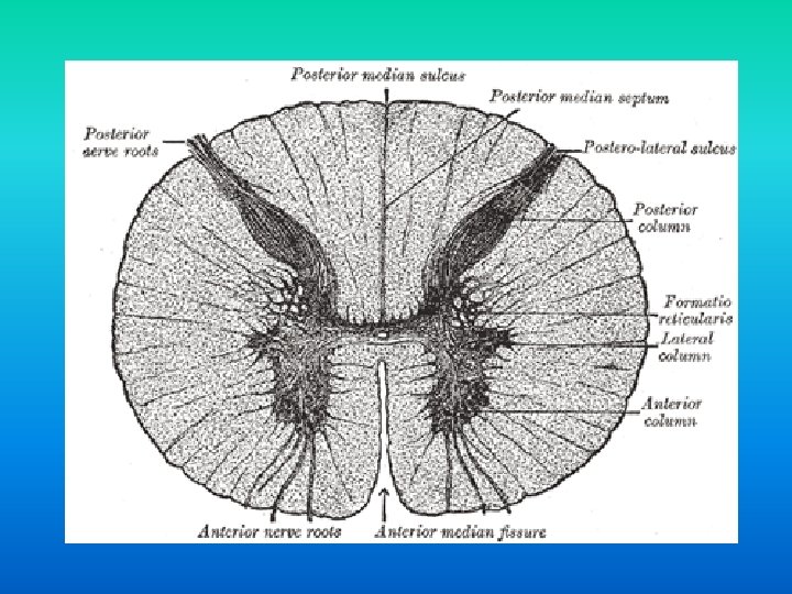
Spinal cord structure • Inner core of grey matter, surrounded by an outer covering of white matter • Grey matter is arranged in an ‘H- shape’, with anterior and posterior horns, joined by a thin grey commissure which contains the central canal • The T 1 -L 3 segments also contain a lateral grey horn
Grey matter of the spinal cord • The anterior horn is divided into medial, central and lateral columns – Medial group is present in most segments innervating the skeletal muscles of the neck and trunk – Central group is the smallest and is present in some cervical and lumbosacral segments – Lateral group is present in the cervical and lumbosacral segments innervates the skeletal muscles of the limbs • Posterior horn has four different groups of nerve cells – Substantia gelatinosa group – Nucleus propius – Nucleus dorsalis – Visceral afferent nucleus • Lateral grey horns contain pre-ganglionic sympathetic fibres
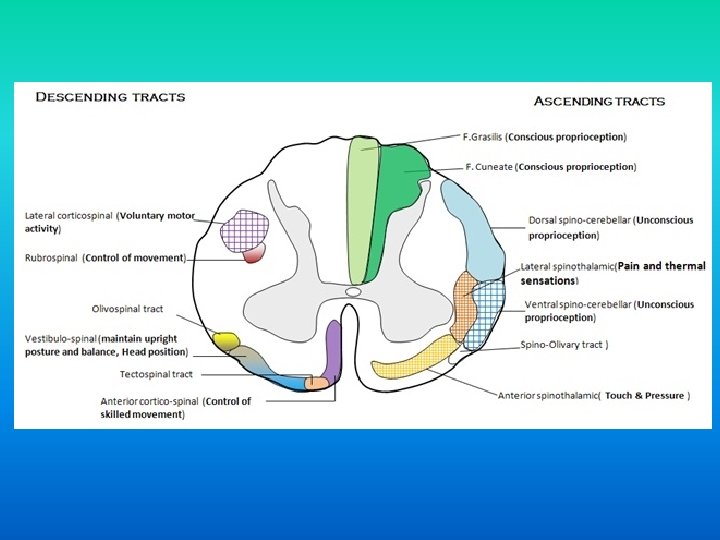
Tracts of the spinal cord • Ascending (sensory): – Dorsal (posterior) columns: deep touch, proprioception, vibration – Lateral spinothalamic: pain and temperature – Anterior spinothalamic: light touch • Descending (motor): – Lateral corticospinal: voluntary motor – Anterior corticospinal: voluntary skilled motor – Rubrospinal: control of movement – Vestibulospinal: posture and balance – Tectospinal: reflex postural movements in response to visual stimuli
Spinal cord injuries • Causes: – Majority caused by MVAs; falls; iatrogenic • Mostly young males, but the other demographic includes older people with concurrent degenerative spinal canal narrowing • Frequently associated with other conditions: – Shock syndromes – Other injuries
A likely story…
Say hello to Jim. • 85 year old male who slipped backwards and hit head on towbar behind car • Presented to Tenterfield Hospital, then T/F to Armidale, where CT showed unstable C 4/5 # • Transferred to JHH for NSx R/V • Conscious and spontaneously breathing • Hard collar in situ, but poorly fitting • GCS 13: E 3 V 4 M 6, PEARL • B/G: metastatic prostate ca, HTN, T 2 DM
Jim’s imaging • Unstable C 4/5 fracture
Further examination… • Normal cranial nerve examination • Decreased strength (2/5) and absent reflexes bilaterally in upper limbs • Decreased pain and temperature sensation bilateral hands • Normal strength (5/5), reflexes and sensation in bilateral lower limbs • Developed urinary retention
American Spinal Injury Association Neurological impairment scale
Classifications • Quadriplegia – Injury to the cervical spine, leading to impairment in the arms, trunk, pelvis and legs • Paraplegia – Injury to the thoracic, lumbar or sacral segments, leading to impairment in the trunk, legs and pelvic organs • Complete – No motor or sensory function below the affect level • Incomplete – Some preserved motor or sensory function below the affected level
Complete injury • • No voluntary anal contraction 0/5 distal motor score 0/2 sensory score Bulbocavernous reflex present
Incomplete spinal cord injuries • • Anterior cord syndrome Central cord syndrome Brown-Sequard syndrome Posterior cord syndrome
Anterior spinal cord syndrome • Injury to the anterior spinal cord caused by either direct compression of the spinal cord, or damage to the anterior spinal artery • Usually from a flexion/compression injury • Bilateral loss of pain, temperature and light touch below the lesion due to disruption of the anterior and lateral spinothalamic tracts • Motor dysfunction due to the disruption of the anterior corticospinal tracts, and damage to the anterior grey horn neurons • Worst prognosis of incomplete SCI – 10 -20% chance motor recovery Anterior
Central spinal cord syndrome • Most common incomplete spinal cord injury • Often in the elderly with extension injury mechanisms, due to anterior osteophytes and posterior infolded ligaments • Motor dysfunction due to disruption of the lateral corticospinal tracts and damage to the anterior grey horn neurons • Bladder and bowel involvement • Bilateral loss of pain, temperature and light touch due to disruption of the spinothalamic tracts • Sacral sparing • Good prognosis, but unlikely to regain full function Anterior
Brown-Sequard syndrome • Caused by complete cord hemitransection • Ipsilateral motor dysfunction, with LMN weakness at the level of the injury, and UMN signs below the injury • Ipsilateral proprioception and vibration loss due to posterior column damage • Contralateral pain and temperature loss 2 -3 segments below the lesion due to disruption of the spinothalamic tracts • Good prognosis
Posterior cord syndrome • Rare syndrome • Most commonly caused by vascular compromise, with occlusion to the posterior spinal artery • Sensory dysfunction with ipsilateral loss of proprioception and vibration, and preservation of pain and temperature Anterior
Cauda equina syndrome • Caused by damage to the cauda equina, a collection of S 1 -L 5 nerves • Technically a peripheral nerve lesion, so will cause lower motor neuron signs • Presentation: – Saddle anaesthesia, bilateral lower limb sensorimotor loss and pain, bowel and bladder symptoms (especially urinary retention) – Absent or reduced lower limb reflexes, decreased rectal tone • MRI best to evaluate nerve compression • Needs urgent surgical decompression within 48 hours
Jim’s progress notes… • Admitted under NSx • Few episodes of vomiting on ward, during which he likely aspirated • RRT on ward for respiratory arrest – intubated and T/F to ICU • Some more stuff happened…. • Improved and ready to trial extubation…. • Unfortunately, he failed extubation due to hypoxia • Why?
Respiratory complications with SCI Spinal cord level Muscle involvement Effect on respiration Clinical consequence C 1 -3 Complete paralysis of all respiratory muscles Vital capacity only 5 -10% of normal; absent cough Apnoea and immediate death C 3 -6 Varied impairment of diaphragmatic contraction Vital capacity 20% of normal; weak and ineffective cough Ventilation necessary in acute stages; majority will be weaned from mechanical ventilation C 6 -8 Diaphragm and accessory cervical inspiratory muscles intact. Intercostals and abdominal muscles intact T 2 -4 Expiration entirely passive. Secretion retention. No respiratory failure unless coexisting lung/chest injury/illness Vital capacity 30 -50% normal, and weak cough
SCI effects on breathing • Loss of intercostal function: – Failure of AP expansion of the ribcage – Chest wall sucked in during diaphragmatic contraction • Loss of lower thoracic segment innervation: – Diaphragm starts in a flatter position, which decreases contraction pressure • Loss of abdominal muscle tone: – As the diaphragm flattens, abdominal contents are pushed outwards and the lower ribcage is pulled inwards, causing paradoxical see-saw breathing – Diaphragm is pulled down by the weight of the abdomen • Inefficient, rapid, shallow breathing results, with more dead space ventilation • Abdominal muscle weakness results in decreased ability to cough and clear secretions
ICU management of SCI patients
Respiratory management • • Airway management Physiotherapy Posture Mucolytics Abdominal binding Monitor for infection Bronchoscopy
Longer term respiratory care • Tracheostomy – More comfortable; minimise laryngeal damage; less dead space compared to ETT; associated with fewer respiratory infections • Weaning from ventilation – Portex sprints are as effective or better than PS weaning, with both superior to SIMV weaning
Cardiovascular complications after SCI • Neurogenic shock – Occurs with lesions above T 6 due to loss of sympathetic tone and unopposed parasympathetic tone – Vasodilation and hypotension; bradycardia • Thromboembolism – Due to immobility and venous stasis • Sympathetic hyperreflexia – Unopposed sympathetic tone below the level of injury, triggered by sensory stimuli
Gastrointestinal complications after SCI • Delayed gastric emptying and ileus – Common and may last 2 -3 weeks – Aperients, early feeding, NGT, prokinetic agents • Gastric stress ulceration – PPI prophylaxis • Constipation
Metabolic system considerations • Temperature regulation – Hypothermic due to vasodilation – Hyperthermic due to inability to sweat below level of injury • Hyperglycaemia – Common due to stress response – Worsens ischaemic neurological injury
Pharmacological treatment of SCI • Steroids – Previously, high dose methylprednisone was standard of care for SCI – Since shown to significantly increase mortality in patients, compared to placebo • NOGO-A antibody – NOGO-A is an inhibitory molecule that prevents neuronal plasticity and axonal regeneration – Current clinical trial to determine effects of an intrathecal infusion of NOGO-A antibody
References • • Snell. Clinical neuroanatomy. 7 th Ed. (2010). Lippincott Williams and Williams: Philadelphia J Patten. Neurological differential diagnosis. 2 nd Ed. (1996). Springer: London M Denton, J Mc. Kinlay. Cervical cord injury and critical care. Continuing education in Anaesthesia and Critical Care. (2009). Vol 9: No. 3 Stahel et al. Management strategies for acute spinal cord injury: current options and future perspectives. Current Opinion Critical Care. (2012). 18: 651 -660 A Neill. Basic neuroanatomy for the critically ill. SMACC. http: //smacc. net. au/2013/02/basic-neuroanatomy-for-thecritically-ill/ C Wheeless. Anterior cord syndrome. Wheeless’ textbook of orthopaedics. . Last updated: 25/4/12. http: //www. wheelessonline. com/ortho/anterior_cord_syndrome Date accessed: 30/6/13 S Hishmeh. Posterior cord syndrome. Orthopaedics: one. Last updated: 22/6/09. http: //www. orthopaedicsone. com/display/Main/Posterior+cord+syndrome Date accessed: 30/6/13 D Moore. Spinal cord injuries. Ortho bullets. Last updated: 20/5/13. http: //www. orthobullets. com/spine/2006/spinalcord-injuries Date accessed: 30/6/13