Spinal Cord Imaging Methods to Evaluate Spine XRAYS


 Often the first diagnostic imaging test Small dose of radiation to visualize")

 Uses radiation Obtain 2 -D images can be processed to")
 Gold standard of imaging for spinal cord disorders No radiation")


 are usually the first series of images to be ordered")


 is a relatively common acquired chronic relapsing demyelinating disease")


- Slides: 31
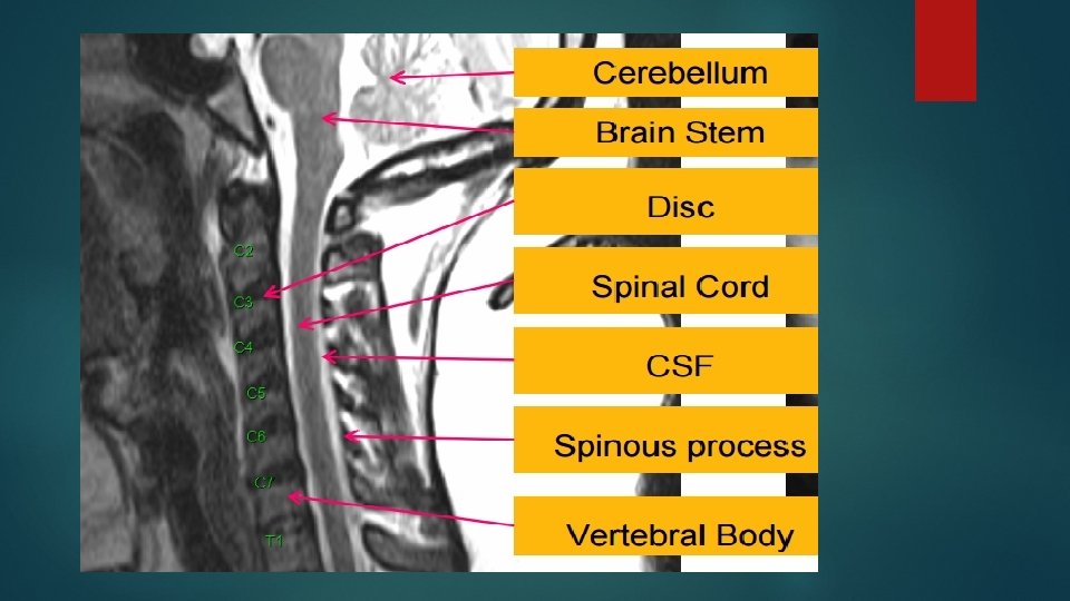
Spinal Cord
Imaging Methods to Evaluate Spine
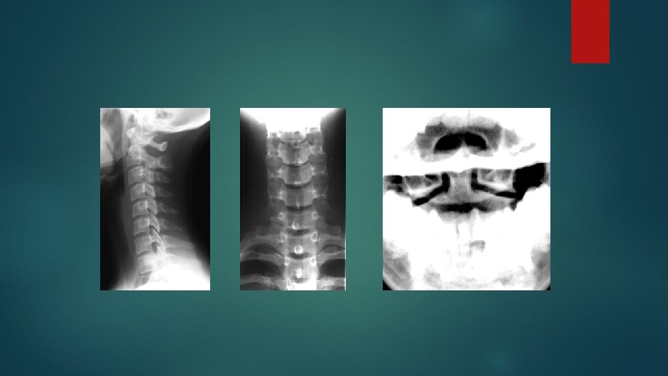
X-RAYS (RADIOGRAPHS) Often the first diagnostic imaging test Small dose of radiation to visualize the bony parts Can detect Spinal alignment and curvature Spinal instability – with flexion and extension views Congenital (birth) defects of spinal column Fractures caused by trauma Moderate osteoporosis (loss of calcium from the bone) Infections Tumors
Is this film an adequate lateral film?
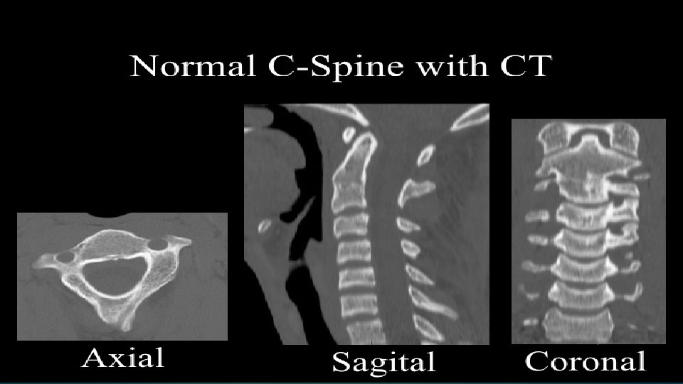
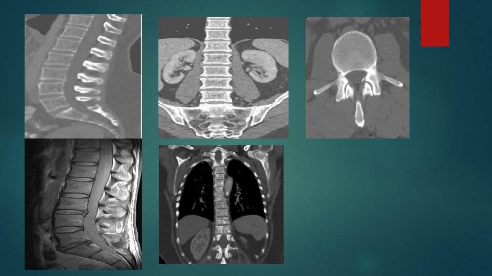
COMPUTERIZED TOMOGRAPHY (CT SCAN) Uses radiation Obtain 2 -D images can be processed to 3 -D images Entire spine can be imaged within a few minutes Detailed information regarding bony structures Limited information about spinal cord & soft tissues
Magnetic Resonance Imaging (MRI) Gold standard of imaging for spinal cord disorders No radiation Can identify abnormalities of bone, soft tissues and spinal cord Claustrophobic patients, uncooperative and children may need sedation or general anesthesia Contraindications include implanted devices e. g. cardiac pacemakers and electromagnetic devices
Indications Advantages Disadvantages X-Ray Trauma Intra-operative localization Inexpensive Widely available Quick Portable Radiation exposure Difficulty in interpretation High rate of false-positive findings CT Trauma Visualization of bony structures Widely available Quick Less useful at visualizing soft tissue structures Radiation exposure Cost MRI Pts with "red flags" case Radiculopathy Tumor Myelopathy Visualization of soft tissue structures (e. g. relationship of disc to nerve) No radiation exposure Contraindications: presence of ferromagnetic implants, cardiac pacemakers, intracranial clips, Claustrophobia Not widely available Cost$$$
Abnormalities Of Spinal Cord • Trauma • Congenital • Demyelination • Tumors
Trauma Plain Radiographs (x-rays) are usually the first series of images to be ordered by the physician. If fractures, or other bony defects, are suspected, CT images can provide very detailed information. When soft tissue injury is suspected, MRI is usually the imaging technology of choice.
Assess four parallel lines. 1. Anterior vertebral line 2. Posterior vertebral line 3. Spinolaminar line 4. Posterior spinous line
Mechanism Of Injury
Compression Fracture
Hangman's Fracture
Hyperflexion
Congenital Defects
Spina bifida
Syringomyelia
Demyelination
Multiple Sclerosis Multiple sclerosis (MS) is a relatively common acquired chronic relapsing demyelinating disease involving the central nervous system. Characteristically disseminated not only in space but also in time
Transverse Myelitis Inflamed cord of uncertain cause Viral infections Immune reactions Idiopathic Myelopathy progressing over hours to weeks
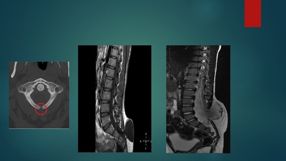
TM VS MS MS lesions in spinal cord are more likely multiple, focal and peripherally located don’t cover the entire section on axial images often < 2 vertebral body heights on sagittal images are disseminated in time and space Transverse myelitis lesions extend over >3 vertebral body heights on axial images often > 4 vertebral body heights on sagittal images no brain lesions
Tumors
Classification Intramedullary lesions its location is determined within the cord. extramedullary lesions May be related to nerve roots and may extend into the foramen (e. g. schwannomas and neurofibromas) or they may have a broad dural attachement (e. g. meningiomas).
Astrocytoma
Ependymoma
Thank you