Spinal cord anatomy Jake Baker and Niamh Mc

Spinal cord anatomy Jake Baker and Niamh Mc. Carville

Contents • • • Features of spinal cord Autonomic vs. somatic nervous systems Comparison of spinal cord segments Spinal cord blood supply Anatomy and function of the meninges Spinal tracts

Spinal cord cross-section

Spinal nerves
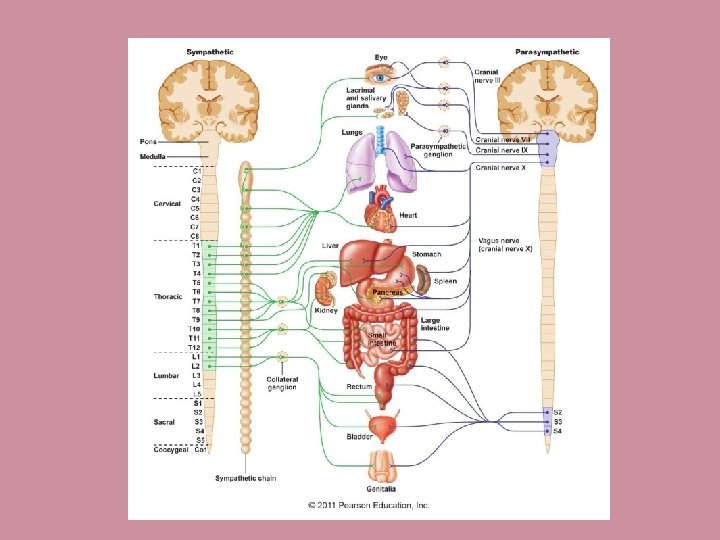

Sympathetic chain

Parasympathetic vs. sympathetic

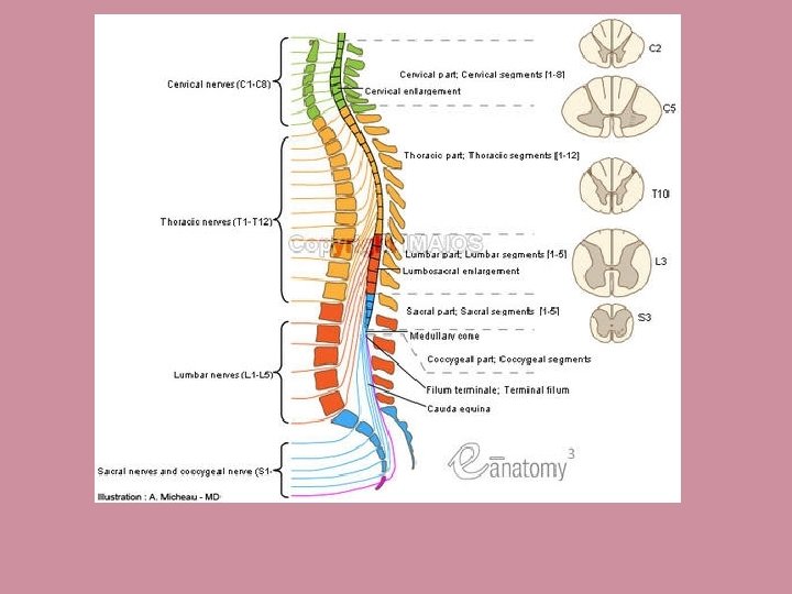
Comparison of Spinal Cord segments

Spinal cord blood supply Artery: Arch of Aorta -> subclavian artery -> vertebral artery -> spinal arteries Vein: Subdural plexus of veins -> hemiazygous -> azygous

Meninges • Subarachnoid space contains CSF. Layer of dura Characteristics Dura mater Creates foldings (tentorium cerebelli, falx cerebri) which contain superior sagittal sinus Arachnoid mater Contains fibroblasts, collagen Pia mater Highly vascularised

The spinal tracts Spinothalamic Dorsal column Corticospinal Sensations Affective sensation, pain, temperature, crude touch Proprioceptive information and fine touch Skilled, voluntary muscle movement First-order First neurone enters spinal cord at spinal nerve. Cell body located in dorsal root ganglion First neurone enters spinal cord at spinal nerve, forming Fasciculus Gracilis and Fasciculus Cuneatus. Terminate in dorsal column nuclei in medulla. Upper Motor Neurone: Motor area of cerebral cortex. Axons pass through internal capsule and brainstem. Desuccate at pyramids. Second-order Axon of second order neurone crosses over to the other side and ascends to thalamus where it terminates. Second order neurons desuccate and ascend to thalamus as a medial lemniscus. Third-order Third order neurone has cell body in thalamus and its axon projects to somatosensory cortex Project from thalamus to somatosensory cortex (post central gyrus) Lower Motor Neurone: From spinal cord level to skeletal muscle Final destination Somatosensory cortex Skeletal muscle Site of crossover Thalamus Medulla oblongata Pyramids
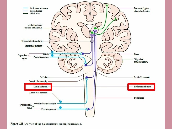


Spinal tracts on a spinal cord segment
- Slides: 15