Spheroids as a model for tumor radiosensitivity and


 Adhesion properties of melanoma cells. All data are given as percentage of adhesion")


- Slides: 34
Spheroids as a model for tumor radiosensitivity and radiolabeling tests Ewa Ł. Stępień, Hanieh Karimi, Bartosz Leszczyński Departament of Medical Physics Marian Smoluchowski Institute of Physics Jagiellonian University 3 rd Jagiellonian Symposium on Fundamental and Applied Subatomic Physics June 23 – 28, 2019, Kraków, Poland
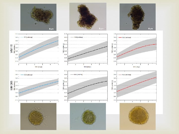
Spheroids are multicellular and tissue –like structured in vitro 3 D models Mimic microenvironment in vivo. Unlike common 2 D in vitro cell models, spheroids reflect the cellular milieu and the pathophysiological conditions inside tumor nodules. Widely used in drug testing and radiation studies. 500 cells 1000 cells 1500 cells
Ross Harrison 1870 – 1959 1910: tedpool neural tube explants cultured several days in „hanging drop” Ross Harrison. The outgrowth of the nerve fiber as a mode of protoplasmic movement. Journal of Experimental Zoology 9: 787 -846, 1910.
1967 1969 1971 1973 1975 1977 1979 1981 1983 1985 1987 1989 1991 1993 1995 1997 1999 2001 2003 2005 2007 2009 2011 2013 2015 2017 2019 600 Pub. Med publications 500 400 300 200 100 0 tumor spheroids 4736 spheroids 4272 spheroids radiotherapy 385
Experimental protocol leading to establishment of melanoma cell lines radial/vertical growth phase https: //histologyblog. com/2014/01/23/histoquarterlysuperficial-spreading-malignant-melanoma/ placed in tissue culture flask for in vitro establishment Cancer Melanoma cell lines primary CM line WM-115 matastatic CM WM-266 -4 lymph node metastasis subcutanous implant (xenograft) in mouse Cancer Melanoma cell lines matastatic CM WM-1205 Lu
MELANOCYTES MELANOMA CELL LINE WM-115 20 L MELANOMA CELL LINE WM-266
3 D tumor models: History, advances and future perspectives Bnien & Swami. Future Oncol. (2014) 10(7), 1311
Flow cytometric analysis of SNA and MAA positive oligosaccharides Kolasińska E et al. Acta Biochim Pol. 2016; 63(3): 533 -41.
Expression of integrin α 5β 1 Kolasińska E et al. Acta Biochim Pol. 2016; 63(3): 533 -41.
(A) Adhesion properties of melanoma cells. All data are given as percentage of adhesion relative to adhesion on poly-L-lysine (taken as 100%). Cell adhesion to BSA-coated wells served as a negative control. (B) Effect of SNA and MAA on the adhesion of melanoma cells to FN. The extent of cell adhesion in the presence of the lectins is presented relatively to cell adhesion in their absence that was considered as 100%. Kolasińska E et al. Acta Biochim Pol. 2016; 63(3): 533 -41.
It is important to know the phenotype of a spheroid model Phenotypic diffrences between WM-115 and WM-266 treat: • α 5β 1 • protein glycation (SNA and MAA positive oligosaccharides)
WM-115 /WM-266 Similarities by Gene mutations NAME TISSUE DISEASE CORRELATION WM-115 skin malignant_melanoma, melanoma 1 WM-266 -4 skin malignant_melanoma, melanoma 0. 97 UACC-62 skin, malignant_melanoma, haematopoietic_and_ melanoma, lymphoid_neoplasm lymphoid_tissue 0. 97 WM-793 skin malignant_melanoma, melanoma 0. 97
WM-115 /WM-266 Similarities by Gene mutations Total number of mutated genes: WM-115: 529 WM-266 -4: 519 WM-793: 613 T. Hulsen, J. de Vlieg and W. Alkema, BMC Genomics 2008, 9 (1): 488
WM-115 /WM-266 Similarities by Gene mutations WM-115 vs WM-793 shared mutations BRAF->ENST 00000288602 ->p. V 600 E WM-115 vs WM-266 -4 shared mutations BRAF->ENST 00000288602 ->p. V 600 E WM-115 vs WM-266 -mel shared mutations BRAF->ENST 00000288602 ->p. V 600 D WM-115 vs UACC-62 shared mutations BRAF->ENST 00000288602 ->p. V 600 E
BRAF mutations in melanoma trancript Exons: 19, Coding exons: 19, Transcript length: 2, 561 bps, Translation length: 806 residues http: //www. ensembl. org/Homo_sapiens/Transcript/Summary? db=core; g=ENSG 00000157764; mr=7: 140753336 -140753337; r=7: 140753270 -140753400; t=ENST 00000288602 Serine/threonine-protein kinase B-raf 1 UWH X-ray 2. 95 Å A/B 448 -723 https: //cancer. sanger. ac. uk/cosmic 3 d/protein/BRAF
Targeted Therapy for Melanoma Skin Cancer Twenty interventions that most often appeared in the ICTRP database from 1971 to 2018 CTLA-4 T cell Wróbel S et al. J Clin Med. 2019 Mar 15; 8(3).
It is important to know the genotype of a spheroid model Genotypic similarities between WM-115 and WM-266 treat: 97% corelations in gene mutations Shared mutations in the crutial gene (BRAF)
Melanoma spheroids - 3 D model CPh FDA PI 250 μm 1500 cells
Melanoma spheroids - 3 D model 3 D melanoma spheroids implanted into a collagen gel matrix https: //www. nature. com/articles/srep 19103
WM-266 spheroids - micro. CT
Radiation response of multicell spheroids-an in vitro tumour model. Changes in radiosensitivity of cells in spheroids were demonstrated due to intercellular contact, cell cycle redistribution and hypoxia. Cells grown in close contact had an enhanced capacity to accumulate and repair sublethal damage. Sutherland RM, Durand RE. Curr Top Radiat Res Q. 1976 Jan; 11(1): 87 -139.
Model of liposome up-take by a cell The three main processes considered are as follows: (a) association and dissociation of vesicles, (b) internalization of bound vesicles, and (c) diffusion of free vesicles. Reprinted by permission from the American Association for Cancer Research: Mok, W. , Stylianopoulos, T. , Boucher, Y. , R. K. Jain, Mathematical modeling of herpes simplex virus distribution in solid tumors: implications for cancer gene therapy, Clinical Cancer Research, 2009, 15: 7, 2352 -2360. DOI: (10. 1177/2211068212458265)
General and multivalent kinetic models of Vesicle Drug Delivery Systems in 2 D DOI: (10. 1177/2211068212458265)
Model in vivo biodistribution of liposomes Pharmacokinetic model used by Arndt et al. Cancer Research and Treatment, Pharmacokinetics of sterically stabilized hexade-cylphosphocholine liposomes versus conventional liposomes and free hexadecyl-phosphocholine in tumor-free and human breast carcinoma bearing mice, 58, 1999, 71 -80, Arndt, D. , Zeisig, R. , Fichtner, I. , Teppke, A. D. , and A. Fahr DOI: (10. 1177/2211068212458265)
Pharmacokinetics The open two-compartment model The central compartment is generally treated as the circulatory system, often referred to as “plasma, ” and the peripheral compartment is referred to as the “tissue” that takes up most of the drug. DOI: (10. 1177/2211068212458265)
Tumor spheroids as an in vitro model for determining therapeutic efficacy against proton beam radiotherapy and doxorubicin encapsulated low temperature sensitive liposome Senavirathna LK et al. Theranostics. 2013; 3(9): 687– 691
Concluding remarks Preclinical study needs to use a model relevant for diseases progress and treatment Spheroids are very well established for basic and translational research of cancer biogenesis and sensitivity micro. CT as a novel aproach for spheroid investigation
Melanoma types 1. 2. 3. 4. Superficial spreading melanoma commonly found on the arms, legs, chest and back. The melanoma cells usually grow slowly at first, and spread out across the surface of the skin. Nodular melanoma - can grow more quickly than other melanomas. It is also more likely to lose its colour when growing, becoming red rather than black. It is more commonly found on the chest, back, head or neck. Lentigo maligna melanoma - usually found in older people, in areas of skin that have had a lot of sun exposure over many years. It is often found on the face and neck. Acral lentiginous melanoma - usually found on the palms of the hands, soles of the feet, or under fingernails or toenails. It is more common in people with black or brown skin. It is not thought to be related to sun exposure.
Molecular background of UV radiation and oxidative stress for the melanoma cause Wróbel S et al. J Clin Med. 2019 Mar 15; 8(3).
Differential dependence of melanoma subgroups on sun exposure and types of gene mutations Wróbel S et al. J Clin Med. 2019 Mar 15; 8(3).
Pie chart demonstrating the percentage distribution of genes with clinicallyrelevant and recurrent driver mutations in individuals with melanoma, including non-V 600 BRAF alterations (left), interrogated on the Vanderbilt melanoma SNa. Pshot assay. BRAF fusions occur in approximately 4– 8% “pan-negative” cases (right).
Molecular subsets of melanoma Anatomic location of the primary tumor Frequency of driver mutations in melanoma Skin without chronic sun damage ~50% BRAF ~20% NRAS Skin with chronic sun damage ~5% BRAF ~10% NRAS ~2% KIT Mucosal surfaces ~5% BRAF ~15% NRAS ~20% KIT Acral surfaces ~15% BRAF ~15% NRAS ~15% KIT Uveal melanoma ~32% GNA 11 ~50% GNAQ ~1% BRAF *Frequencies of MEK 1 and CTNNB 1 mutations according to anatomic location are not known at present