Spectrum of Metabolic Bone Diseases in CKD Pathogenesis

Spectrum of Metabolic Bone Diseases in CKD: Pathogenesis, Clinical Significance, and Diagnosis Tiffani R. Garrett Renal Grand Rounds March 6 th, 2012

Learning Objectives/Relevant Questions • • • Function of bone Review normal bone remodeling Types of bone disease Prevalence Clinical significance ▫ Vascular calcifications ▫ fractures • Diagnosis ▫ Bone Biopsy ▫ PTH ▫ Bone turnover markers in CKD

Function of bone • Structural ▫ Locomotion ▫ Respiration ▫ Protection of internal organs • Metabolic ▫ ▫ Calcium Phosphorus Carbonate Contributes to hydrogen ion concentration

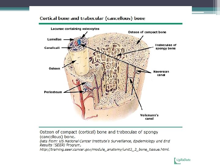
Types of Bone • Cortical bone is dense and compact. ▫ the outer part of all skeletal structures. ▫ The lamellae may be extensive (circumferential) or tightly packed in concentric circles in osteons. ▫ comprises 80 percent of the skeletal weight. • Its major function is to provide mechanical strength and protection, but it can participate in metabolic responses, particularly when there is severe or prolonged mineral deficit.
 bone is found inside the long bones, particularly")
Types of Bone • Trabecular (cancellous) bone is found inside the long bones, particularly at the ends ▫ Located throughout body of vertebrae and inner portions of the pelvis and other flat bones ▫ Important contributor to mechanical support ▫ More metabolically active than cortical bone, supplies initial minerals in acute deficiencies

Normal bone remodeling • Remodeling: replacement of old bone with new bone at the same location. Responsible for complete regeneration of the adult skeleton every 10 years. • Removal of bone (resorption) is the task of osteoclasts. • Formation of new bone is completed by osteoblasts • Both processes are controlled by osteocytes

Cellular Components of Remodeling • Osteocytes 90 -95% of all bone cells ▫ Viable for years (even decades) • Osteoblast <5% of bone cells ▫ Lifespan of weeks • Osteoclast <1 % of bone cells ▫ Lifespan of days
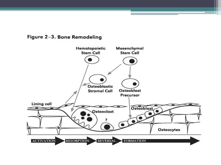

• The purpose of remodeling in the adult skeleton is unclear but most likely it serves to remove ▫ ▫ dead osteocytes maintain oxygen and nutrient supply maintain the appropriate level of matrix hydration and repair fatigue damage, thus preventing excessive aging and its consequences. • Stages of remodeling have different lengths ▫ Resorption 2 weeks ▫ Reversal 4 to 5 weeks ▫ Formation up to 4 months

Osteocytes • choreographers of the remodeling process on the bone surface by virtue of their ability to: ▫ sense worn-out bone ▫ direct osteoclasts to the site that is in need of remodeling ▫ produce the RANKL and sclerostin that regulate osteoclast and osteoblast generation, respectively ▫ control and modify the mineralization of the matrix produced by osteoblasts* • Mechanical forces sustain osteocyte survival • Control and modify the mineralization of the matrix produced by osteoblasts by secreting factors MEPE and FGF 23* ◦ Bonewald L. F. , Ann N Y Acad Sci. 2007 Nov; 1116: 281 -90. Epub 2007 Jul 23.

ANNALS OF THE NEW YORK ACADEMY OF SCIENCES FIGURE 2. Simplified diagram of the interactions between Dmp 1, Phex, and FGF 23, all shown to be expressed in osteocytes. Both Dmp 1 and Phex appear to downregulate FGF 23 expression, which in turn allows reabsorption of phosphate by the kidney thereby maintaining sufficient circulating phosphate to maintain bone mineral content (A). In the absence of either. Dmp 1 or Phex, FGF 23 is highly elevated in osteocytes leading to phosphate excretion by the kidney thereby lowering circulating phosphate leading to osteomalacia and rickets (B). Osteocytes appear to play a major role in mineral homeostasis.

FENG, J. Q. , L. M. WARD, S. LIU, et al. 2006. Loss of DMP 1 causes rickets and osteomalacia and identifies a role for osteocytes in mineral metabolism. Nat. Genet. 38: 1310– 1315.

Renal Osteodystrophy

DEFINITION
 sponsored a Controversies Conference on")
• KDIGO ( Kidney Disease: Improving Global Outcomes) sponsored a Controversies Conference on Renal Osteodystrophy ▫ Develop a clear, clinical revelant, and internationally acceptable definition and classification system ▫ Develop a consenus for bone biopsy evaluation and classification ▫ Evaluate laboratory and imaging markers for the clinical assessment of patients with CKD Moe S, Drueke T, Cunningham J, et al. Definition, evaluation, and classification of renal osteodystrophy: a position statement from Kidney Disease: Improving Global Outcomes ( KDIGO), Kidney Int 2006; 69: 1945

Renal Osteodystrophy “recommended that the term be used exclusively to define alterations in bone morphology associated with CKD, which can be further assessed by histomorphometry, and the results reported based on a unified classification system that includes parameters of turnover, mineralization, and volume” Moe S, Drueke T, Cunningham J, et al. Definition, evaluation, and classification of renal osteodystrophy: a position statement from Kidney Disease: Improving Global Outcomes ( KDIGO), Kidney Int 2006; 69: 1945

CKD-Mineral and Bone Disorder “A broader clinical syndrome that develops as a systemic disorder of mineral and bone metabolism and/or extra- skeletal calcification” Moe S, Drueke T, Cunningham J, et al. Definition, evaluation, and classification of renal osteodystrophy: a position statement from Kidney Disease: Improving Global Outcomes ( KDIGO), Kidney Int 2006; 69: 1945

Background
 have CKD •")
• Approximately 26 million Americans ( 1 in 9 adults) have CKD • Bone abnormalities are common complications • Complications usually start in CKD stage 2 and are found in almost all patients with CKD stage 5 • Increasing evidence that these complications are associated with increased risk of cardiovascular calcification, morbidity, and mortality • In practice bone biopsy is used infrequently because it is invasive and often expensive and procedure that requires special processing not widely available* *Moe S, Drueke T, Cunningham J, et al. Definition, evaluation, and classification of renal osteodystrophy: a position statement from Kidney Disease: Improving Global Outcomes ( KDIGO), Kidney Int 2006; 69: 1945 Malluche HH, Faugere MC. Renal Osteodystrophy in the first decade of the new millennium: analysis of 630 bone biopsies in black and white patients. Journal of Bone and Mineral Research. June 2011 1368 -1376

• Most common forms of renal osteodystrophy are attributable largely to variations in the plasma levels of parathyroid (PTH) • Circulating PTH levels have been used a surrogate indicator of bone turnover, along with serum calcium, phosphorus, and alkaline phosphatase levels to evaluate, diagnosis and guide treatment • Several clinical trials have questioned the specificity of PTH as an indicator of bone turnover Moe S, Drueke T, Cunningham J, et al. Definition, evaluation, and classification of renal osteodystrophy: a position statement from Kidney Disease: Improving Global Outcomes ( KDIGO), Kidney Int 2006; 69: 1945

CLASSIFICATION

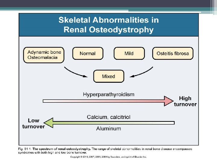
Types of Renal bone disease • • Osteitis fibrosa cystica Adynamic bone disease Osteomalacia Mixed uremic osteodystrophy
 • High bone turnover • Secondary hyperparathyroidism mediated • 3 main")
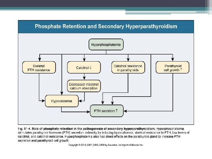
Osteitis fibrosa (OF) • High bone turnover • Secondary hyperparathyroidism mediated • 3 main abnormalites contribute to pathogenesis ▫ Phosphate retention ▫ Decreased free calcium level ▫ Decreased calcitriol level • Increased PTH levels first become evident when GFR drops below 60 ml/min/1. 73 m(2), serum calcium and phosphate levels are normal until e. GFR decreases to approximately 20 ml/min/1. 73 m(2) • Low levels of calcitriol are commom at higher levels of GFR

Phosphorous and Secondary Hyperparathyroidism
: 2534 -40. Phosphorus")
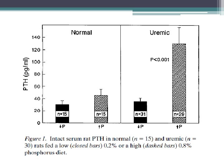
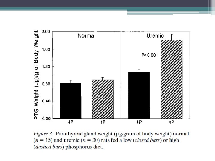
Direct Phosphorus effect on PTH Clin Invest. 1996 Jun 1; 97(11): 2534 -40. Phosphorus restriction prevents parathyroid gland growth. High phosphorus directly stimulates PTH secretion in vitro. Slatopolsky E, Finch J, Denda M, Ritter C, Zhong M, Dusso A Source Department of Internal Medicine, Renal Division, Washington University School of Medicine, St. Louis, Missouri 63110, USA. • Experimental study that examined dietary phosphate restriction in rats with ESRD. Results revealed that normalization of plasma phosphate lowered PTH levels from 130 to 35 pg/ml. Also prevented parathyroid cell growth • serum calcium and calcitriol levels were not altered

Calcitriol and Secondary Hyperparathyroidism

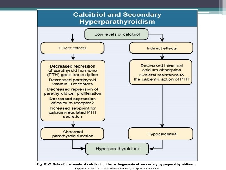
Decreased calcitriol activity • Renal calcitriol production declines due to several mechanisms ▫ Decline in GFR: limits delivery of 25 - hydroxyvitamin D to the site of 1 α-hydroxylase activity in PCT ▫ Phosphate retention: indirectly stimulates FGF-23 which directly decreases activity of 1 α-hydroxylase ▫ PTH fragments directly decrease calcitriol activity • Low levels of calcitriol contribute to the pathogenesis of hyperparathyroidism

Other Factors • Hypocalcemia ▫ Increases PTH m. RNA levels and stimulates proliferation of parathyroid cells over days to wks ▫ Decrease number of calcium-sensing receptors • FGF 23 ▫ Acts to suppress PTH secretion • Skeletal resistance to PTH ▫ Downregulation of PTH receptors • Metabolic acidosis ▫ Stimulates cell mediated bone resorption via osteoclastic activity

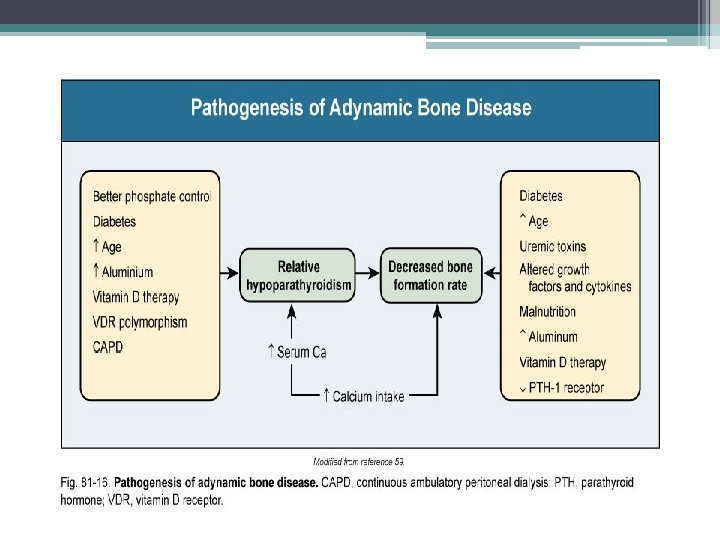
Adynamic bone disease • Bone turnover is markedly reduced • Lack of bone cell activity (osteoblasts and osteoclasts) • No increase in osteoid formation as seen in osteomalacia • Consequence of inadequately low PTH which causes suppression or cessation of both osteoblast and osteoclast • Iatrogenic oversuppression ( high doses of calcium containing phosphate binders, high dialysate calcium concentration, high dose active vitamin D metabolites, and after parathyroidectomy)

Effects of Sevelamer Hydrochloride and Calcium Carbonate on Renal Osteodystrophy in Hemodialysis Patients Ferreira A, Frazao JM, Monier- Faugere MC, et al. • 54 week randomized, open label study • 119 hemodialysis patients received baseline bone biopsy • Assigned to calcium carbonate or sevelamer hydrochloride for 1 year, then second biopsy perfomed • Serum phosphorus, calcium, and i. PTH were controlled • ABD most frequent baseline bone abnormality (59%) • Primary end points (1) changes from baseline in mineralization lag time in lamellar bone and osteoid thickness and (2) changes in bone turnover • Secondary end points were the percentage of patients who developed (1) Osteomalacia, (2) HPBD, and (3) ABD

Ferreira A et al. JASN 2008; 19: 405 -412

Ferreira A et al. JASN 2008; 19: 405 -412

Ferreira A et al. JASN 2008; 19: 405 -412

Results/Discussion • No statistically significant difference in bone turnover or mineralization between the two groups • Sevelamer group showed increased bone formation and trabecular architecture improvement* • Higher use of calcitriol and vitamin d analogs in sevelamer group ( lower calcium levels) • High prevalence of ABD despite most patients being treated in accordance with K/DOQI guidelines ▫ 98% were on calcium carbonate prior to study ▫ 65% had received calcitriol ▫ May explain why baseline mean i. PTH value were low (150 to 300 pg/ml)

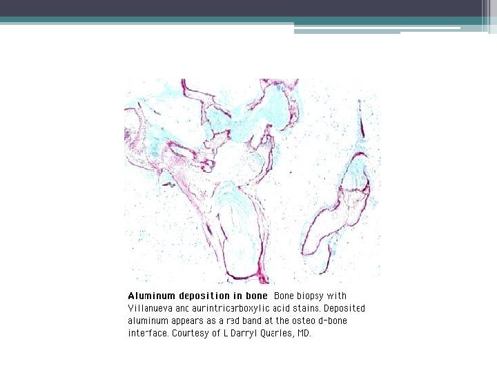
Osteomalacia • Reduction in bone turnover, bone- forming cells and bone-resorbing cells • Marked increase in the volume of unmineralized bone • Historically associated with aluminum toxicity in ESRD patients • Incidence has decreased with abandonment of aluminum- based phosphate binders and more efficient techniques for water treatment

Mixed Uremic Osteodystrophy • Elements of high and low bone turnover • Marrow fibrosis and increased unmineralized osteoid

Histology

Normal Bone

Osteitis Fibrosa

Adynamic Bone Disease

Mixed Uremic Osteodystrophy

Osteomalacia

EPIDEMIOLOGY

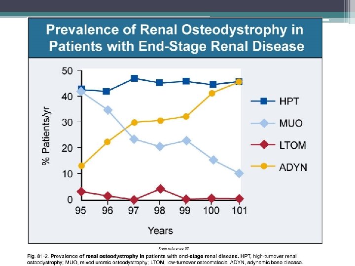
• In hemodialysis patients OF and adynamic bone disease now occur with almost equal frequency • In peritoneal dialysis patients adynamic bone lesion predominates • Currently adynamic bone disease represents the principal bone lesion likely due to an increasing diabetic population • Among CKD patients not yet on dialysis adynamic bone disease is more prevalent
describe the distribution of different types of ROD in patients not")
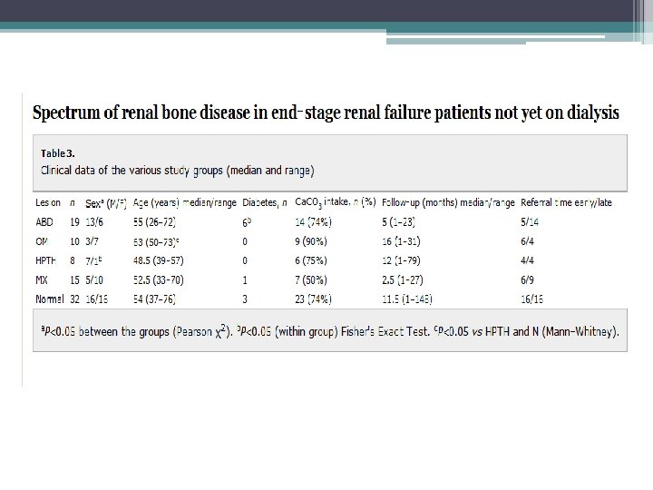
• Purpose: (1)describe the distribution of different types of ROD in patients not yet on dialysis (2) establish risk factors that might influence the development of various types of ROD • 84 ESRD pts underwent transiliac bone biopsies before starting dialysis • Patients were recruited within 10 months • Calcium carbonate was the only prescribed phosphate binder used • None of the patients received vitamin D analogs • 30 % had undergone urgent dialysis session prior to biopsy
 of the various types of ROD of the study population. Spasovski G")
Distribution (%) of the various types of ROD of the study population. Spasovski G B et al. Nephrol. Dial. Transplant. 2003; 18: 1159 -1166
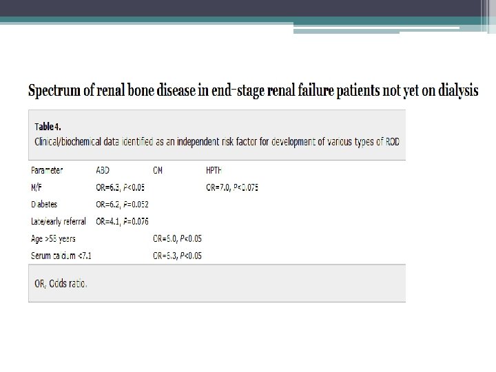

• Purpose of study to provide information about histology abnormalities related to race, gender, diabetes, and treatment with active vitamin d or phosphate binders • bone biopsies were done from 2003 to 2008, only baseline biopsy where included • 316 patients from US, 314 from Europe • All patients volunteered to enter study • All patients received routine dialysis support for at least 6 months ( phosphate binders, active vitamin D, or calcimimetics)
, such as")
• Exclusion • • ▫ Uncontrolled systemic disease ( except DM), such as liver disease, malabsorption, malignancy, thyroid dysfunction ▫ Treatment within last 12 months with bisphonates, fluoride, calcitonin, glucocorticoids, immunosuppressive agents or hormone replacement therapy ▫ Chronic alcoholism and/or drug addiction ▫ Lack of double tetracycline labeling 60 o patients were on HD 30 patients were on PD 87 black patients ( all from US) 141 diabetics 109 treated with active vitamin d analog 429 treated with calcium containing phosphate binder Only 4 patients received calcimimetics
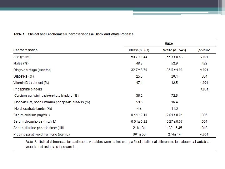
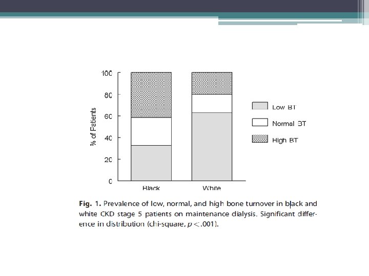
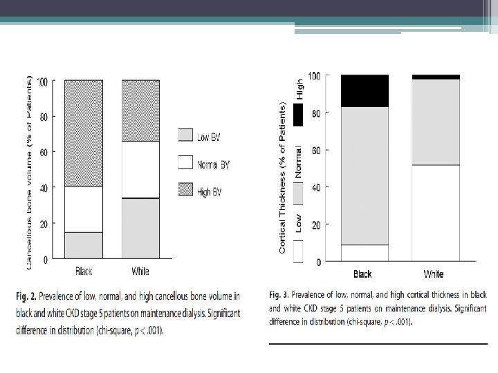
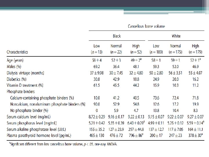

Study findings • Racial differences • TMV system should be expanded to include the architecture of cancellous and cortical bone • Mineralization defects are now rarely observed • Adjustment of current therapeutic paradigm that takes into consideration risk for low bone turnover and volume given association with increased vascular calcifications

Limitations of study • Relatively low number of white patients treated with active vitamin d ( 12. 5% white vs. 47. 1% blacks) • Patients volunteered to participate possible bias favoring clinically uncomplicated patients • no information included on 25 vitamin d or FGF 23

Clinical Implications of ROD
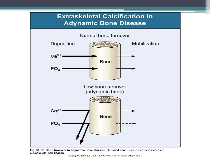
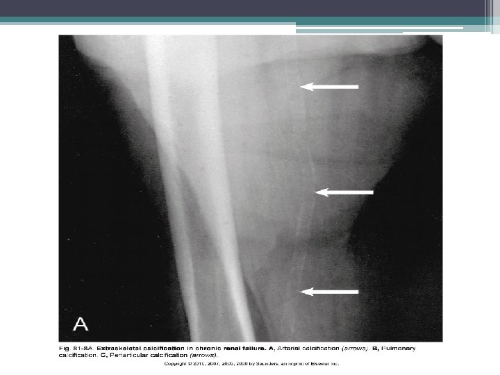

Extraosseous calcification • Results from changing effect of PTH on phosphate balance as renal failure progresses • PTH causes calcium and phosphate release from bone • High PTH levels induce vascular calcification, in dialysis patients markedly high levels may enhance coronary artery calcification

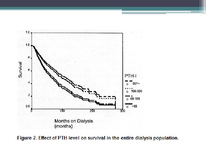
• There is a general opinion that low levels of serum PTH in hemodialysis patients are associated with increased vascular and cardiac calcium deposits • This opinion is based on the concept that low bone turnover, associated with low serum PTH levels, is a condition of decreased calcium phosphate buffering capacity of bone • Therefore, increased serum levels of calcium and phosphate, associated with low bone turnover, may induce increased severity of vascular calcifications. • increased cardiovascular mortality is difficult interpret since low levels of PTH may also be associated to malnutrition and inflammation, known causes of vascular damage, anemia and cardiovascular death

• The study aimed to evaluate the association between PTH serum levels and coronary calcifications, in favor of a direct association between PTH serum levels and coronary calcium deposits. • cohort of 197 hemodialysis patients. • 133 males and 64 females. 22 patients had DM • Average was 58. 6 years. • Patients were divided into groups of intact PTH levels, 0– 150 (A), 150– 300 (B), 300– 600 (C) and >600 (D) pg/ml.
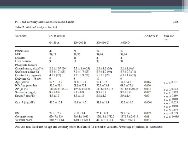
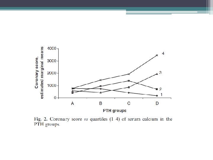

Limitations/ Discussion • Limitations ▫ cross-sectional nature, with patients selected from different dialysis units, with possibly somewhat different therapeutic approaches ▫ Single biochemical assay being related to long term arterial lesions • the finding of no major association between low turnover bone disease and the severity of coronary calcium deposits in hemodialysis patients bears practical implications • Special attention should be paid to the control of elevated levels of secondary hyperparathyroidism and the related calcium phosphate derangements.

Arterial Calcifications and Bone Histomorphometry in End-Stage Renal Disease Gérard M. London*, Caroline Marty†, Sylvain J. Marchais*, Alain P. Guerin*, Fabien Metivier* and Marie-Christine de Vernejoul† *Service d’Hémodialyse, Hôpital F. H. Manhès, Fleury-Mérogis, †Hôpital Lariboisière, INSERM Unité 606, Paris, France. • evidence that low bone turnover is associated with increased arterial calcifications • They studied 58 hemodialysis patients subjected to bone biopsy and evaluated for arterial calcifications with an X-ray survey of some other indicative arterial sites. The extent of arterial calcifications was inversely related to histomorphometric bone turnover. • Limitations: the cohort of patients included a relatively large number of patients with aluminum deposition, and also previously parathyroidectomized cases, with low turnover induced by surgery.

Fractures • The morbidity and mortality associated with hip fractures have been well established in the general population • Coco and Rush examined the incidence of hip fractures in dialysis patient over a 10 year period • They found an association of lower PTH levels and hip fractures • The 1 year mortality rate from the hip fracture event was nearly two and half times greater than the general population

Methods • Hospital admission list and medical records of patients treated for at least 6 months at outpatient dialysis unit affiliated with Montefiore Medical Center (NY), 1, 272 patients reviewed, 56 hip fractures documented • Hip fracture was defined as an event diagnosed by radiology • Pathologic fractures secondary to metastatic malignancies were excluded • Chemistry test were performed monthly except PTH which was measured quarterly • Chemistry test results of patients who sustained a hip fracture were included only up to time of fracture • Patients divided into four groups based on their average PTH value
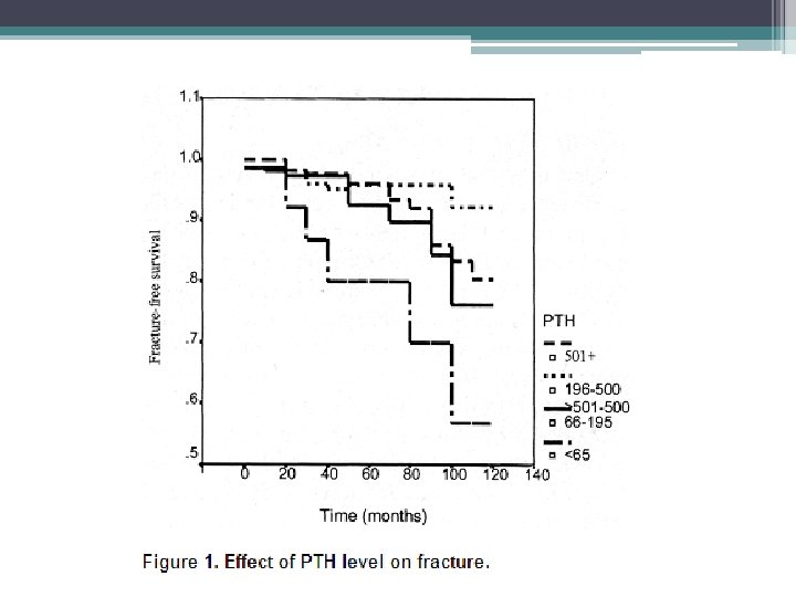
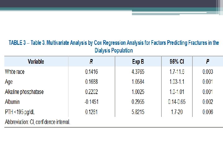
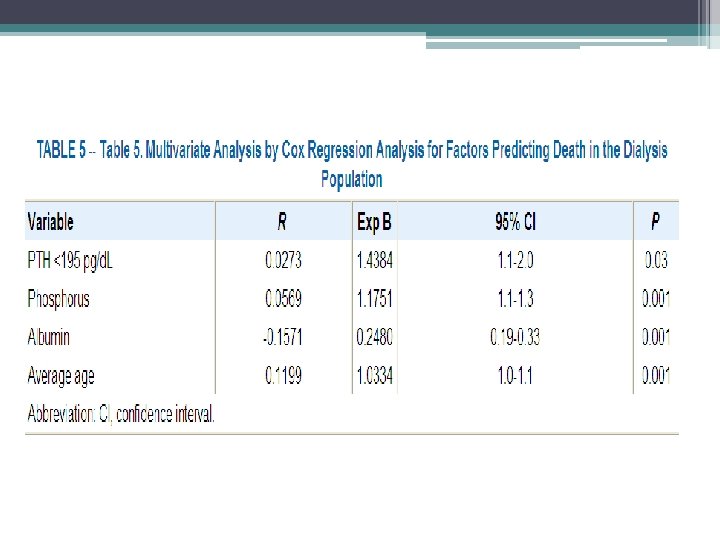

• Similar trends with respect to race and gender, however overall incidence of hip fractures in the dialysis population was 17. 4 times greater than that in the general population • Age at time of fracture in dialysis population was 11 to 15 years younger than their counterparts in the general population • 1 year mortality rate 64% after hip fracture in dialysis patients compared to 15% to 20% in general population • Bone biopsy were not systemically performed, can not exclude a contribution from aluminum bone disease

Bone Biopsy

Guidelines • 2003 K/DOQI guidelines ▫ Bone biopsy is not necessary for settings, it can be considered in patients with ESRD and the following Fractures with minimal or no trauma i. PTH levels between 100 and 500 pg/ml in association with unexplained hypercalcemia, severe bone pain, or unexplained increases in bone alkaline phosphatase activity Suspected aluminum bone disease • 2009 KDIGO ▫ Bone biopsy is reasonable in the following settings: Unexplained fractures, unexplained hypercalcemia, and /or unexplained hypophosphatemia Persistent bone pain Possible aluminum toxicity Before therapy with bisphonates

Additional implications • Establish ABD in symptomatic patients with serum PTH levels below 100 pg/ml • Determine the extent of bone aluminum accumulation prior to chelation therapy with DFO ▫ Also prior to parathyroidectomy, since low bone turnover disease with enhanced aluminum deposition may be precipitated if there is significant aluminum overload • Direct therapy in patients with intermediate PTH levels (100 -450 pg/ml)

Barriers to performing bone biopsies • Cost • Invasive • Lack of local resources to properly procure, process, and reliably interpret the bone biopsy • Reimbursement • Can other noninvasive test reliably aid in diagnosis of renal bone disease?

PTH • Intact serum PTH values below 100 pg/m. L are associated with a decreased likelihood of osteitis fibrosa and an increased incidence of adynamic bone disease. • An intact serum PTH level above 450 pg/m. L is typically associated with hyperparathyroid bone disease and/or mixed uremic osteodystrophy. • Intermediate PTH levels between 100 and 450 pg/m. L may be associated with normal, elevated bone remodeling, or even reduced bone remodeling. • Circulating intact PTH levels predict the presence and severity of hyperparathyroidism; they may not, however, predict underlying bone disease, particularly when PTH levels are only moderately elevated • Second generation assays may be the optimal test for assessing abnormal parathyroid hormone dynamics, all current data is based on intact PTH, however the NKF/DOQI guidelines currently state that is premature to rely clinically on the newer assays

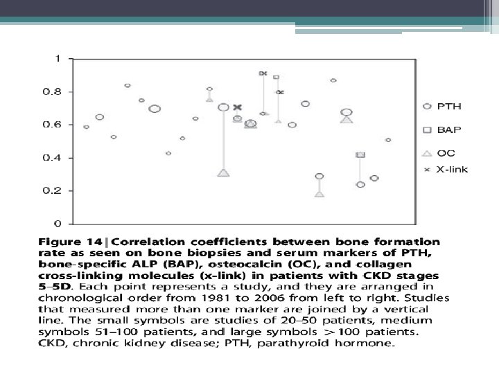
New worldwide trends in presentation of renal osteodystrophy and its relationship to parathyroid hormone levels. Clinical Nephrology 2005 Apr; 63(4): 284 -9. Gal- Moscovici A, Popovtzer MM • study was undertaken to evaluate the current prevalence of different forms of bone disease in a large population on chronic hemodialysis and its relationship to PTH levels • 96 chronic hemodialysis patients underwent double tetracyclinelabeled bone biopsy • Serum PTH levels were obtained in 52 patients at the time of biopsy. • 40% of all patients were affected by osteitis fibrosa , in the remaining 60%, various forms of low-turnover bone disease were observed • There was no correlation between PTH and BFR (Bone formation rate) in all patients and in subgroups whose PTH levels ranged between 150 - 500 and 500 - 1, 200 pg/ml (r = 0. 027, r = 0. 21) • A close correlation between PTH and BFR (r = 0. 84, p < 0. 05) was found only in the subgroup with a PTH level (0 -150 pg/ml) ranging low-turnover bone disease.

KDIGO clinical practice guidelines for the diagnosis, evaluation, prevention, and treatment of chronic kidney disease-mineral and bone disorder (CKD-MBD)
 Osteocalcin Tartrate-resistant acid phosphatase")
Bone Turnover Markers • • • Bone-specific alkaline phosphatase (b-ALP) Osteocalcin Tartrate-resistant acid phosphatase (TRAP) Collagen degradation products FGF-23 Osteoblasts secrete b-ALP, osteocalcin, and collagen based bone markers • Osteoclasts secrete TRAP • Osteocytes secrete FGF-23 in response to high serum phosphorous levels
![KDIGO 2009 recommendations • In patients with CKD stage 3 -5, [we] suggest that](http://slidetodoc.com/presentation_image_h/a6d55ed0710ef302b80199798f5c88ff/image-92.jpg "KDIGO 2009 recommendations • In patients with CKD stage 3 -5, [we] suggest that")
KDIGO 2009 recommendations • In patients with CKD stage 3 -5, [we] suggest that measurements of serum PTH or bone- specific alkaline phosphatase can be used to evaluate bone disease because markedly high or low values predict underlying bone turnover (2 B)* • • Level 2 weak recommendation Grade B moderate quality of evidence • Bone biopsy is not practical in the majority of clinical patients, when PTH and b-ALP are above or below thresholds, they can be used to estimate bone turnover. Large discrepancies between serum PTH and b-ALP should prompt further investigation •

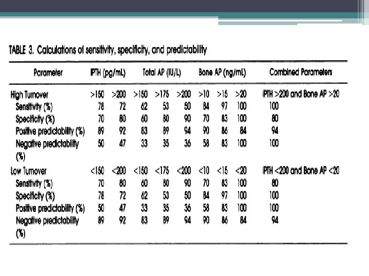
Plasma total versus bone alkaline phosphatase as markers of bone turnover in hemodialysis patients Urena P, Hruby M, Ferreira A, et al. Service de Néphrologie et d'Hémodialyse, Clinique de l'Orangerie, Aubervilliers, France. JASN 1996 • This study surveyed 42 hemodialysis patients who underwent a systematic transiliac bone biopsy for histomorphometry study. • Plasma b. AP values were compared with those of two other plasma markers of bone metabolism, t. AP and intact parathyroid hormone (i. PTH), for the correlations with bone histomorphometric parameters. • Patients with high-turnover bone disease (N = 32) had significantly higher plasma b. AP levels than patients with normal or low bone turnover (N = 10) • Bone formation and resorption were highly correlated in patients with high bone turnover, and plasma b. AP levels were positively correlated with bone resorption parameters. • The bone formation rate was better correlated with plasma b. AP levels than with either plasma t. AP or i. PTH concentrations. • Plasma b. AP level equal or higher than 20 ng/m. L, either alone or combined with plasma i. PTH of 200 pg/m. L, had the highest sensitivity, specificity, and predictability values for the diagnosis of high-turnover bone disease, and formally excluded patients with normal or LTBD

Total alkaline phosphatase bone specific alkaline phosphatase Intact PTH

KDIGO clinical practice guidelines for the diagnosis, evaluation, prevention, and treatment of chronic kidney disease-mineral and bone disorder (CKD-MBD)
![KDIGO 2009 recommendations • In patients with CKD stages 3 -5 D, [we] suggest](http://slidetodoc.com/presentation_image_h/a6d55ed0710ef302b80199798f5c88ff/image-98.jpg "KDIGO 2009 recommendations • In patients with CKD stages 3 -5 D, [we] suggest")
KDIGO 2009 recommendations • In patients with CKD stages 3 -5 D, [we] suggest not to routinely measure bone- derived turnover markers of collagen synthesis and breakdown. (2 C) • Level 2 weak recommendation • Grade c low quality of evidence • Collagen-based markers of bone turnover, measured in the serum, have not been extensively evaluated in patients with CKD stages 4– 5. • The available studies show that these markers do not predict clinical outcomes or bone histology any better than does circulating PTH or b-ALP. • Therefore, at this time, they are not recommended for diagnostic purposes in patients with later stages of CKD–MBD.

K/DOQI practice guidelines PTH levels • 35 to 70 pg/m. L for patients with an estimated GFR of 30 to 59 m. L/min per 1. 73 m 2 (stage 3 chronic kidney disease) • 70 to 110 pg/m. L for patients with an estimated GFR of 15 to 29 m. L/min per 1. 73 m 2 (stage 4 chronic kidney disease) • 150 to 300 pg/m. L for patients with an estimated GFR less than 15 m. L/min per 1. 73 m 2 (stage 5 chronic kidney disease)

Summary • Mineral and bone disorders are complex abnormalities that cause morbidity and decreased quality of life in patients with CKD. • The prevalence of the different types of bone disease has changed with adynamic bone disease being the most common, likely due to an increasing diabetic population and more aggressive PTH therapeutic parameters • The principal factor underlying ABD appears to be oversuppression of PTH release, which may be induced by the relatively high doses of vitamin D analogues and possibly calcium-based phosphate binders • Patient with ABD are at increased risk of fractures and cardiovascular calcification • Bone biopsy remain gold standard for diagnosing bone disease • Intact PTH levels predict the presence and severity of hyperparathyroidism but not necessarily the presence of underlying bone disease • Bone specific alkaline phosphatase may provide useful information in conjunction with PTH measurements • Additional evidence-based evaluation is required to determine the correlation of outcomes with various biochemical parameters

References • • • Pictures: L. Darryl Quarles, MD Up To Date American Society of Bone and Mineral Research Internal Society of Nephrology National Kidney Foundation Journal of American Society of Nephrology Dialysis and Transplantation American Journal of Kidney Disease Comprehensive Clinical Nephrology 4 th ed. ▫ Floege J, Johnson R, and Feehally J.
- Slides: 101