Spectrophotometry Xray Fluorescence XRF Xray Fluorescence Spectrophotometry is


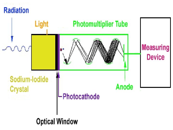
 Scintillation detector 1. When an X-ray photon enters the Na. I crystal")

- Slides: 17
Spectrophotometry X-ray Fluorescence
XRF • X-ray Fluorescence Spectrophotometry is a non-destructive method in which samples are bombarded with X-ray radiation and the residual X-rays that fluoresce are measured.
Advantages • X-ray spectra are unaffected by the physical and chemical state of an element with minimal spectral interference. • The technique can be performed with bulk solids, powders, slurries and liquids. • All elements above atomic number 17 can be analysed. Those between 9 and 17 require some adaptation of procedure. • Concentrations from fractional ppm to 100%, with precision of 0. 1 - 0. 3% attainable. • Typical measurement times are about 20 to 60 seconds per element for a sequential instrument whereas multichannel instruments can measure 30 elements in a couple of minutes. • Running costs are low despite high initial costs of equipment and the replacement costs of X-ray tubes.
Disadvantages • The inability to measure elements below atomic number 9. • Because of the short penetration power of X-rays, there is a need for homogeneous sample and standard preparation to minimise the effects of surface texture and particle size. • More sensitive methods are available through ICP-OES and ICP-MS. • In spite of rapid analysis time the preparation of samples, standards and implementation of standard addition is time consuming. • Standards need to be similar to unknowns in all respects, which may be hard to achieve. • XRF analysis requires highly trained staff to operate the equipment.
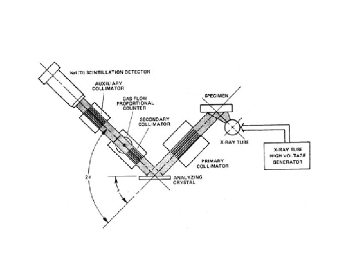
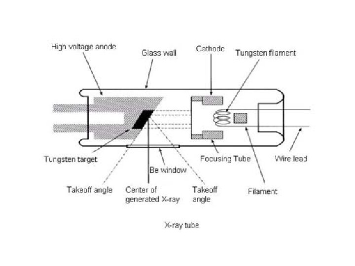
Components • An X-ray spectrometer consists of five main components: – The X-ray tube – The specimen chamber – The collimators – The crystal – The detector
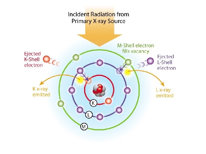
X-ray Spectra
X-ray Spectra
Crystal • Is used as a diffraction grating for the analysis which disperses the X-ray beam so it can be read by the detector.
Na. I(Tl) Scintillation detector 1. When an X-ray photon enters the Na. I crystal inside the detector, the I atoms are placed in an excited state. 2. As the atoms return to lower energy states, they give off a flash of light. 3. The light strikes a photocathode, which releases photoelectrons that are amplified in a photomultiplier tube. 4. The voltage produced by the photomultiplier tube is proportional to the energy of the detected X-ray.
Applications Soil surveys Mining – Iron Ore Cement production Ceramic and Glass Manufacturing Metallurgy Petroleum Industry