Specialized Connective Tissues Specialized Connective Tissues this group


")

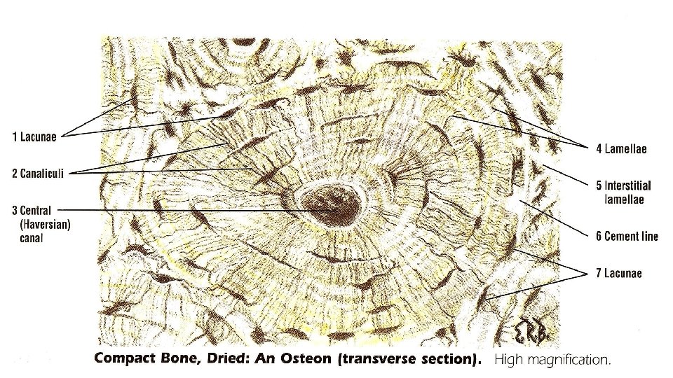
 surround the central canals")
 - basically bone tissue with many spaces/struts and is")


- Slides: 33
Specialized Connective Tissues
Specialized Connective Tissues -- this group includes cartilage, bone, and blood. Cartilage and bone form the skeletal framework of the body while blood is the vascular (transport) tissue.
Cartilage is a very rubbery tissue made by cells called chondroblasts. Cartilage does not have any blood vessels. There are three types of cartilage in the human body and they classified depended the different types of protein fibers within them and how they are arranged. .
1 -Hyaline - most common, found in the ribs, nose, larynx, trachea. Is a precursor of bone. 2 -Fibro- is found in invertebral discs, joint capsules, ligaments. 3 -Elastic - is found in the external ear, epiglottis and larynx.
All three types of cartilage contain the same cells, chondrocytes. Chondroblasts are called chondrocytes after they are done producing the cartilage and are just sitting within the rubbery tissue
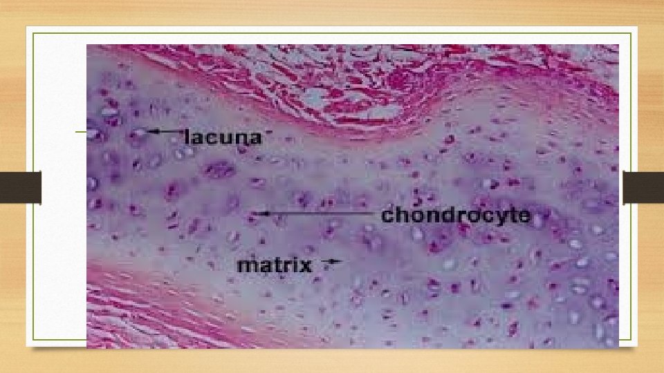
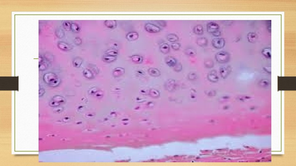
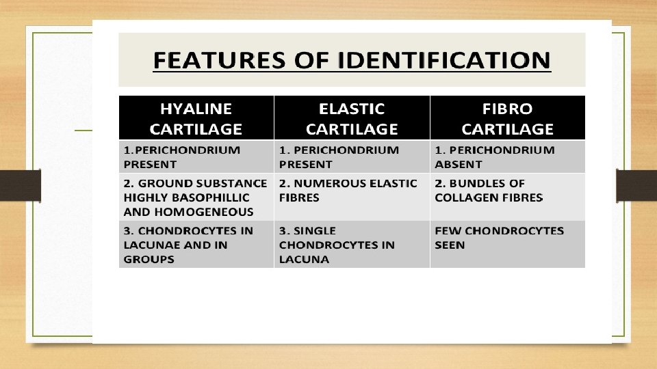
- Hyaline cartilage - very rubbery, soft cartilage - Hyaline cartilage is covered externally by a fibrous membrane, called the perichondrium. This membrane contains vessels that provide the cartilage with nutrition. it will be found to consist of cells (chondrocytes) of a rounded or bluntly angular form, lying in groups of two or more in a granular or almost homogeneous matrix. Chondrocytes arranged in groups of two or more, The cells are contained in cavities in the matrix, called cartilage lacunae ,
The matrix appears as a smooth, solid, blue or pink-coloured substance. Fine protein fibres, are embedded in the matrix, but they are not visible with the light microscope since they do not stain well.
Hyaline cartilage
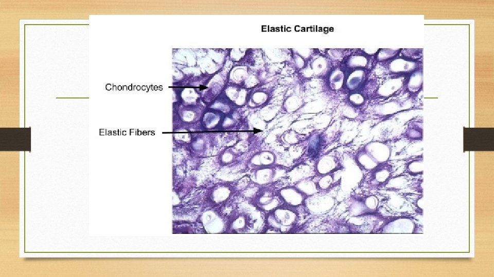
- Elastic cartilage - this is basically hyaline cartilage that contains lots of elastic protein fibers (elastin) - this type of cartilage allows for flexibility and makes structures elastic
Elastic cartilage is histologically similar to hyaline cartilage but contains many yellow elastic fibers lying in a solid matrix. These fibers form bundles that appear dark under a microscope. The chondrocytes lie between the fibers
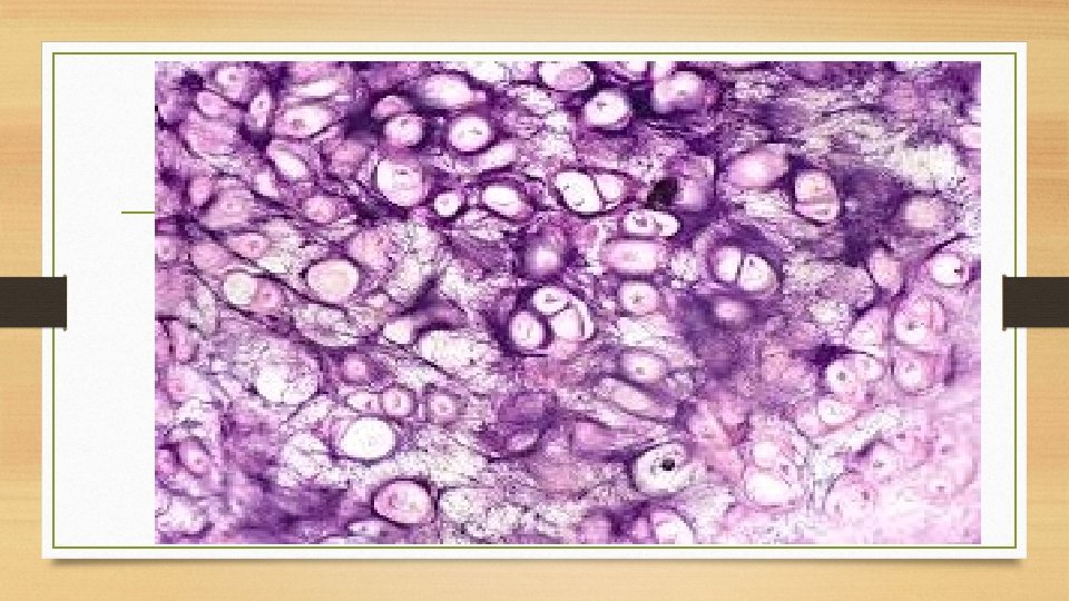
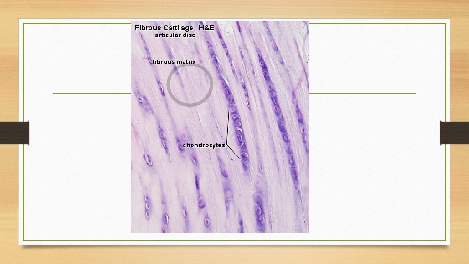
Fibrocartilage - This is the most tough of the three - it is very densely packed with collagen fibers - it is very useful for resisting compressive forces and physical shock
Bone tissue can be classified in several ways, including texture, matrix arrangement, maturity, and developmental origin. There are three key cells of bone tissue. Osteoblasts, Osteocytes and Osteoclasts They each have unique functions and are derived from two different cell lines.
The bones themselves are formed from several different connective tissues, including: Bone (called "Osseous") tissue, Periosteum, Red Bone Marrow, Yellow Bone Marrow, and Endosteum.
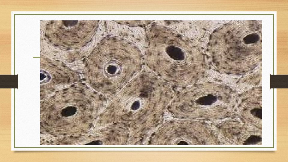
Bone tissue can be divided into the following categories: A-Compact bone ; mainly serves a mechanical function. This is the area of bone to which ligaments and tendons attach. It is thick and dense in texture without cavities; it is the shell of many bones and surrounds the trabecular bone in the center; it consists mainly of haversian systems or secondary osteons
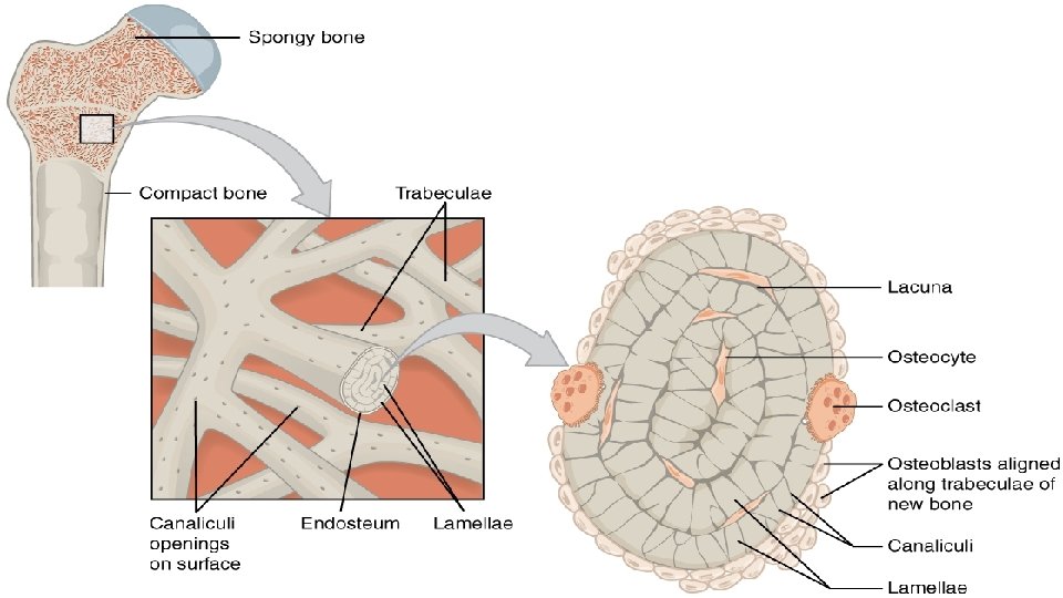
Compact bone - filled with central canals - cells (osteocytes) surround the central canals - the circle of cells that are formed by each central canal and the cells surrounding it are called the osteon - we use bone for structural support, protecting soft organs and mineral storage
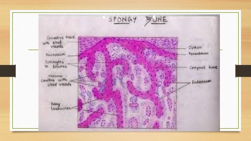
B-Spongy bone(trabecular bone, cancellous bone) - basically bone tissue with many spaces/struts and is covered by compact bone - this is where you find red bone marrow - spongy bone is found on the ends of long bones and within flat and in some irregular bones.
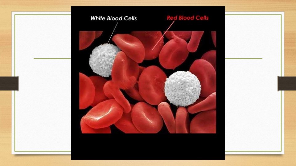
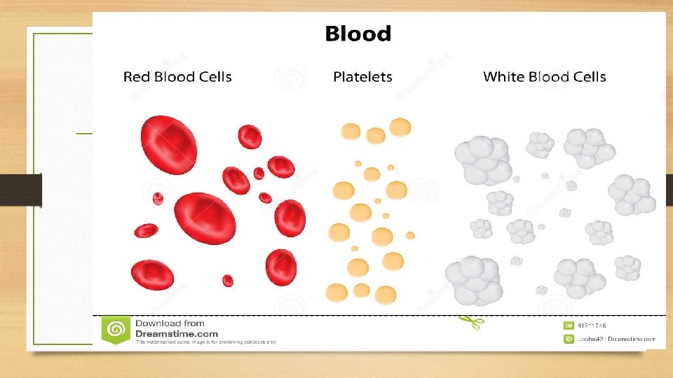
Blood is a highly specialized tissue composed of many different kinds of components. Blood is a constantly circulating fluid providing the body with nutrition, oxygen, and waste removal. Blood is mostly liquid, with numerous cells and proteins suspended in it. Four of the most important ones are red cells, white cells, platelets.
Red cells, or erythrocytes, They lack a cell nucleus and most organelles. Mature red blood cells are flexible and oval biconcave disks.
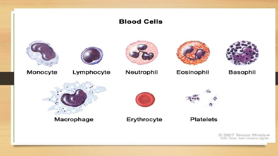
White cells, or leukocytes, All white blood cells have nuclei, which distinguishes them from the other blood cells, Types of white blood cells can be classified in standard ways. Two pairs of broadest categories classify them either by structure (granulocytes or agranulocytes). These broadest categories can be further divided into the five main types: neutrophils, eosinophils, basophils, lymphocytes, and monocytes.
Platelets have no cell nucleus: they are fragments of cytoplasm that are derived from the megakaryocytes of the bone marrow, and then enter the circulation. These unactivated platelets are biconvex discoid (lens-shaped) structures, 2– 3 µm in greatest diameter