SPECIAL STAINS FOR NUCLEIC ACID Histochemistry Based on

SPECIAL STAINS FOR NUCLEIC ACID

Histochemistry: Based on chemical reactions between cell components and stains. • The end products of the reaction are permanent, colored precipitates that can be viewed under the microscope. • There are stains specific to each component of the cell, based on the basic or acidic nature of the dye.

Basic Principles of Histochemistry � Histochemistry combines the methods of histology with those of chemistry or biochemistry, biochemistry to reveal the biochemical composition of tissues and cells beyond the acid-base distribution shown by standard staining methods (H & E), without disrupting the normal distribution of the chemicals.

Application � Identify, quantify, and localize �Chemical substances �Gene expression �Biological structures, organelles �Specific cell types � Clarify cell and tissue structure and morphology.

Limitations Of the Current Methods � Cannot be used for real time in vivo analysis of any tissue (requires the removal and killing of the tissue). tissue � Uses in humans limited to biopsied tissues � Tissue preparation and histo-chemical analysis may alter specimen morphology or chemistry depending on the methods and materials used

The goal of Histochemistry � Presentation of Normal Chemical Distribution. � Presentation of Normal Chemical Composition. � Specificity of the Reaction. � Detectability of the Reaction Product. � Insolubility of the Reaction Product

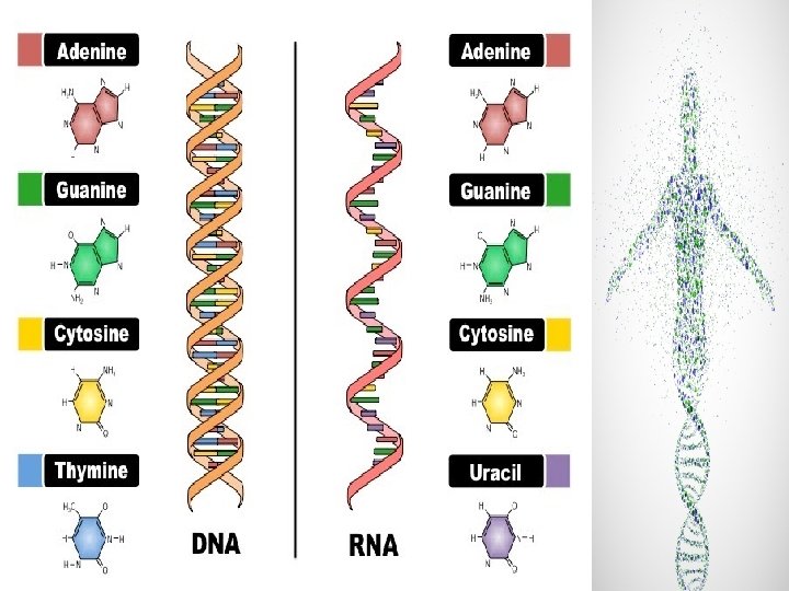
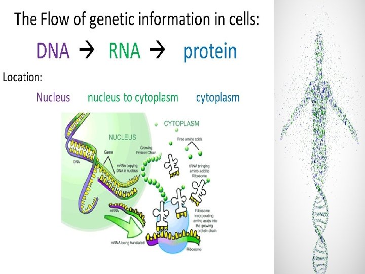
Nucleic Acids � The nucleic acids, DNA and RNA, RNA can be localized by specific and non specific methods � DNA is found mainly in nuclei, nuclei and its amount is much the same in every cell. � RNA is found both in nuclei and in cytoplasm, cytoplasm and its amount varies widely, depending on a cell's functional state

Cont. � DNA STAINING TECHNIQUES need to be improved to achieve more accurate data. � Conventional histological procedures can preserve tissue for morphological characterization, but not necessarily for macromolecules. � Analysis of DNA plays an important role in molecular biology, pathology, histochemistry, and immunohistochemistry. � A cell can be quantified by measuring total DNA content, normal or abnormal, abnormal in its nucleus

Objectives � Explain the methods used in demonstrating nucleic acids. � Describe the techniques and principles of the methods used.

Staining used for N. A � Feulgen's reaction: determine the amounts of DNA. � Methyl Green Pyronin Stain to determine DNA and RNA � Acridine orange: The fluorescence is yellow green if the complex contains DNA and redorange if it contains RNA.

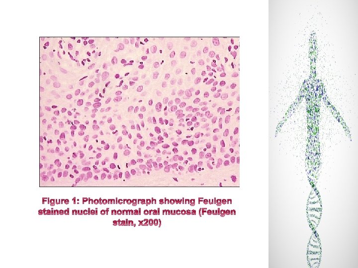
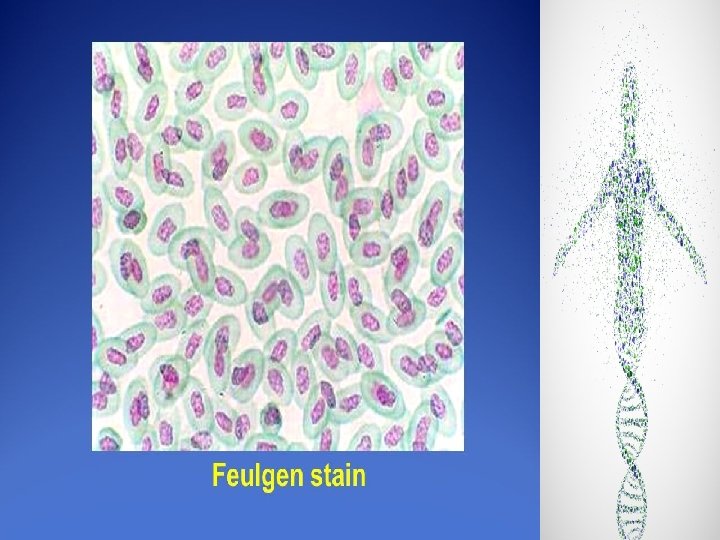
Feulgen Stain � The Feulgen nuclear reaction for the specific staining of DNA in cytohistochemical samples in situ was introduced by Feulgen and Rossenbeck in 1924. � These authors devised a chemical treatment, HCI hydrolysis, to produce free aldehyde groups in the DNA backbone structure that could be detected by a colored reaction for aldehydes.
 1 M HCL acid - used for")
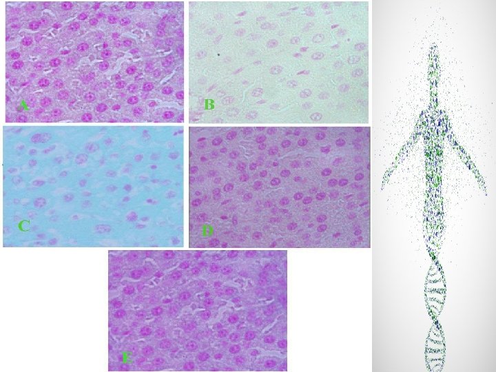
FEULGEN STAIN SOLUTIONS USED ARE : A) 1 M HCL acid - used for acid hydrolysis to break the purine-deoxyribose bond and yield an aldehyde. - Done at 60 C (HCL should be preheated to 60 C) - Time (minutes) depends upon the fixative used - For carnoy’s and formalin – 8 minutes used B) Schiff reagent - The aldehydes are then demonstrated by schiff’s reagent C) Bisulfite solution RESULT DNA : red-purple CYTOPLASM : green

DNA BY FEULGEN STAIN

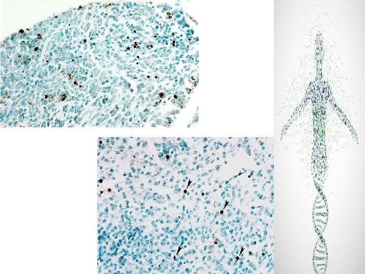
METHYL GREEN PYRONIN Y � Methyl Green-Pyronin Stain is for identification of Plasma Cells in Tissue Sections. Methyl Green-Pyronin Stain is for “In Vitro Diagnostic use. ” � Methyl Green-Pyronin (MGP) (MGP is used to demonstrate plasma cells and RNA in tissue sections and cytologic preparations. The procedure is a simplified method for use with formalin fixed tissue or alcohol-ether fixed smears. � Pyronin stains the cytoplasm of plasma cells and most nucleoli red.

METHYL GREEN PYRONIN METHOD Reagents : 1. Methyl green - impure dye contains methyl violet – removed by washing with chloroform - pure methyl green specific for DNA - NH 2 of dye reacts with phosphate of DNA 2. Pyronin - binds to any negatively charged tissue constituent - apart from RNA, binds to acid mucins and cartilage RESULTS – DNA & cytoplasm: green-blue RNA : red

Depapffinization by Xylene Hydration by Alcohol Staining by MGP for 5 -30 min. Dehydration by n-butyl alcohol for 5 min x 2. Clear by toluene for 5 min.
- Slides: 24