Special Senses Eye Ear and Skin Functions of


 • Made up of keratin cells:")


: A burn only injuring the epidermis.")
: A burn that destroys some epidermis")
 burn: A burn that destroys the epidermis, and")


- Slides: 54
Special Senses Eye, Ear, and Skin
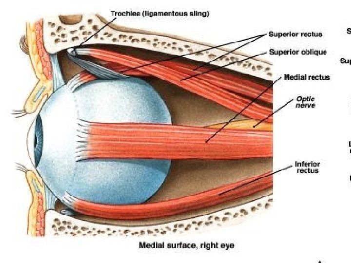
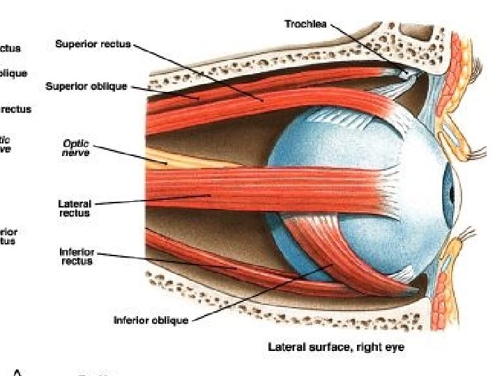
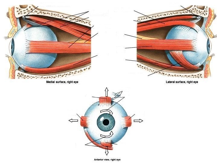
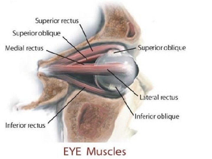
Functions of eye muscles • 1. Superior rectus: Rotates the eye upward and toward the midline (oculomotor) • 2. Inferior rectus: Rotates the eye downward and toward the midline (oculomotor) • 3. Medial rectus: Rotates the eye toward the midline (oculomotor) • 4. Lateral rectus: Rotates the eye away from the midline (abducens) • 5. Superior oblique: Rotates the eye downward and away from the midline (trochlear) • 6. Inferior oblique: Rotates the eye upward and away from the midline (oculomotor)
2. 1. 3. 6. 5. 4.
• • 7. What are the 6 eye muscles? 8. What is myopia? 9. What is hyperopia? 10. What is astigmatism? 11. Explain what causes motion sickness. 12. What are the auditory ossicles? 13.
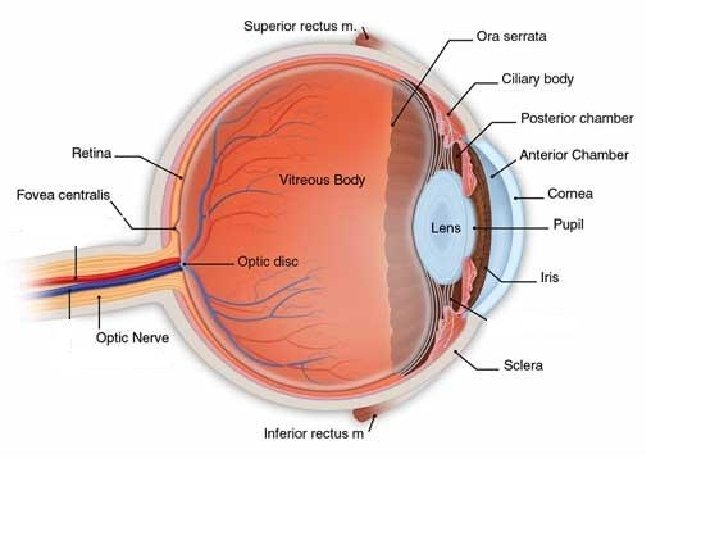
Eye functions • 3 Layers: Outer tunic, middle tunic, and inner tunic. • Outer tunic: • Cornea- “window of the eye”, focuses entering light rays • Sclera- white part of eye, attachment for eye muscles • Optic nerve-Transmits info. to brain (second cranial nerve)
Eye functions Middle tunic: Ciliary body-Holds the lens in position Lens-Focuses on objects near and far Iris-Colored part of eye, separates anterior and posterior chambers • Ciliary body-Secretes a watery fluid used to give the eye nutrients and form • Ora Serrata- The separation between the photosensitive region of the retina and the non-photosensitive region. • Pupil-Circular opening in the center of the iris where light passes through. • •
Eye functions • Inner tunic: • Retina-Captures images seen in the environment and sends them to certain parts of the brain, like film in a camera. • Fovea centralis-The region of the retina that produces the sharpest vision. • Optic disc-Area where nerve fibers from the retina pass through to the optic nerve, often called the “blind spot” of the eye.
Lacrimal sac Superior/inferior
Lacrimal apparatus • Secretes tears and carries them into the nasal cavity • Located on the superior and lateral side of the eye • Tears are secreted continuously and pass out through tiny tubules and flow downward and medially across the eye. • There, they’re collected in the superior/inferior lacrimal canals and empty into the lacrimal sac (which lies in the groove of the lacrimal bone). • It empties into the nasolacrimal duct and then into the nasal cavity. • The cells of the conjunctiva also secrete a tear like fluid that works with the fluid of the lacrimal glands to lubricate the eye and eye lids. Tears contain an emzyme (proteins that cause chemical reactions in the body) called lysozyme that has antibacterial properties that reduce eye infections.
Rods/cones/fovea centralis • There are 2 photoreceptors in the eye, rods and cones: • Rods: Provide vision in dim light (due to extreme sensitivity), produce colorless vision, and produce general outlines of objects. • Cones: Detect color (red, green, blue), provide sharp images (because rods sometimes have light hit many of them simultaneously). • Fovea Centralis: Has no rods and has many cones, so it produces sharper images. We will position an object so it falls on our fovea centralis when we’re trying to focus on something.
• Myopia: Also known as near-sightedness, happens when the eye is too long causing the light waves to focus in front of the retina, blurring the image.
• Hyperopia: Also known as far-sightedness, happens when the eye is too short causing the light waves to focus behind the retina, blurring the image.
• Astigmatism: When the curvature of the cornea or lens has a “bump” in it causing some parts of an image to be clear and others to be blurred. This causes a person to constantly focus in and out, leading to headache.
External ear Helix Scapha Antihelix Pinna Concha Earlobe
Middle ear Malleus Incus Stapes Tympanic membrane Auditory canal
Process of interpreting sound • When sound enters the auditory canal (also known as the external acoustic meatus) the sound waves alter the pressure on the tympanic membrane. , the tympanic membrane creates vibrations in response to the sound waves. • The tympanic membrane is connected by the auditory ossicles which is a name for the three small bones: malleus, incus, and stapes (sometimes called the hammer, anvil, and stirrup by the less intelligent) *attached in that order
Inner ear Semicircular canals Vestibulocochlear nerve Cochlea
Equilibrium • Equilibrium is attained by multiple sources working together. • Eye sight: to see the environment around you and adapt to changes • Cerebellum: Tells body to shift to maintain balance and which muscles need to be used • Semicircular canals: Contains fluid that helps the brain detect balance. Usually, when the head/torso turn the fluid remains stationary because of inertia (resistance of change in motion).
Motion sickness? • The cause is unknown but it is believed to be a contradiction by the brain where the eyes tell the brain the body is in motion but the fluid in the ears say the body is motionless. This results in the brain sending a signal to the medulla oblongata to vomit.
Steps in generating impulses from the ear • 1. sound waves enter auditory canal • 2. waves change pressure of tympanic membrane thus producing vibrations • 3. auditory ossicles amplify and transmit vibrations • 4. Vibrations are transmitted to cochlea • 5. Different frequencies stimulate receptor cells in the inner ear • 6. Receptor cells release neurotransmitters to relay signals to sensory neurons • 7. Sensory impulses are carried to vestibulocochlear nerve • 8. The temporal lobe interprets the sensory impulses • …and they think we EVOLVED that ability? !? !
The whole shabang!
Epidermis
Epidermis Dermis
Epidermis Dermis Subcutaneous layer
Epidermis Stratum corneum
Epidermis Stratum corneum Stratum lucidum
Epidermis Stratum corneum Stratum lucidum Stratum granulosum
Epidermis Stratum corneum Stratum lucidum Stratum granulosum Stratum spinosum
Epidermis Stratum corneum Stratum lucidum Stratum granulosum Stratum spinosum Stratum basale
Epidermis Stratum corneum Stratum lucidum Stratum granulosum Stratum spinosum Stratum basale Basement membrane
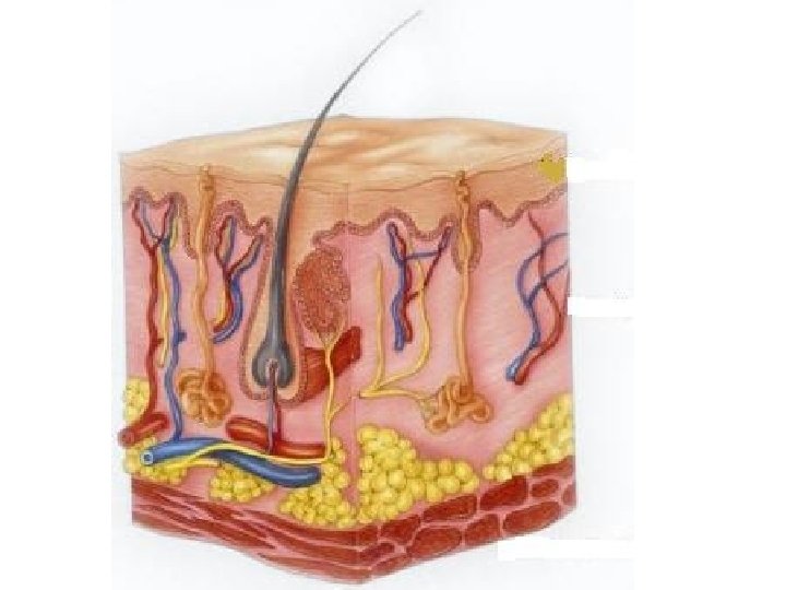
Epidermis • No blood vessels (except stratum basale) • Made up of keratin cells: protein cells in the hair, skin, and nails. • Stratum corneum: Strands of tough, waterproof, dead skin cells. • Stratum lucidum (found mostly in thick skin such as palms/soles of feet): • Stratum granulosum: 3 -5 layers of flattened granular shaped cells that contain shrunken fibers of keratin. • Stratum spinosum: Many layers of cells with developing keratin fibers • Stratum basale: Deepest layer of epidermis made up of cube shaped cells that divide and grow. • Basement membrane: Separates epidermis from dermis.
Hair shaft
Hair shaft Pore
Hair shaft Pore Sebaceous gland
Hair shaft Pore Sebaceous gland Arrector pili muscle
Hair shaft Pore Sebaceous gland Arrector pili muscle Hair root
Hair shaft Pore Sebaceous gland Arrector pili muscle Hair root Hair follicle
Hair shaft Pore Sebaceous gland Arrector pili muscle Hair root Hair follicle Sweat glands
Hair shaft Pore Sebaceous gland Arrector pili muscle Hair root Hair follicle Sweat glands Adipose tissue Muscle layer
Accessory structures of skin • Adipose tissue: Helps conserve body heat and controls body temp. • Hair follicle: Deep depression in the dermis where cells form hair • Hair root: The portion of the hair embedded in the skin • Hair shaft: The portion of the hair outside the skin which has become keratinized (hardened and will die) • Arrector pili muscle: Makes hair stand (controlled by nervous impulses; cold, emotions) • Sebaceous gland: Produce sebum (collection of fat and cellular waste). Used to keep hair soft, pliable, and waterproof. Also cause acne. • Sweat glands: originates with a ball-shaped coil in the deep dermis layer. Reach the surface of skin through openings called pores.
Burns • Superficial partialthickness burn (1 st degree): A burn only injuring the epidermis. Common cause for this type of burn is sunburn. Healing occurs within a few days to a few weeks with no scarring.
Burns • Deep partial-thickness burn (2 nd degree): A burn that destroys some epidermis as well as some underlying dermis. A blister usually develops. Common cause is exposure to hot objects, liquids, or flames. Healing depends on severity and location of burn, but usually will take two to four weeks.
Burns • Full-thickness (3 rd degree) burn: A burn that destroys the epidermis, and the accessory structures of the skin. The skin will become dry and leathery and may be red, black, or white. Usually caused by immersion of hot liquid, prolong exposure to fire, or corrosive chemicals. Healing does not occur and scarring is likely.
Treatment of serious burns • Autograft: Taking a thin layer of skin from another part of your body and transplanting it to the burn site. • Homograft: Taking skin from a cadaver and transplanting it to the burn site. After a period of time, an autograft can replace the homograft.
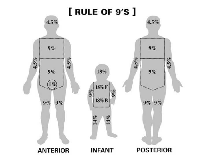
Rule of Nines • The rule of nines is a way physicians measure the severity of burns on a victim. It only applies to 2 nd and 3 rd degree burns. • Each area of the body is subdivided into regions, each representing 9% (or some multiple of 9) • The estimated survival chances are determined by the following formula: • 100% - [Age + % Skin lost]= % survival chance
Rule of Nines
Questions • 1. If you burned your face with 2 nd and 3 rd degree burns, what would be your chances of survival? • 2. If your mother or father burned their face, chest, and front of arms, what would be their chances of survival? • 3. If a 80 year old burned their back, what would be their chances of survival? • 4. Ask 5 people to randomly select portions of the body and calculate what their survival chances would be if they had 2 nd and 3 rd degree burns there.