Solid liver lesions Differential diagnosis and evaluation www


 Mass/nodule at imaging <1")

")
- Slides: 13
Solid liver lesions: Differential diagnosis and evaluation www. uptodate. com
Επιτήρηση σε ασθενείς υψηλού κινδύνου Recommendations Level of evidence Grade of recommendation Cirrhotic patients, Child–Pugh stage A and B Low Strong Cirrhotic patients, Child–Pugh stage C awaiting LT Low Strong Non-cirrhotic HBV patients at intermediate or high risk of HCC* (according to PAGE-B† classes for Caucasian subjects, respectively 10 17 and ≥ 18 score points) Low Weak Non-cirrhotic F 3 patients, based on an individual risk assessment Low Weak n Interval should be dictated by rate of tumour growth and tumour incidence in target population n 6 -month interval is reasonable and cost-effective • • 3 months: no clinical benefit 12 months: fewer early stage diagnoses and shorter survival *Patients at low HCC risk left untreated for HBV and without regular 6 -month surveillance must be reassessed at latest on a yearly basis to verify progression of HCC risk. †PAGE-B score is based on decade of age (16– 29 = 0, 30– 39 = 2, 40– 49 = 4, 50– 59 = 6, 60– 69 = 8, ≥ 70=10), gender (M = 6, F = 0) and platelet count (≥ 200, 000/µl = 0, 100, 000– 199, 999µl = 1, <100, 000 = 2): a total sum of ≤ 9 is considered at low risk of HCC (almost 0% HCC at 5 years) a score of 10– 17 at intermediate risk (3% incidence HCC at 5 years) and ≥ 18 is at high risk (17% HCC at 5 years) EASL CPG HCC. J Hepatol 2018; doi: 10. 1016/j. jhep. 2018. 03. 019
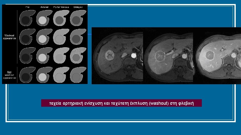
Διάγνωση σε κιρρωτικό ήπαρ (σε μη κιρρωτικούς, επιβεβαίωση με βιοψία) Mass/nodule at imaging <1 cm >1 cm Repeat US at 4 months Multiphasic contrast-enhanced CT or MRI, * or gadoxetic-enhanced MRI † Growing/changing pattern Stable‡ 1 positive technique: HCC imaging hallmarks No Biopsy unclear: Consider re-biopsy ‖ Yes Use other modality: multiphasic contrast-enhanced CT or MRI, * or gadoxetic-enhanced MRI, † or contrast-enhanced ultrasound § 1 positive technique: HCC imaging hallmarks Non-HCC malignancy/ benign No Yes Biopsy HCC *Using extracellular MRI contrast agents or gadobenate dimeglumine; †Diagnostic criteria: APHE and washout on the portal venous phase; ‡Lesion <1 cm stable for 12 months (three controls after 4 months) can be shifted back to regular 6 -month surveillance; §Diagnostic criteria: APHE and mild washout after 60 seconds; ‖Optional for centre-based programmes EASL CPG HCC. J Hepatol 2018; doi: 10. 1016/j. jhep. 2018. 03. 019
Modified BCLC staging system and treatment strategy HCC in cirrhotic liver Prognostic stage Very early stage (0) Intermediate stage (B) Advanced stage (C) Terminal stage (D) Early stage (A) Single <2 cm Multinodular, Portal invasion/ Not transferable HCC Solitary or Preserved liver unresectable extrahepatic spread End-stage 2– 3 nodules <3 cm function* Preserved liver function Preserved liver PS 0 function* PS 3– 4 function* † PS 0 PS 1 – 2 PS 0 2– 3 nodules ≤ 3 cm Solitary Optimal surgical candidate‡ Yes No Yes Transplant candidate No Treatment§ Ablation Resection Transplant Ablation Chemoembolization Systemic therapy‖ BSC Survival >5 years >2. 5 years ≥ 10 months 3 months *Child–Pugh A without ascites. Applies to all treatment options apart from LT; †PS 1; tumour-induced modification of performance capacity; ‡Multiparametric evaluation: compensated Child–Pugh class A liver function with MELD score <10, matched with grade of portal hypertension, acceptable amount of remaining parenchyma and possibility to adopt a laparoscopic/minimally invasive approach; §The stage migration strategy applies; ‖Sorafenib has been shown to be effective in first line, while regorafenib is effective in second line in case of radiological progression under sorafenib. Lenvatinib has been shown to be non-inferior to sorafenib in first line, but no effective second-line option after lenvatinib has been explored. Cabozantinib has been demonstrated to be superior to placebo in 2 nd or 3 rd line with an improvement in OS. Nivolumab has been approved in second line by FDA but not EMA based on uncontrolled Phase 2 data. EASL CPG HCC. J Hepatol 2018; doi: 10. 1016/j. jhep. 2018. 03. 019
Eastern Cooperative Oncology Group (ECOG)