Sole of the Foot Dr Qudsia Sultana Skin


- Slides: 35
Sole of the Foot Dr. Qudsia Sultana
• Skin is thick & hairless, lacks pigmentation • The subcutaneous tissue contains a lot of fat, especially in the heel • Extremely sensitive to touch due to a high concentration of nerve endings.
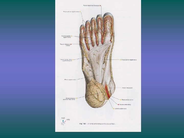
Cutaneous Nerve Supply v Medial plantar nerve v Lateral plantar nerve v Saphenous nerve v Sural nerve v Medial calcaneal branch of the tibial nerve
DEEP FASCIA ØPlanter aponeurosis ØDeep transverse metatarsal ligament ØFibrous flexor sheath ØSeptae
Deep Fascia • Lies beneath the subcutaneous tissue • Much thicker in the central part and thinner at the margins • The central thicker part forms triangular plantar aponeurosis
Plantar Aponeurosis • Protects the underlying nerves, blood vessels, and muscles. • Maintains the longitudinal arches of the foot. • Origin –muscles first layer of sole
Plantar Aponeurosis • Apex is attached to the medial and lateral tubercles of the calcaneum. • Base divides into five slips that pass into the toe • Each slip further divides into the: • Superficial band • Deep band
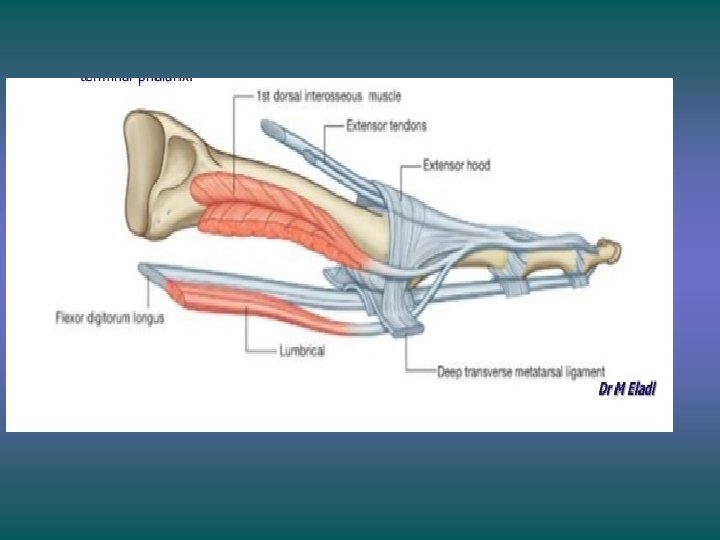
Muscles of the Sole • The sole contains both Extrinsic & Intrinsic muscles • These muscles: § Help to flex, extend, abduct, and adduct the toes § Support the arches of the foot § Are supplied by branches of tibial nerve § Are supplied by branches of posterior tibial artery § Are arranged in four layers
• The muscles of the first layer are: § Abductor hallucis § Flexor digitorum brevis § Abductor digiti minimi
• The muscles of the second layer are: • Tendons of the flexor hallucis longus • Tendons of the flexor digitorum longus from which the lumbricals arise • flexor digitorum Accessorius (quadratus plantae) • Lumbricals
• The muscles of the third layer are: • Flexor hallucis brevis • Adductor hallucis § oblique head § transverse head • Flexor digiti minimi brevis
• The muscles of the fourth layer are: • Dorsal interossei • Plantar interossei • Tendon of the peroneus longus • Tendon of the tibialis posterior
The muscles of the fourth layer • 4 Dorsal interossei • 3 Plantar interossei
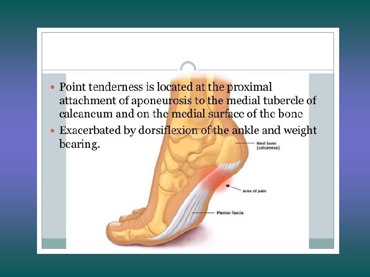
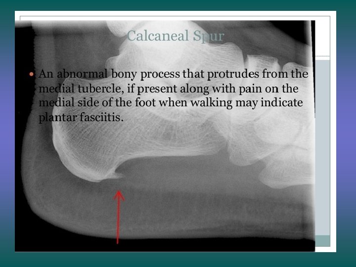
APPLIED ANATOMY • PLANTAR FASCITIS: -strainining & inflammation of plantar aponeurosis -pain over heel & med. aspect of foot; increases with passive extension of great toe& by dorsiflexion of ankle jt. -results from running & high-impact aerobics (most common hindfoot problem in runners). -calcaneal spur
Thank you
Arteries of the Sole of the Foot
• Posterior tibial artery enters the foot: § Medially under the medial malleolus § Deeper to flexor retinaculum • Divides to give the medial and lateral plantar arteries which supply the sole
Medial Plantar Artery • Arises beneath the flexor retinaculum and passes forward deep to the abductor hallucis • Ends by supplying the medial side of the big toe Gives numerous muscular, cutaneous & articular branches
Lateral Plantar Artery • Arises beneath the flexor retinaculum • Passes forward deep to the abductor hallucis and the flexor digitorum brevis • On reaching the base of the fifth metatarsal bone, the artery curves medially to form the plantar arch • At the proximal end of the first joins with the dorsalis pedis artery Gives numerous muscular, cutaneous & articular branches and plantar digital arteries
Planter Arch • Formed by lateral plantar artery and dorsal pedis artery • The arch gives rise four plantar metatarsal arteries • Three proximal perforators
Veins of the Sole of the Foot • Medial and lateral plantar veins accompany the corresponding arteries, and they unite behind the medial malleolus to form the posterior tibial venae comitantes.
Nerves of the Sole of the Foot • Tibial nerve enters the foot medially § Deeper to flexor retinaculum • Divides to give the medial and lateral plantar nerves which supply the sole
Medial Plantar Nerve • Runs forward deep to the abductor hallucis, with the medial plantar artery • Comes to lie in the interval between the abductor hallucis and the flexor digitorum brevis muscles
Branches • Muscular branches : § Abductor hallucis § Flexor digitorum brevis § Flexor hallucis brevis § First lumbrical muscle • Cutaneous branches: § Plantar digital nerves- medial 3½ toes § The nerves extend onto the dorsum and supply the nail beds and the tips of the toes.
Lateral Plantar Nerve • Runs forward deep to the abductor hallucis and the flexor digitorum brevis, in company with the lateral plantar artery • On reaching the base of the fifth metatarsal bone, it divides into: § Superficial § Deep branches
Branches From the main trunk § Muscular branches: § Quadratus plantae § Abductor digiti minimi § Cutaneous branches: § skin of the lateral part of the sole
• From the superficial terminal branch § Muscular branches to the: § Flexor digiti minimi brevis § Interosseous muscles of the fourth intermetatarsal space § Cutaneous branches § Skin lining the fourth interdigital cleft.
• From the deep terminal branch § Muscular branches to the: • Adductor hallucis • Second, third, and fourth lumbricals • All the interossei, except those in the fourth intermetatarsal space
Thank You