Sole and Arches of Foot Dr Anita Rani


- Slides: 35
Sole and Arches of Foot Dr Anita Rani Anatomy Lecture 17 th December 2016
Lesson Plan • • Introduction Skin & Superficial fascia Deep Fascia Layers Muscles Nerves & Arteries Bones: Arches Applied Anatomy
Introduction • Homologous to palm BUT • Is an organ of support and locomotion • Great toe : lost its power of mobility & prehension • Lesser four toes markedly reduced in size • Tarsal bones and Ist Metatarsal forms broad base for better support
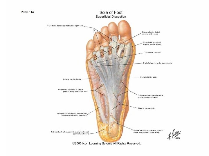
Skin • Thick for protection • Firmly adherent to underlying plantar aponeurosis • Creased
Innervation
Plantar Reflex • Babinski’s Sign
Superficial fascia • Fibrous and dense • Fibrous bands bind the skin to plantar aponeuposis • Divide the fat in to tight compartments: water cushion • Reinforce spring effect to arches • Contains superficial nerve and vessels • Forms superficial transverse metatarsal ligament
Superficial and deep transverse metatarsal ligaments
Deep Fascia • Plantar aponeurosis • Deep transverse metatarsal ligament • Fibrous flexor sheath of toes
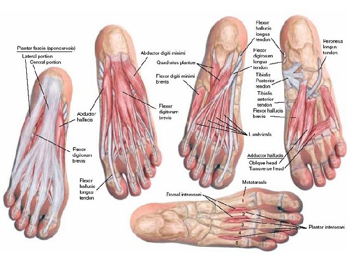
Muscles of sole of foot • 18 intrinsic + 4 Extrinsic muscles • Arranged in 4 layers • First layer : 3 [2 small abductors & 1 small flexor] • Second layer: 7 (5 +2) [ 2 long tendons of toes – FHL & FDL] + FD Accessorius & 4 lumbricals] • Third Layer: 3 [ 2 small flexors & 1 adductor] • Fourth layer : 9 ( 7+2) [2 long tendons of TP & PL + 3 PI + 4 DI]
First layer Abductor Hallucis FDB Abductor digiti minimi
Second layer
Third Layer 2 small flexors & 1 adductor Adductor Hallucis FHB FDMB
Fourth Layer
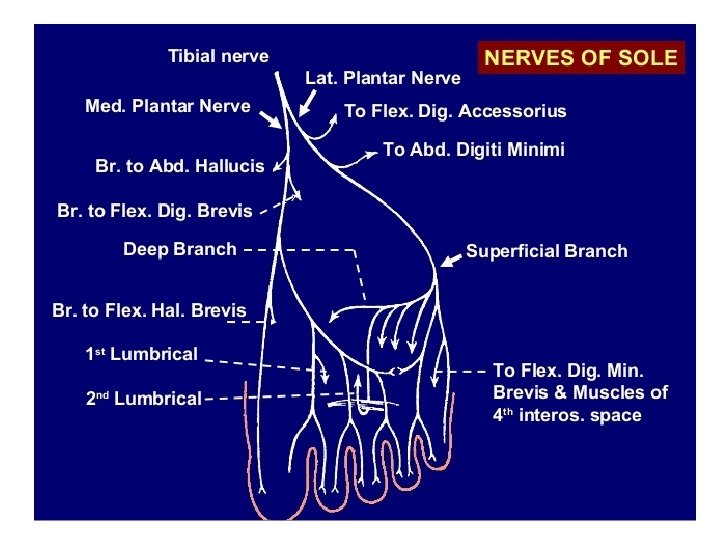
Nerves of sole
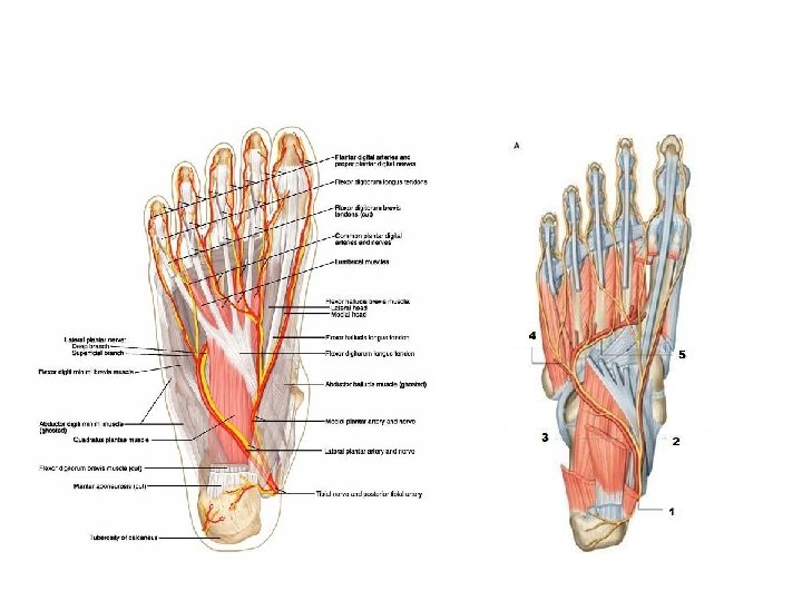
Arteries of Sole
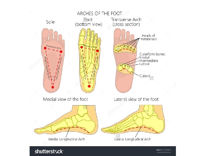
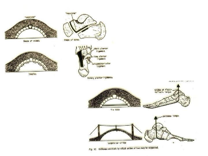
Arches of foot Foot Support the body weight & Serve as a lever to propel the body Segmented Arches help to sustain stresses of thrusts and weights.
Arches of foot serve as elastic springs for efficient walking
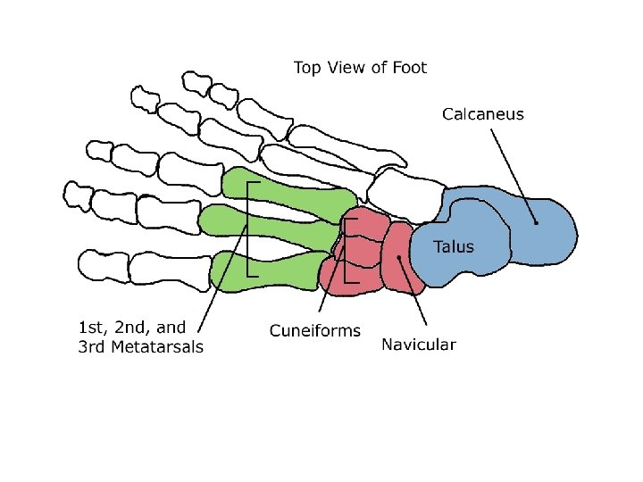
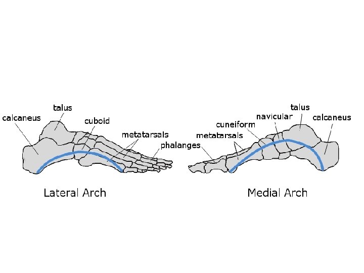
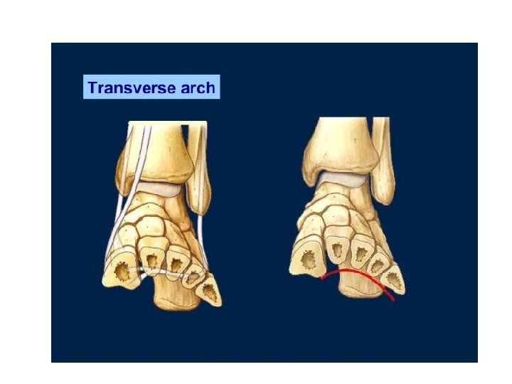
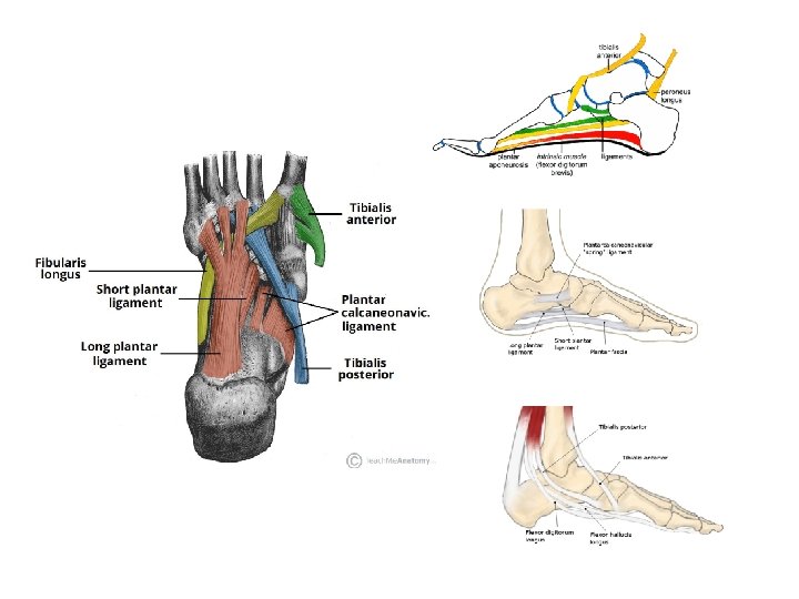
Identify the bony components….
Maintenance of Arches • Bony factor • Intersegmental ties • Tie beams/ bow strings • Slings
Pes Planus & Pes Cavus
Claw foot
Talipes deformities
Club Foot