
Smooth muscles Dr MADIHA NAZIR 10 of the
- Slides: 65
Smooth muscles Dr. MADIHA NAZIR
� 10% of the body smooth and cardiac muscles
Types of smooth muscles �Two main types 1 - multi unit smooth muscles 2 - single unit smooth muscles(unitary smooth muscles)
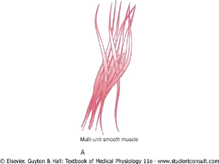
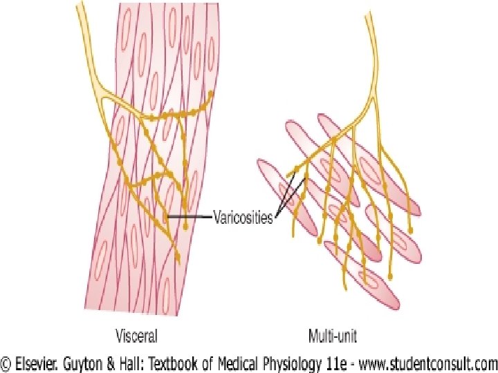
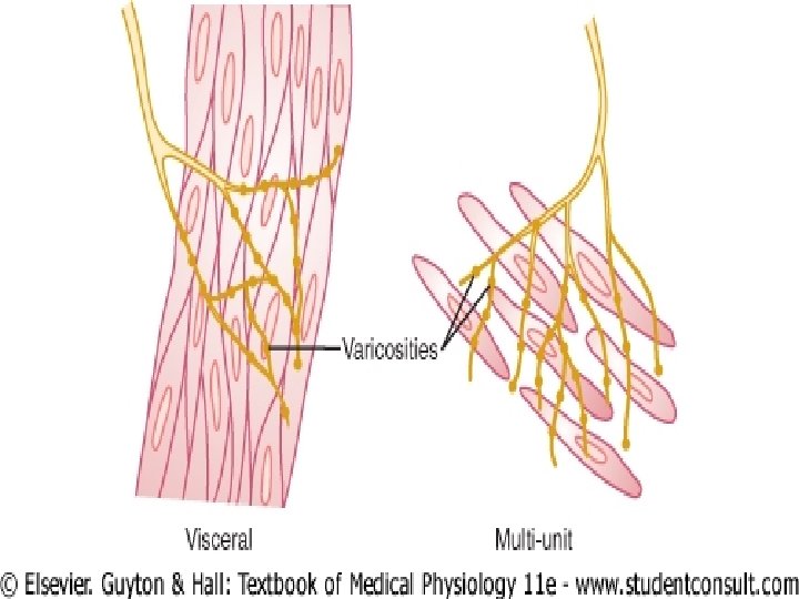
Multi unit smooth muscles �Composed of separate and discrete smooth muscle fibers �Each smooth muscle fiber is innervated by one nerve ending �Can contract independently
�Examples ciliary muscle of eye iris muscle of eye piloerector muscles
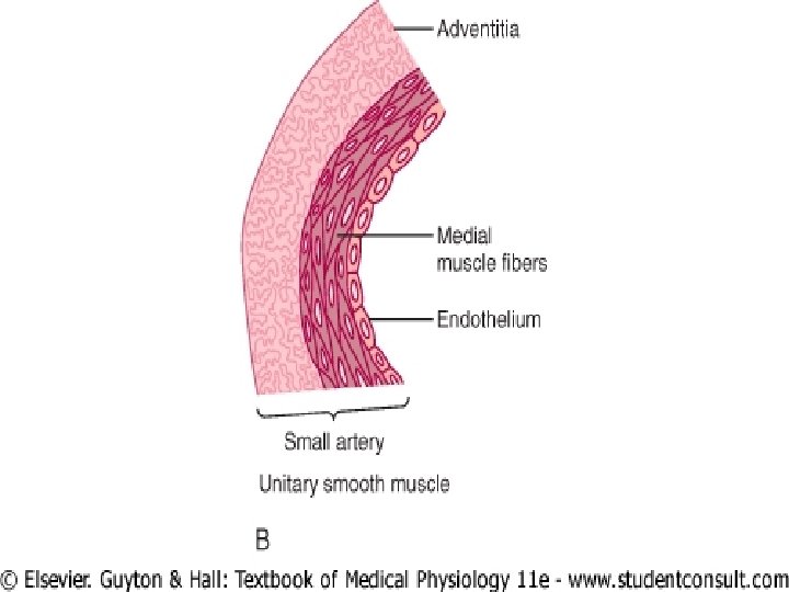
Single unit smooth muscle �Smooth muscle fibers are arranged in sheeths or bundles �When there are many layers of muscle cells , the nerve fiber only innervate the outer layer �Syncytial smooth muscle
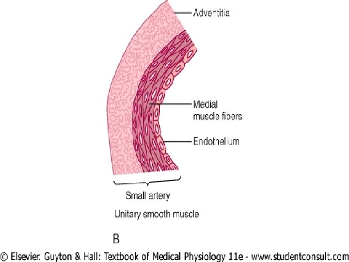
�Examples visceral smooth muscles
Physiologic anatomy �Diameter range from 1 -5 micrometer �Length range from 20 -500 micrometer
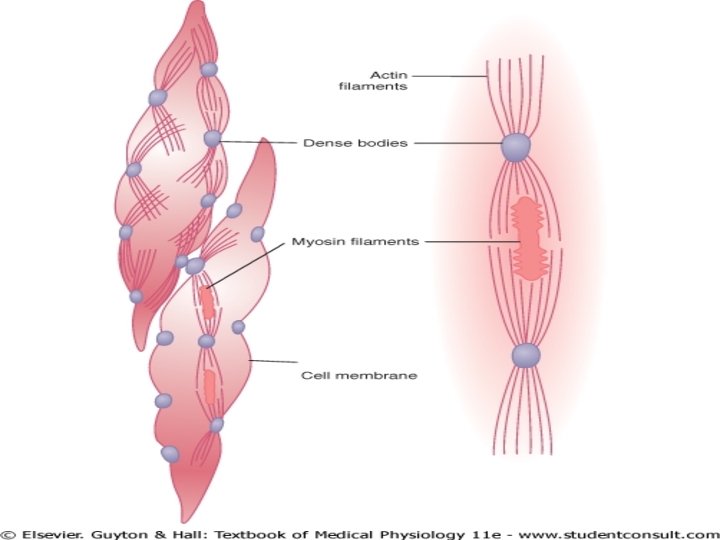
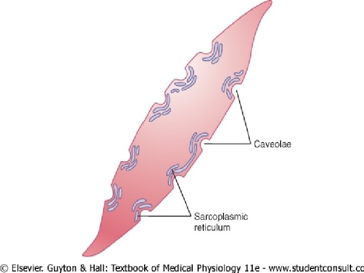
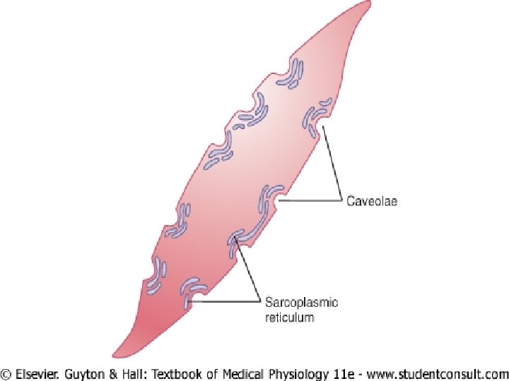
�Each smooth muscle fiber has cell membrane �It contains myosin filaments actin filaments cytoplasm sarcoplasmic reticulum caveolae
Cytoplasm �Contains enzymes – myosin phosphatase regulatory protein - calmodulin
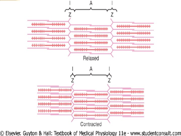
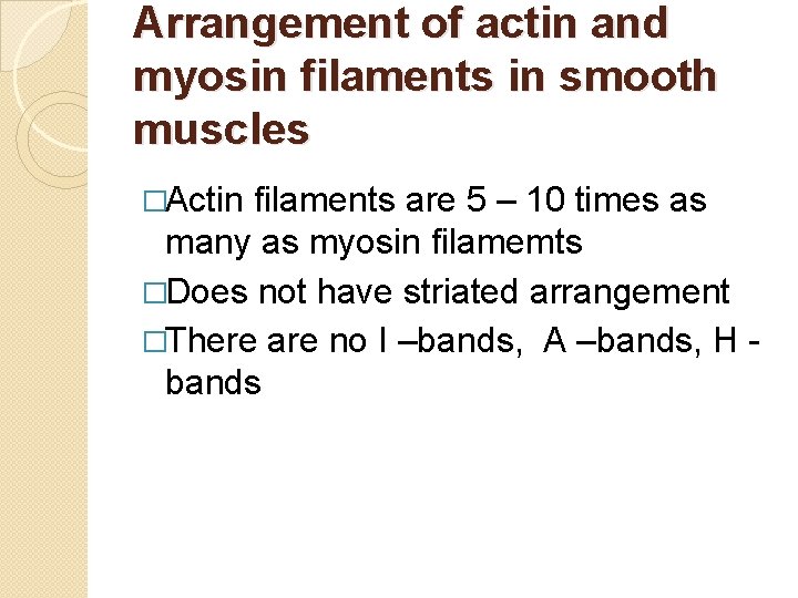
Arrangement of actin and myosin filaments in smooth muscles �Actin filaments are 5 – 10 times as many as myosin filamemts �Does not have striated arrangement �There are no I –bands, A –bands, H bands
Dense bodies �May be attached to the cell membrane , others are dispersed inside the cell �Function of dense bodies 1 -actin filaments are attached 2 -bound together by inter cellular protein bridges
Individual contractile unit in smooth muscles �Between two dense bodies �Large no. of actin filaments radiate from dense bodies �The ends of actin filaments overlap a myosin filament
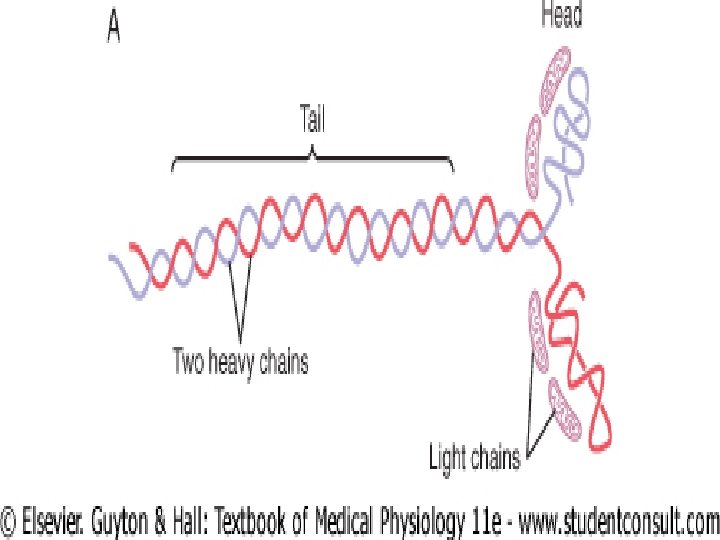
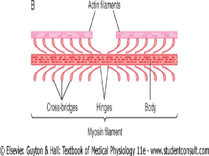
Structure of myosin filament �Have side – polar cross –bridges �It allows smooth muscles to contract as much as 80% of their length instead of being limited to less than 30% as in skeletal muscles
Structure of actin filament �Does not contain troponin �Calmodulin – it initiates contraction by activating the myosin cross - bridges
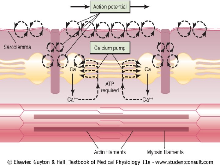
Sarcoplasmic reticulum �Only slightly developed �Lie near the cell membrane in some large smooth muscles �They have only tubular part �Most of calcium enters into smooth muscles by diffusion through cell membrane
Caveolae �Small invaginations of cell membrane �Rudimentary analog of T-tubules in skeletal muscles �Function
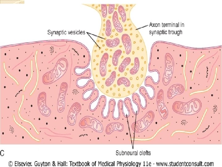
Neuro muscular junctions �Innervated by autonomic nerves �Varicosities dilated terminal parts of axons multiple , distributed along their axis contains vesicles cytoplasm mitochondria voltage gated calcium channels
�For cholinergic nerve endings – acetylcholine esterase �For adrenergic nerve endings – noradrenaline is removed by three ways 1 -50 -80% reuptake by active transport 2 -diffusion into surrounding body fluids 3 -destruction by monoamine oxidase and catechol-o-methyl transferase
Receptors on smooth muscle membrane �Ion –channel linked receptor �G –protein linked receptor �Alpha and beta receptors
Two types of neuromuscular junctions �Diffuse junctions �Contact junctions
Diffuse junctions �Distance between nerve ending and smooth muscle membrane—few nm to few micro m �In single unit smooth muscles- all �In multi unit smooth muscles- mostly
Contact junctions �In few cases of multi unit type �Distance between varicosities and membrane is 20 -30 nm �More rapid to contract
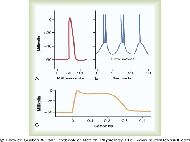
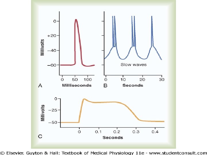
Action potential in smooth muscles �Resting membrane potential is -50 to - 60 mv �Three types 1 -spike potential 2 -action potential with plateau 3 -slow wave potential with spontaneous generation of action potential
Spike potentials Stimuli �Electrical stimulus �Hormones �Nerve fibres �Stretch �Spontaneous generation due to slow waves
�Duration sec of spike potential is 10 -50 m
Action potential with plateaus �Occurs in ureter , uterus under some conditions, certain vascular smooth muscles �Importance of plateau is for prolong contraction �It is because of voltage gated calcium channels
Slow wave potential with spontaneous generation of action potentials �Occurs in gut �Membrane potential has slow wave rhythm �Causes 1 -waxing and waning of sodium pumps 2 -conductances of ion channelsincrease or decrease rhythmically
Contraction in smooth muscles �Stimuli for contraction are 1 -autonomic nerve stimulation 2 -hormonal stimulation 3 -stretch of the fiber 4 -Change in chemical environment of fiber
Autonomic nerve stimulation �Depolarization reach to varicosities �Opening of voltage gated calcium channels �Attractive force on vesicles �If cholinergic nerve ending – acetylcholine �Bind to receptors �Alters permeability of ions �Action potential in smooth muscles �Influx of calcium into smooth muscles
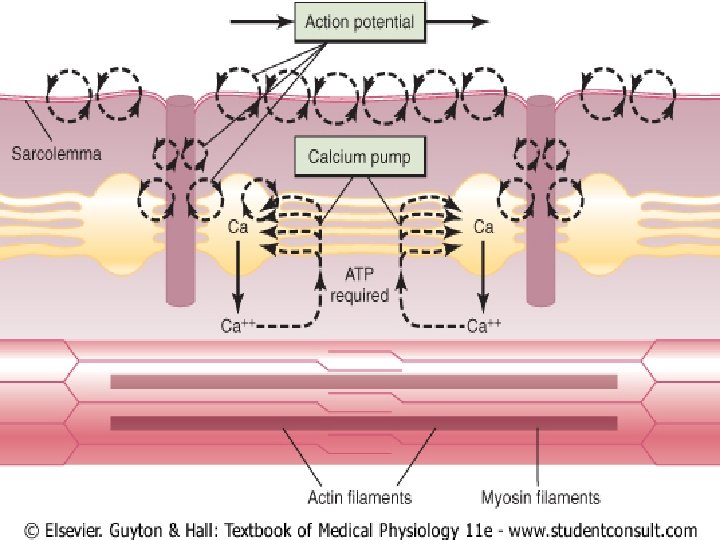
Sources of calcium in smooth muscles 1 -from extra cellular fluid – by opening of voltage gated calcium channels in smooth muscle membrane 2 -very little from sarcoplasmic reticulum
Sources of calcium in smooth muscles 1 -from extra cellular fluid – by opening of voltage gated calcium channels in smooth muscle membrane 2 -very little from sarcoplasmic reticulum
�Calcium – calmodulin complex �Activates myosin kinase �Phosphorylates one of the light chain of each myosin head �Increase myosin ATPase activity �Sliding of actin and myosin
Relaxation of smooth muscles �Activation of calcium pumps �Myosin phosphatase �Endothelial derived relaxation factor(EDRF) – nitric oxide(NO) activates Guanylate cyclase C GMP vasodilation
Latent period �Time required for diffusion of calcium from extracellular fluid is about 200 to 300 m sec � 50 times as great for smooth muscles as for skeletal muscles
Latch bridge mechanism �Prolong holding of contraction of smooth muscles(sustained contraction) �Occurs in visceral smooth muscles
�May be because of decrease activity of myosin phophatase �Advantages 1 -energy economy of body 2 -little continuous excitatory signals
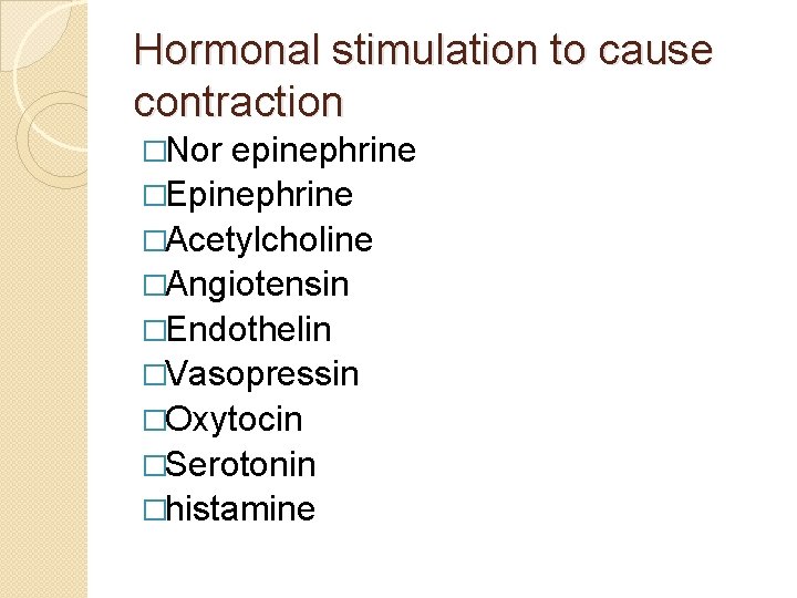
Hormonal stimulation to cause contraction �Nor epinephrine �Epinephrine �Acetylcholine �Angiotensin �Endothelin �Vasopressin �Oxytocin �Serotonin �histamine
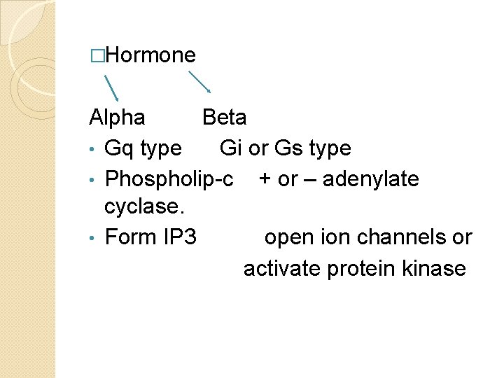
�Hormone Alpha Beta • Gq type Gi or Gs type • Phospholip-c + or – adenylate cyclase. • Form IP 3 open ion channels or activate protein kinase
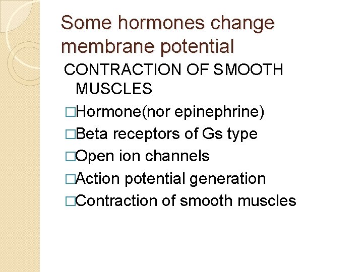
Some hormones change membrane potential CONTRACTION OF SMOOTH MUSCLES �Hormone(nor epinephrine) �Beta receptors of Gs type �Open ion channels �Action potential generation �Contraction of smooth muscles
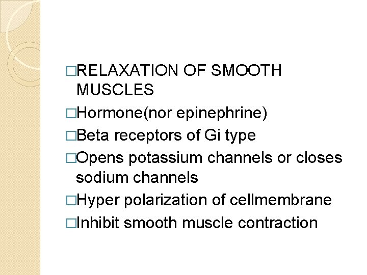
�RELAXATION OF SMOOTH MUSCLES �Hormone(nor epinephrine) �Beta receptors of Gi type �Opens potassium channels or closes sodium channels �Hyper polarization of cellmembrane �Inhibit smooth muscle contraction
Some hormones does not change membrane potential CONTRACTION OF SMOOTH MUSCLES �Hormone alpha receptors of Gq type stimulate phospholipase C in cell membrane formation of IP 3 opens calcium channels
�RELAXATION OF SMOOTH MUSCLES �Hormone Gi protein linked receptor inhibit adenylate cyclase or gaunylate cyclase decrease c AMP or c. GMP stimulate emzyme which inhibit contraction stimulate calcium pumps inhibit contraction
Change in chemical environment of muscle fibres for contraction �Arterioles , met arterioles and pre capillary sphincter have little or no nerve supply �In normal resting state , many of these blood vessels remain contracted �Auto regulation of blood flow
�Lack of oxygen �Excess carbondioxide �Increase hydrogen ion concentration �Lactic acid, adenosine, increase body temperature �NO released by endothelial cells
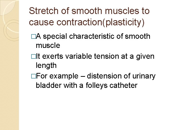
Stretch of smooth muscles to cause contraction(plasticity) �A special characteristic of smooth muscle �It exerts variable tension at a given length �For example – distension of urinary bladder with a folleys catheter
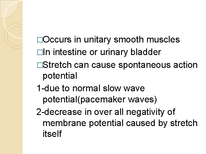
�Occurs in unitary smooth muscles �In intestine or urinary bladder �Stretch can cause spontaneous action potential 1 -due to normal slow wave potential(pacemaker waves) 2 -decrease in over all negativity of membrane potential caused by stretch itself
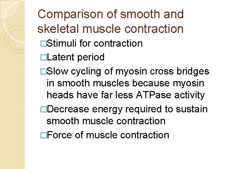
Comparison of smooth and skeletal muscle contraction �Stimuli for contraction �Latent period �Slow cycling of myosin cross bridges in smooth muscles because myosin heads have far less ATPase activity �Decrease energy required to sustain smooth muscle contraction �Force of muscle contraction
�Stress relaxation of smooth muscles(plasticity) �Sources of calcium �Action potential