Small Molecules and Ions as Second Messengers The
 that")
 is one of the most widely used")


 • Calcium ions act as a second")
 G protein DAG GTP G protein-coupled receptor Phospholipase")
 G protein DAG GTP G protein-coupled receptor Phospholipase")


- Slides: 19
Small Molecules and Ions as Second Messengers • The extracellular signal molecule (ligand) that binds to the receptor is a pathway’s “first messenger” • Second messengers are small, non-protein, watersoluble molecules or ions that spread throughout a cell by diffusion • Second messengers participate in pathways initiated by G protein-coupled receptors and receptor tyrosine kinases • Cyclic AMP and calcium ions are common second messengers
Cyclic AMP • Cyclic AMP (c. AMP) is one of the most widely used second messengers • Adenylyl cyclase, an enzyme in the plasma membrane, converts ATP to c. AMP in response to an extracellular signal ATP c. AMP
• Receptor tyrosine kinases are membrane receptors that attach phosphates to tyrosines • A receptor tyrosine kinase can trigger multiple signal transduction pathways at once Ligand-binding site Signaling molecule (ligand) Signaling molecule Helix Tyrosines Tyr Tyr Tyr Tyr Tyr Receptor tyrosine kinase proteins CYTOPLASM Dimer 1 2 Activated relay proteins Tyr Tyr Tyr P P 6 ATP Activated tyrosine kinase regions 6 ADP P Tyr Tyr Tyr P P P Tyr Tyr P P Fully activated receptor tyrosine kinase Inactive relay proteins 3 4 Cellular response 1 Cellular response 2
• Many signal molecules trigger formation of c. AMP • Other components of c. AMP pathways are G proteins, G proteincoupled receptors, and protein kinases • c. AMP usually activates protein kinase A, which phosphorylates various other proteins • Further regulation of cell metabolism is provided by G-protein systems that inhibit adenylyl cyclase First messenger Adenylyl cyclase G protein-coupled receptor GTP ATP c. AMP Second messenger Protein kinase A Cellular responses
Calcium Ions and Inositol Triphosphate (IP 3) • Calcium ions act as a second messenger in many pathways • Calcium is an important second messenger because cells can regulate its concentration EXTRACELLULAR FLUID Plasma membrane Ca 2+ pump ATP (Ca 2+) Mitochondrion Nucleus CYTOSOL Ca 2+ pump Endoplasmic reticulum (ER) ATP Key High [Ca 2+] Low [Ca 2+] Ca 2+ pump
EXTRACELLULAR FLUID Signaling molecule (first messenger) G protein DAG GTP G protein-coupled receptor Phospholipase C PIP 2 IP 3 (second messenger) IP 3 -gated calcium channel Endoplasmic reticulum (ER) CYTOSOL Ca 2+ • A signal relayed by a signal transduction pathway may trigger an increase in calcium (2 nd messenger) in the cytosol • Pathways leading to the release of calcium involve inositol triphosphate (IP 3) and diacylglycerol (DAG) as additional second messengers
EXTRACELLULAR FLUID Signaling molecule (first messenger) G protein DAG GTP G protein-coupled receptor Phospholipase C PIP 2 IP 3 (second messenger) IP 3 -gated calcium channel Endoplasmic reticulum (ER) CYTOSOL Ca 2+ (second messenger)
Cell signaling leads to regulation of transcription or cytoplasmic activities EXTRACELLULAR FLUID Signaling molecule (first messenger) G protein DAG GTP G protein-coupled receptor • The cell’s response to an extracellular signal is sometimes called the “output response” PIP 2 Phospholipase C IP 3 (second messenger) IP 3 -gated calcium channel Endoplasmic reticulum (ER) CYTOSOL Various proteins activated Ca 2+ (second messenger) Cellular responses
Nuclear and Cytoplasmic Responses • Ultimately, a signal transduction pathway leads to regulation of one or more cellular activities • The response may occur in the cytoplasm or may involve action in the nucleus • Many signaling pathways regulate the synthesis of enzymes or other proteins, usually by turning genes on or off in the nucleus • The final activated molecule may function as a transcription factor Growth factor Reception Receptor Phosphorylation cascade Transduction CYTOPLASM Inactive transcription factor Active transcription factor P Response DNA Gene NUCLEUS m. RNA
• Other pathways regulate the activity of enzymes Reception Binding of epinephrine to G protein-coupled receptor (1 molecule) Transduction Inactive G protein Active G protein (102 molecules) Inactive adenylyl cyclase Active adenylyl cyclase (102) ATP Cyclic AMP (104) Inactive protein kinase A Active protein kinase A (104) Inactive phosphorylase kinase Active phosphorylase kinase (105) Inactive glycogen phosphorylase Active glycogen phosphorylase (106) Response Glycogen Glucose-1 -phosphate (108 molecules)
RESULTS • Signaling pathways can also affect the physical characteristics of a cell, for example, cell shape Wild-type (shmoos) ∆Fus 3 ∆formin CONCLUSION 1 Mating factor G protein-coupled receptor Shmoo projection forming Formin P Fus 3 GDP GTP Phosphorylation cascade 2 Actin subunit P Formin P 4 Fus 3 P 3 Microfilament 5
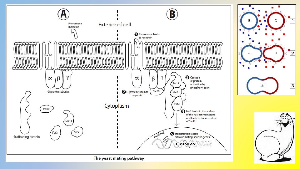
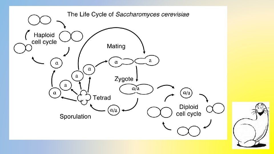
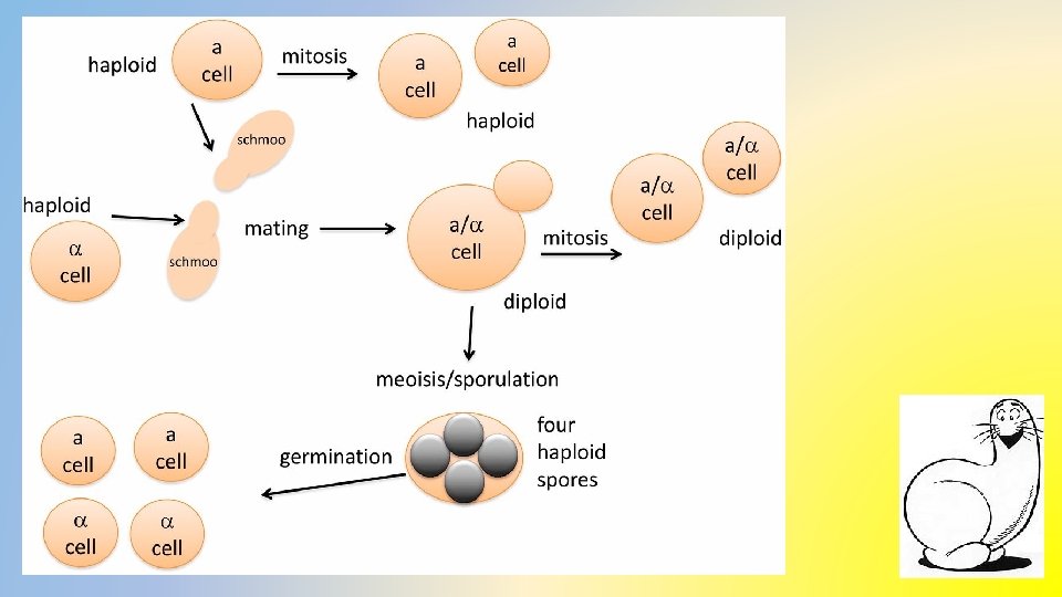
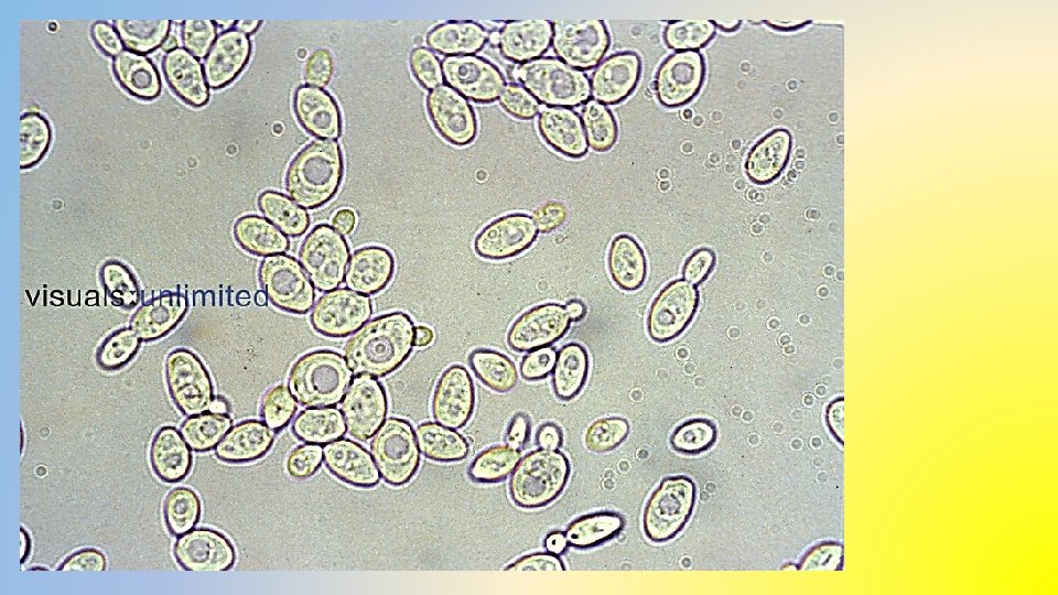
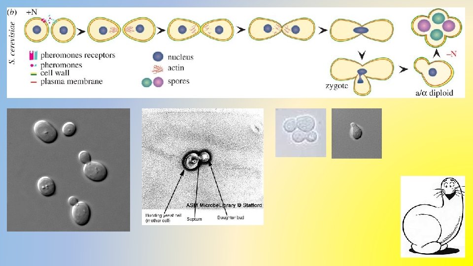
Questions to set your compass… What evidence is there that S. cerevisiae is communicating? How do the S. cerevisiae monocultures differ from the mixed culture observations? What pathway are we investigating that is involved with this process and how does it work? How do S. cerevisiae cells communicate?
Yeast cell types observed. 1. Single haploid 2. Budding haploid 3. Shmoos 4. Single zygotes 5. Budding zygotes 6. Asci (cluster of 4 haploid spores)