SMALL LARGE INTESTINES Color index Slides Important Notes


:")


 Simple tubular glands that open between villi. Composed of 5 cell")
")


- Slides: 14
SMALL & LARGE INTESTINES Color index: Slides. . Important. . Notes. . Extra. .
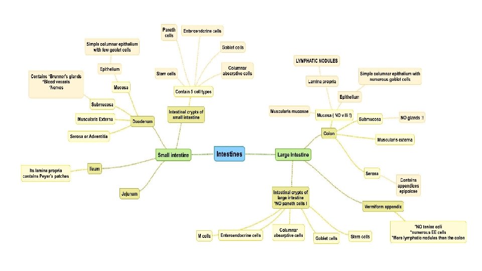
Objectives : • By the end of this lecture, the student should be able to discuss the microscopic structure in correlation with the function of the following structures : üDuodenum üJejunum üIleum üColon üAppendix Definitions: Plicae circulares: transverse/horizontal folds of mucosa and submucosa. Crypts of Lieberkuhn: invaginations of epithelium into the lamina propria between the villi to form glands.
SMALL INTESTINE To increase surface area the mucosa has: - Plicae circulares (circular folds): Permanent folds of the mucosa and submucosa. (Increase the rate of absorption 3 -2 times) - Villi. (Increase the rate of absorption 10 times) - Intestinal crypts (crypts of Lieberkühn). Secretes intestinal - Microvilli (Brush border). (Increase the rate of absorption 20 times) juice (includes enzymes)
Wall of the duodenum 1 - Mucosa: • Shows villi and crypts. • A- Epithelium: simple columnar epithelium with few goblet cells. ( the goblet cells gradually increase) • B- Lamina propria: loose areolar C. T. • C- Muscularis mucosae: 2 layers of smooth muscle cells. 2 - Submucosa: • Connective tissue containing blood vessels & nerves. • Contains Brunner’s glands (secrete mucus). ( in the first inch of the upper part of the duodenum) 3 -Muscularis externa: • 2 smooth muscle layers: • 1 - Inner circular layer. • 2 - Outer longitudinal layer. 4. Serosa or Adventitia: • Duodenum is invested by a serosa or adventitia. All parts of the small intestine has the same features of the 4 layers except some differences will be discussed later on. Other than that it’s all the same.
Intestinal villi: Each Villus is a finger-like projection of small intestinal mucosa and it is formed of: I-Central core of loose areolar C. T. : containing: Lymphocytes, Plasma cells, Fibroblasts, Smooth muscle cells, Capillary loops, Lacteal (blindly ending lymphatic channels). Important in absorption of fat droplets II- Villus- covering epithelium Loose areolar CT with lymphocytes as the lamina propria Cells Covering the Villi: 1 -Surface columnar absorptive cells: -They have brush border (microvilli). (Increase the rate of absorption 20 times) -They are covered with thick glycocalx that has digestive enzymes. -They have Junction complex (tight, adhering and desmosome junctions). 2 -Goblet cells: Increase toward the ileum. 3 -Enteroendocrine (EE) cells (DNES cells). 4 -M cells (Membrane or Microfold epithelial cells).
Intestinal Glands (Crypts) Simple tubular glands that open between villi. Composed of 5 cell types: 1. 2. 3. 4. Columnar absorptive cells. Goblet cells: secrete mucus. Enteroendocrine (EE) (DNES) cells: secrete hormones. Paneth cells: secrete Lysozyme (antibacterial). are found in the base of the crypts. That’s why small intestine has no bacteria 5. *Stem cells: are regenerative cells. are found in the base of the crypts. *Important for MCQs - Stem cells and paneth cells are special for the crypts Columnar Absorptive cells EE (DNES) cells 1. 2. 3. 4. 5. 6. EC cells: secrete endorphin and serotonin. S cells: secrete secretin. D cells: secrete somatostatin. A cells: secrete glucagon. Mo cells: secrete motilin. Regulates motility CCK-PZ cells: secrete cholecystokinin (pancreozymin). Hormonal secreting cells are found on the base ‘near to vessels on C. T. ’ Paneth cell 1, 2, 3 are also found in cells covering the villi. - They are mainly found within the intestinal epithelium overlying lymphatic nodules of lamina propria. - Each is a dome-shaped cell (or specialized squamous cell) with a basal concavity that contains intraepithelial lymphocytes and macrophages. - They phagocytose and transport antigens present in the intestinal lumen to the underlying lymphoid tissue cells to initiate the immune response to these antigens leading to the secretion of Ig. A.
Intestinal Glands ( crypts )
Regional differences of small How to differentiate between them? intestine The duodenum Brunner’s glands The ileum Peyer's patches The jejunum By exclusion of the other 2 parts. Jejunum: • Its submucosa has Brunner’s glands. • Secretes alkaline mucous • It is invested by serosa or adventitia Duodenum: • has neither Brunner’s glands nor Peyer’s patches. • It is invested by serosa. • Its lamina propria, opposite the attachment of the mesentery, has lymphoid nodules (Peyer's patches) that extend to the submucosa. (Anti- Brunner’s glands mesenteric area) • It is invested by serosa. Ileum: Peyer's patches
Large intestine Appendix ascending Cecum: right side Divided anatomically into: transverse Colon descending Rectum sigmoid Anal canal
Wall of Colon 1 - Mucosa the mucosa is thinner than that of small int. o Shows only crypts (NO villi) Ø Epithelium: o Simple columnar epithelium with numerous goblet cells. (more numerous goblet cells than small intestine, and increase near the rectum) Ø Lamina propria: o C. T containing numerous crypts. ( Glands ) o Cells of the crypts are the same as in small intestine but WITHOUT Paneth cells. o Lymphatic nodules (solitary): frequent. The ileum starts in the antibacterial process with having peyer’s patches, and bacteria really appear in the colon. Ø Muscularis mucosae: o 2 layers of smooth muscle. 2 - Submuosa o NO glands. (esophagus + duodenum) ﺍﻟﻘﻼﻧﺪﺯ ﺑﺲ ﻣﻮﺟﻮﺩﺓ ﺑﺎﻟـ o Meissner’s nerve plexus. 3 - Muscularis Externa: o Inner circular & outer longitudinal smooth muscle layers. o The outer longitudinal layer is not continuous but in the form of 3 ribbons (teniae coli). o Auerbach’s nerve plexus. Serosa: o C. T. covered by mesothelium. o Has fat-filled pouches ( pendolous masses) called appendices epiploicae.
Intestinal Crypts of Colon Cells lining the crypts are: 1. Surface columnar absorptive cells. 2. Goblet cells. 3. Enteroendocrine cells. 4. Stem cells. 5 - M-cells. The only difference between here and small intestine is here there is no paneth cell. Vermiform Appendix Similar to the colon, but with much smaller diameter, shallow crypts, more lymphoid nodules (aggregated lymphoid nodules, all around, in lamina propria and extending into submucosa), Few goblet cells and more EE (DNES) cells. Muscularis mucosae: Not continuous. Muscularis externa: No teniae coli. (continuous, no 3 ribbons) Serosa: no appendices epiploicae (fat) In crypts: Goblet cells less than the colon It is mainly an immune organ.
MCQs 1 -B 2 -B 3 -B 4 -D 5 -A 6 -C 7 -D 8 -B 9 -A 101112 - 1 - Brunner’s glands are found in which of following ? A-Colon B- Duodenum C-Jejunum D- Ileum 2 - Brunner’s glands are found in which of following layers ? A-Mucosa B-Submucosa C-Muscularis mucosae D-Serosa. 3 -Peyer’s patches may found in Jejunum ? a-true B-false 4 - A cells and D cells secrete? A-Secretin and Motilin B-Endorphin and Glucagon C-CKK and somatostatin D-Glucagon and somatostatin 5 - Which cell of the following secretes antibacterial material? A- Paneth cells B-Enteroendocrine C-Stem cells D-Goblet cells 6)-Which cell of the following secretes Ig. A and transports antigens in intestinal lumen to the lymphoid tissue? A. S cells B. Mo cells C. M cells D. EC cells 7 -Which one of these layers contains fat-filled pouches called appendices epiploicae? a. Mucosa b. Submucosa c. Muscularis externa d. Serosa a. b. c. d. 8 - Muscularis mucosae in Vermiform appendix are? Continuous non continuous not formed contain M cells 9 -Lymphatic nodules (solitary) are found in which layer of the Colon? A-Mucosa B-Submucosa C-Serosa D-Muscularis externa
Thank you & good luck - Histology team Done by: ü Ahmed Badahdah ü Mutasem Alhasani ü Omar Turkistani ü Nawaf Aldarweesh ü Mohammed Khojah ü Shahad Al. Anzan References: ü ü Females’ and Males’ slides. Doctors’ notes Team leaders: ü Rana Barasain ü Faisal Alrabaii