Small bowel obstruction By Nebal Atallah Mahd AlFoqhaa

Small bowel obstruction By: • Nebal Atallah • Mahd Al-Foqhaa • Thara Anwer

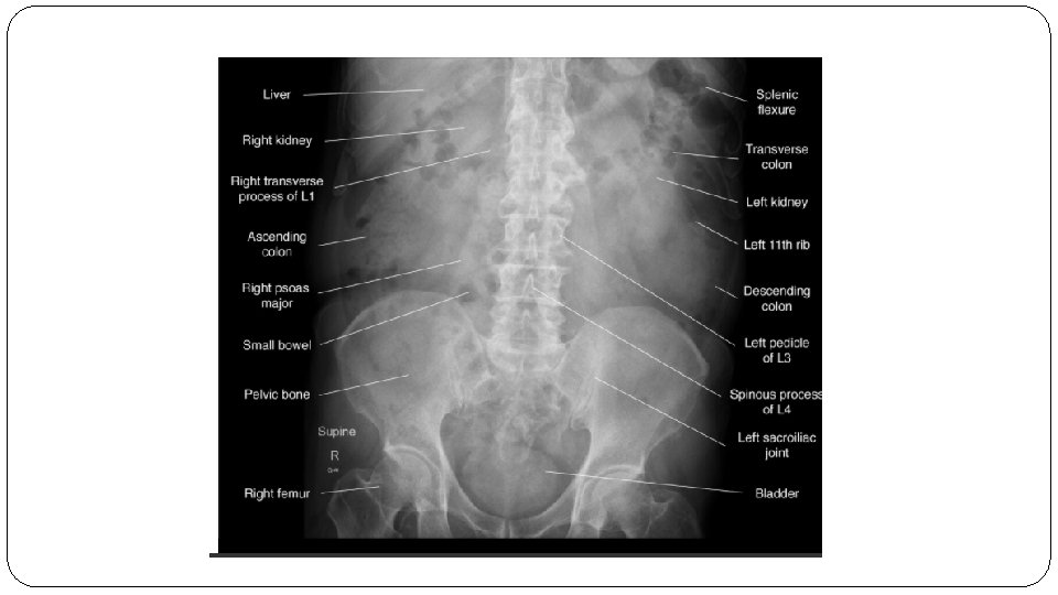
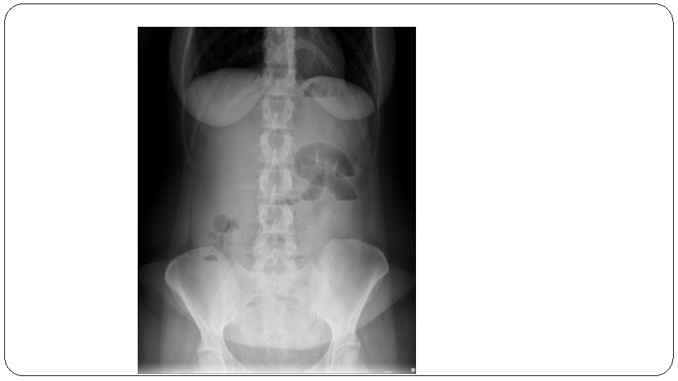
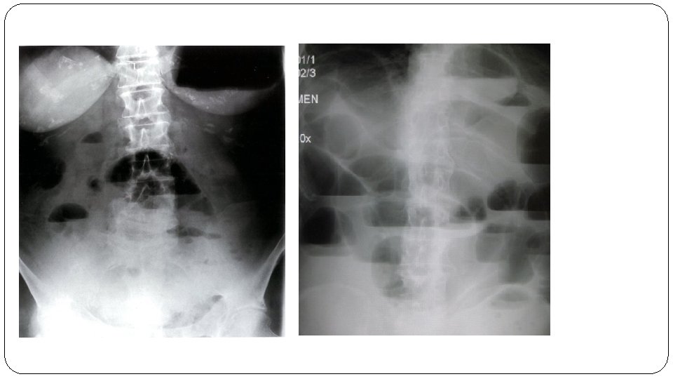
Normal abdominal X-ray

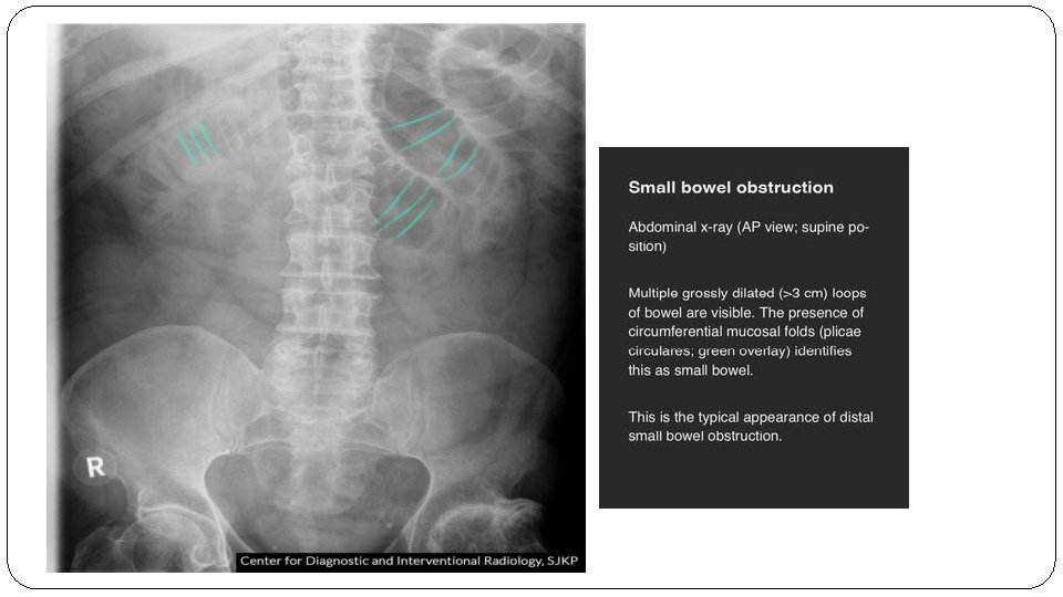
Location Small bowel Large bowel Central Peripheral Wall Pattern Encircling valvulae conniventes visible depending on degree of air filling/distention. Haustral folds interspaced with Plicae semilunaris Size 6 cm diameter (caecum 9 cm) 3 cm diameter

Small bowel obstruction • Inability of the intestinal contents to pass distally in the lumen of intestine either from a mechanical barrier or absence of peristalsis without any mechanical barrier. • Depending upon the nature of obstruction: 1 -Dynamic obstruction 2 - Adynamic obstruction

adhesion “common” Volvulus Hernia Intussusception • Strictures • Crohn’s disease Intraluminal • • intramural Extraluminal Causes : • • • Gall stones ileus Food Round worm Mass Foreign body
 cramps and pain 2. Bloating 3. Nausea")
Clinical presentation : - 1. Abdominal (stomach) cramps and pain 2. Bloating 3. Nausea & vomiting 4. Dehydration 5. Malaise (an overall feeling of illness) 6. Lack of appetite 7. Severe constipation: In cases of complete obstruction, a person will not be able to pass stool (feces) or gas. 8. Absence of bowel sound: (initially it is increased to overcome the obstruction)

Imaging 1. Abdominal X-ray 2. CT abdomen and pelvis 3. MRI abdomen and pelvis (with and/or without IV contrast) 4. Abdominal ultrasound 5. Barium or water-soluble contrast enema
 : �Air under")
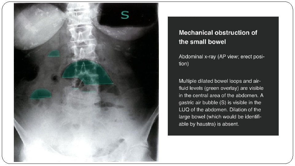
Abdominal x-ray : 1. The upright abdominal X-ray (AP or PA) : �Air under diaphragm(CXR could be used) �air–fluid levels �may demonstrate dilated loops of bowel �Radiopaque foreign bodies 2. left lateral decubitus abdominal X-ray: �Patient cant stand or sit to do PA view �Left? ?

3. The supine AP abdominal X-ray : �dilated loops of bowel suggesting obstruction may be seen. �air–fluid levels are not seen on the supine view
 (Dynamic obstruction) • Interruption in the normal passage due")
Mechanical obstruction (small bowel obstruction) (Dynamic obstruction) • Interruption in the normal passage due to a structural barrier •

Etiology : 1 -Bowel adhesions

2 -Hernia

Findings on imaging. . . � 1 -Dilated bowel loops proximal to obstruction. � 2 -Collapsed bowel loops distal to obstruction � 3 -No air within rectum � 4 -Multiple air-fluid levels � 5 -Cause of obstruction (e. g. , tumor)

Diagnosis. . . �In the workup of suspected mechanical bowel obstruction, imaging allows for quick confirmation of the diagnosis as well as detection of conditions requring immediate surgery (e. g. , perforation). � Small bowel dilatation if > 3 cm �n SBO: The dilated loops are predominantly central

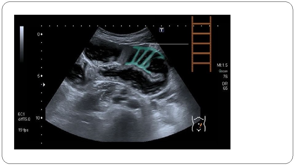
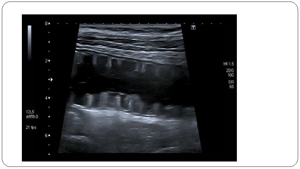
Ultrasound
 �Definition : Temporarily impaired peristalsis of the gastrointestinal")
Functional obstruction ( PARALYTIC ILEUS ) �Definition : Temporarily impaired peristalsis of the gastrointestinal tract in absence of mechanical obstruction �Causes : 5 P’s Peritonitis , Postoperative , Potassium level decrease , Pelvic and spinal fractures , Parturition

Types of adynamic ileus Generalized Affect all loop of small & large bowel Localized Affect only one or two loops of small intestine

�Pathophysiology : Ø stressful stimulation of the bowel Ø Inflammation release NO locally �Imaging : activation of the sympathetic system decrease peristalsis relaxation of the intestinal smooth muscle decrease peristalsis Ø generalized small and large bowel distention Ø visible gas shadows in rectum Decrease peristalsis will cause bowel distention which progress to mechanical obstruction

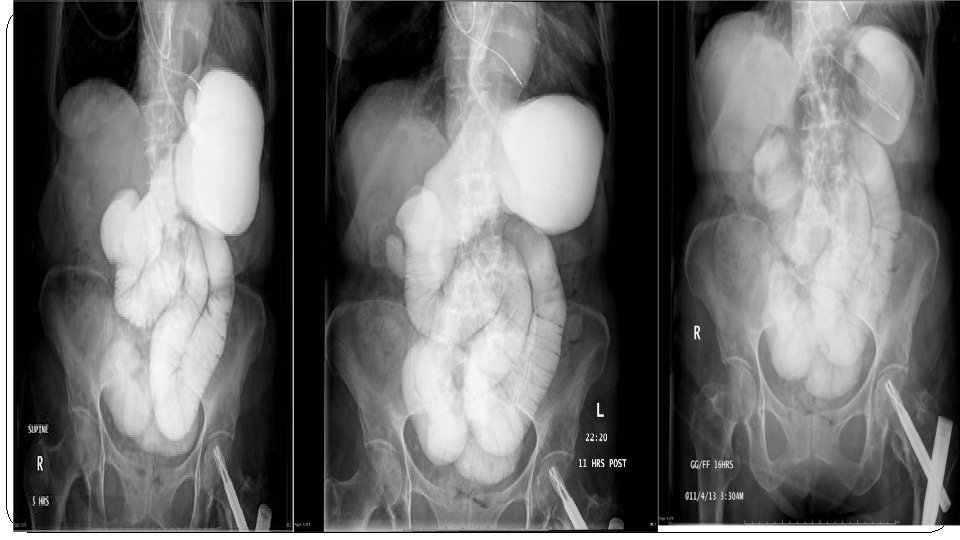
PARALYTIC ILEUS SECONDARY TO MESENTRIC ISCHEMIA • Abdominal X-ray AP view : 1 - multiple dilated loop of bowel 2 - several air fluid level

• Abdominal X-ray left lateral view : Significantly distended intestinal loops with air fluid levels are visible

• The fluid levels long and at the same level. • Gases in both colon and small bowel loops.

• Generalized adynamic ileus : Ø Dilated loop of large bowel ( white arrows ) Ø Dilated loops of small bowel ( dotted white arrows ) Ø Gases in rectum ( black arrows )

THE END
- Slides: 31