
SMALL ANIMAL ABDOMEN CHAPTER 18 Brown Lavins Radiography
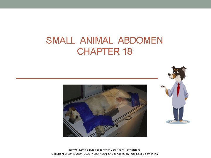
SMALL ANIMAL ABDOMEN CHAPTER 18 Brown: Lavin’s Radiography for Veterinary Technicians Copyright © 2014, 2007, 2003, 1999, 1994 by Saunders, an imprint of Elsevier Inc.
CHAPTER OUTLINE • Objectives • Key terms • Overview • Radiographic concerns • Positions • • • Lateral Ventrodorsal Further Views • Normal anatomy • Alternate mode images • Key points • Review questions • Bibliography Brown: Lavin’s Radiography for Veterinary Technicians Copyright © 2014, 2007, 2003, 1999, 1994 by Saunders, an imprint of Elsevier Inc. 2
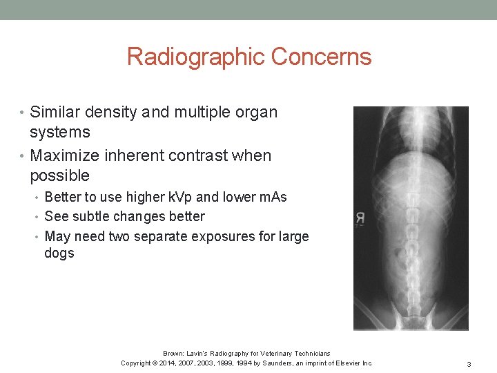
Radiographic Concerns • Similar density and multiple organ systems • Maximize inherent contrast when possible • Better to use higher k. Vp and lower m. As • See subtle changes better • May need two separate exposures for large dogs Brown: Lavin’s Radiography for Veterinary Technicians Copyright © 2014, 2007, 2003, 1999, 1994 by Saunders, an imprint of Elsevier Inc. 3
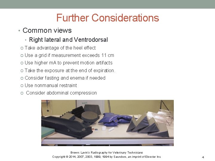
Further Considerations • Common views • Right lateral and Ventrodorsal Take advantage of the heel effect Use a grid if measurement exceeds 11 cm Use higher m. A to prevent motion artifacts Take the exposure at the end of expiration. Consider fasting and enema if needed Use nonmanual restraint Consider abdominal compression Brown: Lavin’s Radiography for Veterinary Technicians Copyright © 2014, 2007, 2003, 1999, 1994 by Saunders, an imprint of Elsevier Inc. 4
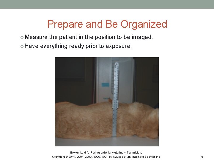
Prepare and Be Organized Measure the patient in the position to be imaged. Have everything ready prior to exposure. Brown: Lavin’s Radiography for Veterinary Technicians Copyright © 2014, 2007, 2003, 1999, 1994 by Saunders, an imprint of Elsevier Inc. 5
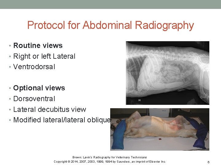
Protocol for Abdominal Radiography • Routine views • Right or left Lateral • Ventrodorsal • Optional views • Dorsoventral • Lateral decubitus view • Modified lateral/lateral oblique Brown: Lavin’s Radiography for Veterinary Technicians Copyright © 2014, 2007, 2003, 1999, 1994 by Saunders, an imprint of Elsevier Inc. 6
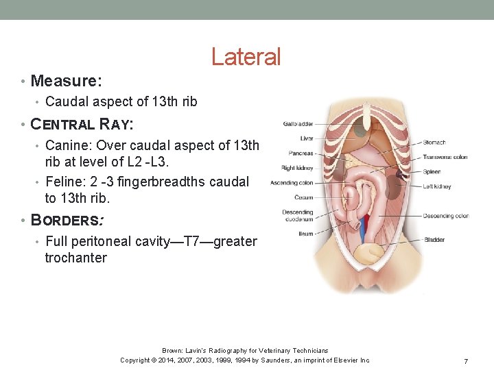
Lateral • Measure: • Caudal aspect of 13 th rib • CENTRAL RAY: • Canine: Over caudal aspect of 13 th rib at level of L 2 -L 3. • Feline: 2 -3 fingerbreadths caudal to 13 th rib. • BORDERS: • Full peritoneal cavity—T 7—greater trochanter Brown: Lavin’s Radiography for Veterinary Technicians Copyright © 2014, 2007, 2003, 1999, 1994 by Saunders, an imprint of Elsevier Inc. 7
Checklist • Settings correct • Image receptor/machine/grid in position • Proper location of markers and identification (ID) • Correct body part and view • Properly centered • Borders correct and collimated • Thickest part to the cathode • Patient properly prepared, positioned, and restrained • Image will be perpendicular to the beam and parallel to the film • Full expiration Brown: Lavin’s Radiography for Veterinary Technicians Copyright © 2014, 2007, 2003, 1999, 1994 by Saunders, an imprint of Elsevier Inc. 8
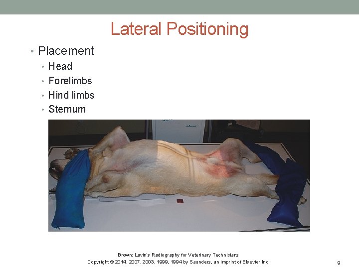
Lateral Positioning • Placement • Head • Forelimbs • Hind limbs • Sternum Brown: Lavin’s Radiography for Veterinary Technicians Copyright © 2014, 2007, 2003, 1999, 1994 by Saunders, an imprint of Elsevier Inc. 9
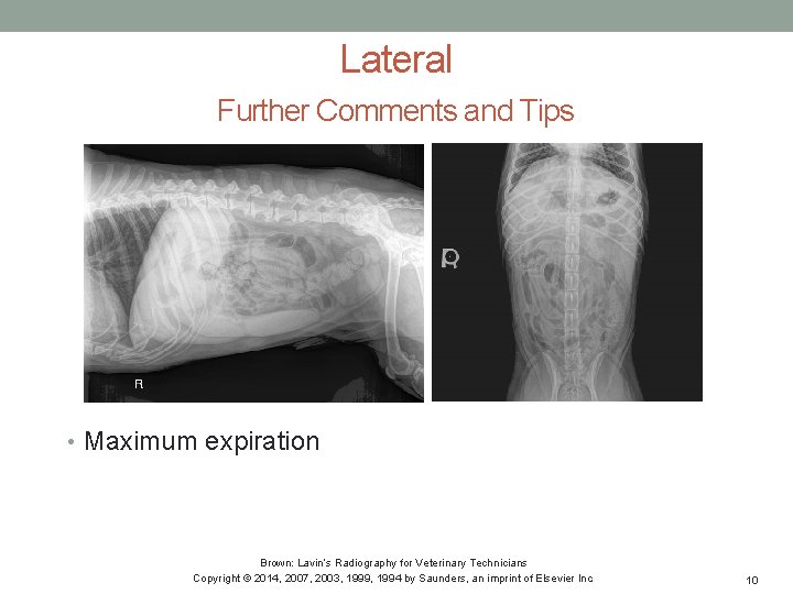
Lateral Further Comments and Tips • Maximum expiration Brown: Lavin’s Radiography for Veterinary Technicians Copyright © 2014, 2007, 2003, 1999, 1994 by Saunders, an imprint of Elsevier Inc. 10
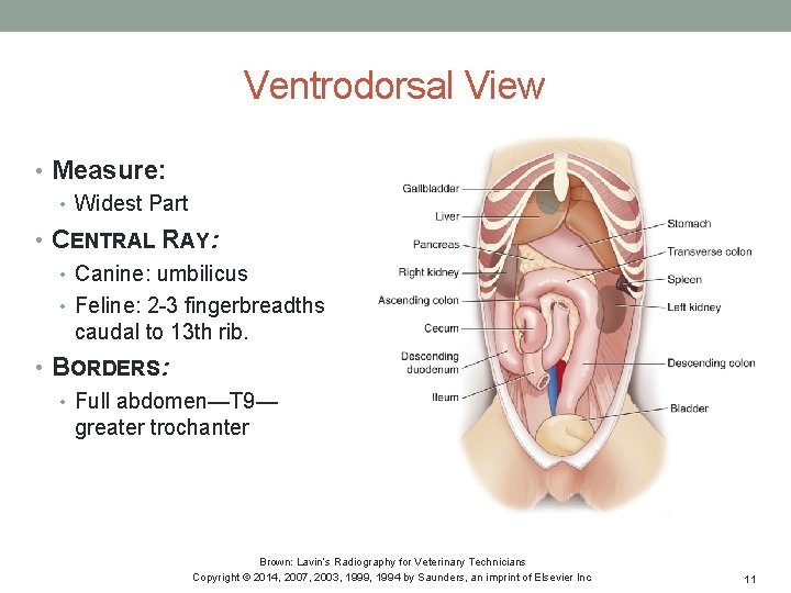
Ventrodorsal View • Measure: • Widest Part • CENTRAL RAY: • Canine: umbilicus • Feline: 2 -3 fingerbreadths caudal to 13 th rib. • BORDERS: • Full abdomen—T 9— greater trochanter Brown: Lavin’s Radiography for Veterinary Technicians Copyright © 2014, 2007, 2003, 1999, 1994 by Saunders, an imprint of Elsevier Inc. 11

Ventrodorsal Positioning • Placement • Head • Forelimbs • Hind limbs • Maximum expiration Brown: Lavin’s Radiography for Veterinary Technicians Copyright © 2014, 2007, 2003, 1999, 1994 by Saunders, an imprint of Elsevier Inc. 12
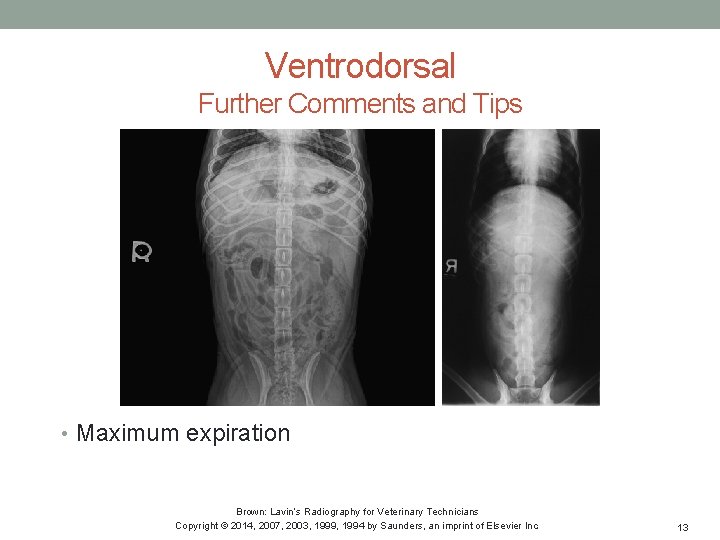
Ventrodorsal Further Comments and Tips • Maximum expiration Brown: Lavin’s Radiography for Veterinary Technicians Copyright © 2014, 2007, 2003, 1999, 1994 by Saunders, an imprint of Elsevier Inc. 13
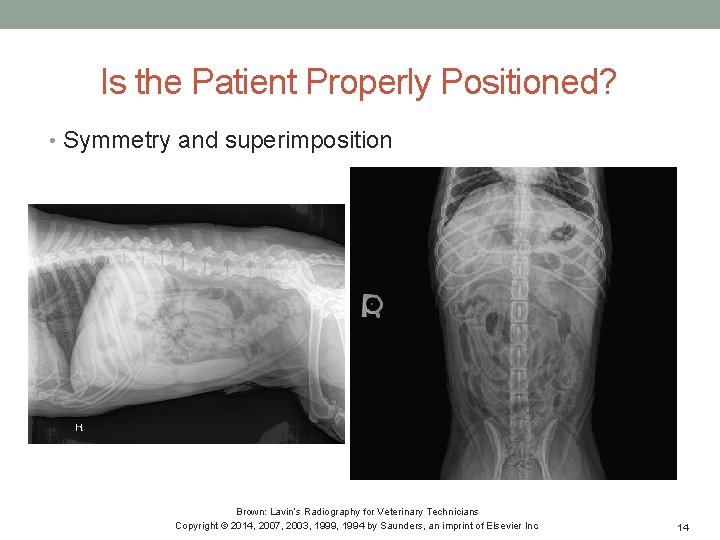
Is the Patient Properly Positioned? • Symmetry and superimposition Brown: Lavin’s Radiography for Veterinary Technicians Copyright © 2014, 2007, 2003, 1999, 1994 by Saunders, an imprint of Elsevier Inc. 14

Further Views Dorsoventral View • Measure, center, and borders as per VD • Placement • Head • Forelimbs • Hind limbs • Maximum expiration Image courtesy of Seneca College, Veterinary Technician Program Brown: Lavin’s Radiography for Veterinary Technicians Copyright © 2014, 2007, 2003, 1999, 1994 by Saunders, an imprint of Elsevier Inc. 15
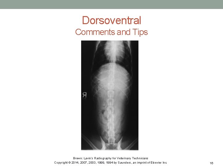
Dorsoventral Comments and Tips Brown: Lavin’s Radiography for Veterinary Technicians Copyright © 2014, 2007, 2003, 1999, 1994 by Saunders, an imprint of Elsevier Inc. 16

Further Views Lateral Decubitus View • Measure, center, and borders as per VD • Placement • Head • Forelimbs • Hind limbs • Sternum • Comments and Tips Brown: Lavin’s Radiography for Veterinary Technicians Copyright © 2014, 2007, 2003, 1999, 1994 by Saunders, an imprint of Elsevier Inc. 17
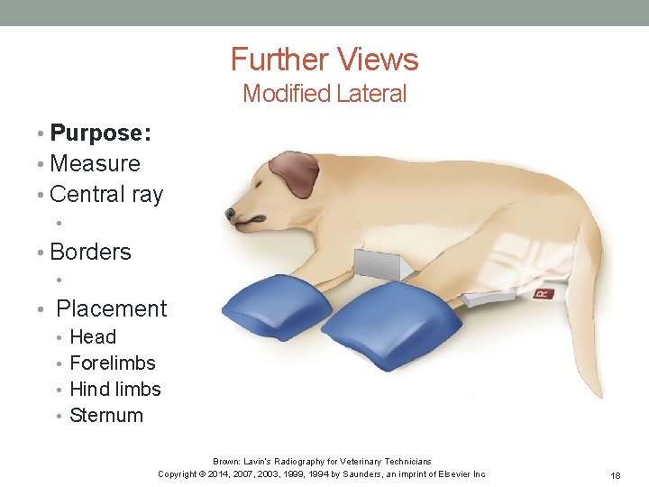
Further Views Modified Lateral • Purpose: • Measure • Central ray • • Borders • • Placement • Head • Forelimbs • Hind limbs • Sternum Brown: Lavin’s Radiography for Veterinary Technicians Copyright © 2014, 2007, 2003, 1999, 1994 by Saunders, an imprint of Elsevier Inc. 18
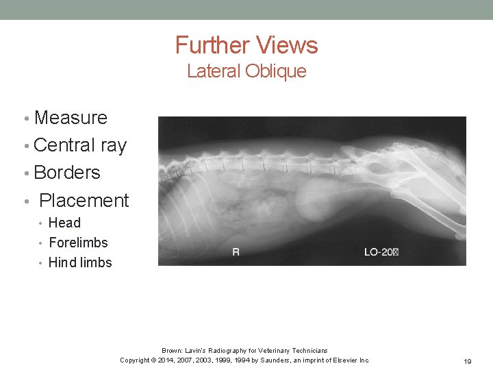
Further Views Lateral Oblique • Measure • Central ray • Borders • Placement • Head • Forelimbs • Hind limbs Brown: Lavin’s Radiography for Veterinary Technicians Copyright © 2014, 2007, 2003, 1999, 1994 by Saunders, an imprint of Elsevier Inc. 19
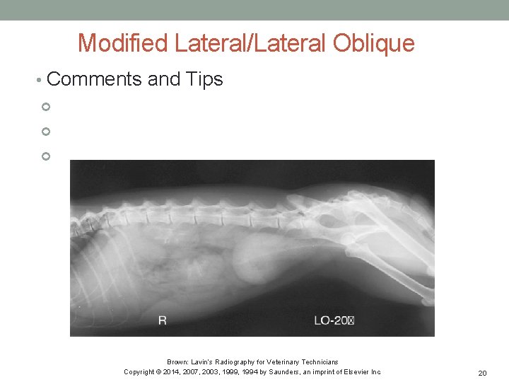
Modified Lateral/Lateral Oblique • Comments and Tips Brown: Lavin’s Radiography for Veterinary Technicians Copyright © 2014, 2007, 2003, 1999, 1994 by Saunders, an imprint of Elsevier Inc. 20
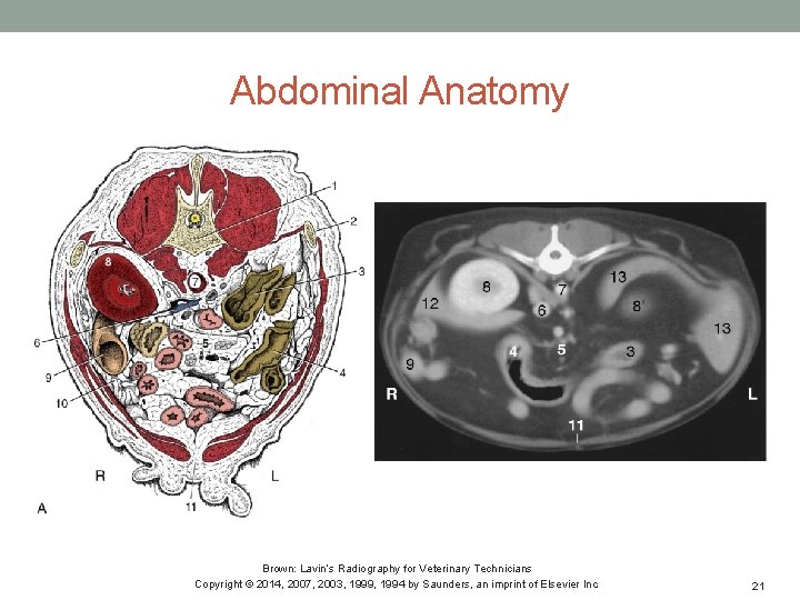
Abdominal Anatomy Brown: Lavin’s Radiography for Veterinary Technicians Copyright © 2014, 2007, 2003, 1999, 1994 by Saunders, an imprint of Elsevier Inc. 21
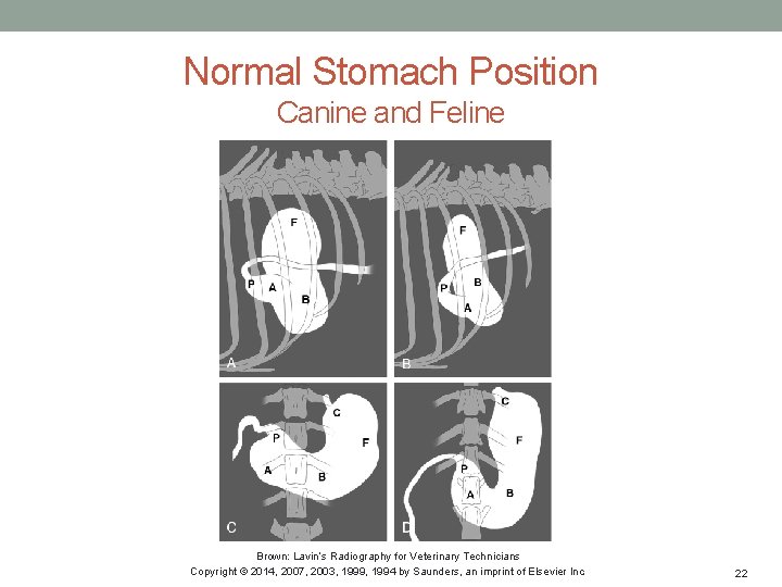
Normal Stomach Position Canine and Feline Brown: Lavin’s Radiography for Veterinary Technicians Copyright © 2014, 2007, 2003, 1999, 1994 by Saunders, an imprint of Elsevier Inc. 22
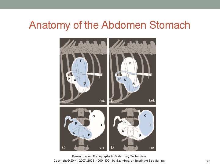
Anatomy of the Abdomen Stomach Brown: Lavin’s Radiography for Veterinary Technicians Copyright © 2014, 2007, 2003, 1999, 1994 by Saunders, an imprint of Elsevier Inc. 23
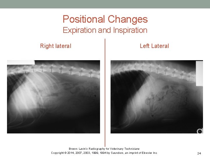
Positional Changes Expiration and Inspiration Right lateral Left Lateral Brown: Lavin’s Radiography for Veterinary Technicians Copyright © 2014, 2007, 2003, 1999, 1994 by Saunders, an imprint of Elsevier Inc. 24

Positional Changes Expiration and Inspiration VD View DV View Brown: Lavin’s Radiography for Veterinary Technicians Copyright © 2014, 2007, 2003, 1999, 1994 by Saunders, an imprint of Elsevier Inc. 25
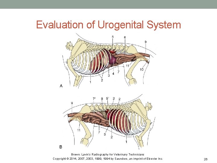
Evaluation of Urogenital System Brown: Lavin’s Radiography for Veterinary Technicians Copyright © 2014, 2007, 2003, 1999, 1994 by Saunders, an imprint of Elsevier Inc. 26
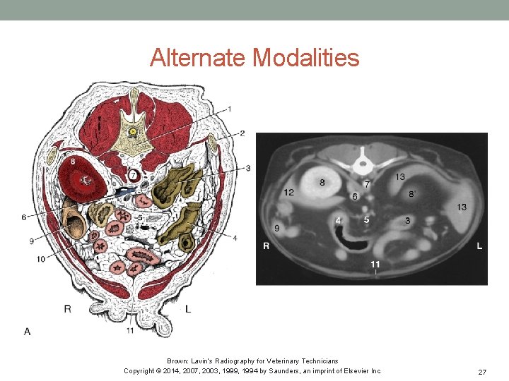
Alternate Modalities Brown: Lavin’s Radiography for Veterinary Technicians Copyright © 2014, 2007, 2003, 1999, 1994 by Saunders, an imprint of Elsevier Inc. 27
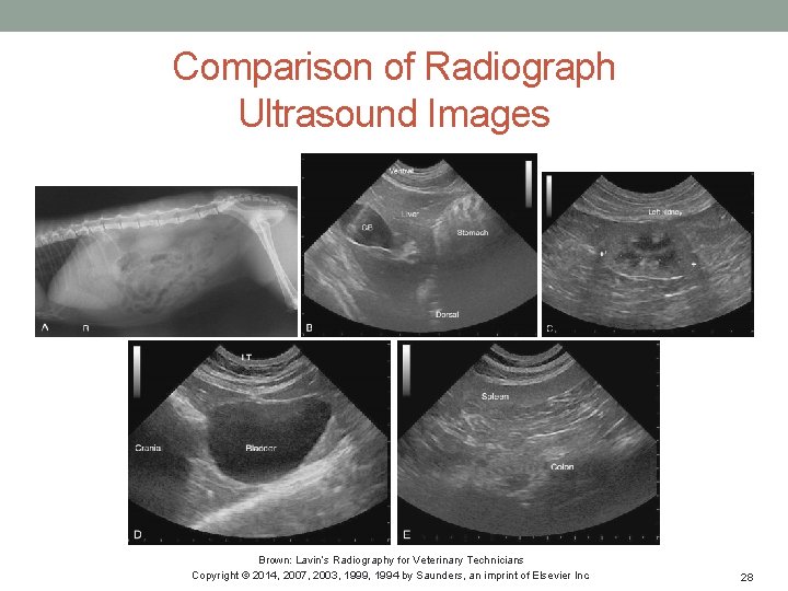
Comparison of Radiograph Ultrasound Images Brown: Lavin’s Radiography for Veterinary Technicians Copyright © 2014, 2007, 2003, 1999, 1994 by Saunders, an imprint of Elsevier Inc. 28
Key Points • Use proper exposure • Utilize proper technique and positioning • Right lateral and ventrodorsal are standard • Understand basic anatomy • Other modalities may be best to visualize further structures Brown: Lavin’s Radiography for Veterinary Technicians Copyright © 2014, 2007, 2003, 1999, 1994 by Saunders, an imprint of Elsevier Inc. 29
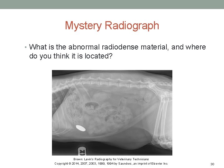
Mystery Radiograph • What is the abnormal radiodense material, and where do you think it is located? Brown: Lavin’s Radiography for Veterinary Technicians Copyright © 2014, 2007, 2003, 1999, 1994 by Saunders, an imprint of Elsevier Inc. 30
- Slides: 30