Sleep Apnea C Tyler Sleep Apnea Kaiser SF


- Slides: 30

Sleep Apnea: • C Tyler

Sleep Apnea Kaiser SF Sleep Lab a. k. a. ‘apnea clinic’ Part 2 C Tyler, Sep 2016 Medical Director Kaiser, San Francisco

Control of Sleep and Waking • Two intrinsic processes – Sleep homeostasis (Process ‘S’) • dependent on sleep-wake cycle • Sleep ‘pressure’ and the sleep bank account – related to duration of wakefulness – relieved by sleep – Circadian rhythm (Process ‘C’) • independent of sleep-wake cycle • Primary function: promoting daytime wakefulness • Peaks and troughs in firing over 24 -hour cycle

Neural Mechanisms of Circadian Rhythm • Suprachiasmatic Nucleus (SCN) • Ablation: • Random distribution of sleep • Reduction in duration of waking periods – Transcription feedback loop: • CLOCK, Per 1, BMAL 1 – Entrainment: ‘Zeitgebers’ (Light, Melatonin)

Blindness: • Loss of ‘timing’ function – Retinal Ganglion Cells – RHT (retino-hypothalamic tract) – SCN • Melatonin – the ‘darkness hormone’ – Mild soporific at big doses – ‘Zeitgeber’ at small doses • ‘Non-24’

Sleep Homeostasis: • Linear Increase in ‘Sleep Pressure’ with wakefulness – Only relieved by SLEEP – replenishing the bank • Extracellular Brain Adenosine – Brain metabolism: • ATP – ADP+P+energy – Accumulates during wake – Causes ‘sleep’

What causes Wakefullness? Neural systems generating wakefulness: Outlined areas: pontine midbrain reticular formation basal forebrain caudal diencephalon (posterior hypothalamus, subthalamus, ventral thalamus) ac, anterior commissure; Ach, acetylcholine; CB, cerebellum; cc, corpus callosum; DA, dopamine; Glu, glutamate; H, histamine; Hi, hippocampus; NE, norepinephrine; OB, olfactory bulb; ot, optic tract; Orx, orexin; SC, spinal cord.

What causes Sleep? ac, anterior commissure; CB, cerebellum; cc, corpus callosum; Hi, hippocampus; OB, olfactory bulb; oc, optic chiasm; Ser, serotonin.

Activity of deep brain structures:

Brain Regions Involved in Sleep

Pathways SCNdm, dorsomedial suprachiasmatic nucleus (shell); SCNvl, ventrolateral suprachiasmatic nucleus (core); PAG, periaqueductal gray matter; PVH, paraventricular hypothalamic nucleus; PVT, paraventricular thalamic nucleus; RCA, retrochiasmatic area; SPZ, subparaventricular zone. BSTL, bed nucleus of the stria terminalis; IGL, intergeniculate leaflet; MPOA, medial preoptic area; OPT, olivary pretectal nucleus; MRN, median raphe nucleus; DMH, dorsomedial hypothalamic nucleus; Neuro-Transmitters 5 -HT, 5 -hydroxytryptamine (serotonin); GABA, gamma-aminobutyric acid; Glu, glutamate; NPY, neuropeptide Y; PACAP, pituitary adenylate cyclase–activating polypeptide The Suprachiasmatic Nucleus (SCN)

NREM pathways

REM sleep: A peculiar state of being: Dreams Paralysis

Narcolepsy: REM breaking bad Gelineau 1880: – Hypersomnia: – Cataplexy: Intrusion of REM atonia into wake – Hypnogogic hallucination: Intrusion of ‘dream’ – • HLA DQB 1*0602 • 1998, two hypothalamic peptides identified: – hypocretin (Hcrt)-1 and Hcrt-2 – aka orexin-A and orexin-B

Neuro-chemestry of Narcolepsy

Consequences of Sleep Deprivation • Sleep Deprivation: – Behavioral – Physiological – Molecular Vigilance • Behavioral: – Sleepiness – Loss of ‘energy’ and motivation – Learning - Remembering – Task performance: Driving

Sleep Deprivation Vulnerability to sleep deprivation varies differ depending on chronic vs acute sleep restriction Individuals underestimate neg impact on cognition and performance. The autonomic nervous system incr sympathetic activity Cognitive performance diminished attention working memory executive functioning information processing decision making Response time is slowed Children - hyperactivity • Other Consequences – – – – – greater morbidity increased mortality sleepiness diminished vigilance decreased pain tolerance lower seizure threshold hyperactive gag and DTR’s nystagmus and ptosis sluggish corneal reflexes decreased cerebral metabolism • subcortical frontal and midbrain regions.

Psychomotor Vigilance Task (PVT) Performance and sleep restriction

Sleep Latency and Vigilance

Micro-sleep • Momentary sleep = loss of consciousness – Sleep onset is imperceptible – Duration of sleep is completely unconscious – Return to consciousness • may be smooth and imperceptible • … or startling • Judgement about risk of sleep is POOR

I’m listening… no you’re not…

Micro-Sleep:

The Self-awareness of Sleepiness • 2007 Kaplan, Dement, et al. – 41 partially sleep-deprived subjects – 30 consecutive two-minute intervals – Self prediction of likelihood of sleep – Physiological and cognitive signs of sleepiness • involuntary eye closure, head-nodding, • wandering thoughts, yawns, • instances of sleep documented on EEG

• Dement, et al: 55% of subjects predicted low likelihood of sleep onset before their first sleep event In a separate experiment: a light was flashed in front of sleepy subjects were asked to push a button for every flash

Can you predict if you will nod off in the next 2 minutes?

Ready for Sleep Apnea?

Respiration and Sleep

Neurological Control of Respiration • Medulla and pons – Pons • Pneumotaxic center – rostral pons • Apneustic center – dorso-lateral caudal pons – Medulla • Dorsal Respiratory Group (DRG) – Nucleus tractus solitaries • Ventral Respiratory Group (VRG) – Nucleus ambiguous – Nucleus retroambugualis

The Muscles of Respiration Figure brainstem (cerebellum removed) respiratory neurons dorsal respiratory group (DRG) ventral respiratory group (VRG). expiratory (E) inspiratory (I) neurons Bötzinger complex (BC), pre-Bötzinger complex (PBC), rostral retroambigualis (R-RA), caudal retroambigualis (C-RA) cervical inspiratory neurons (CIN) respiratory-related neurons in the lateral reticular formation (RF) Projecting to the hypoglossal motor nucleus (XII) inspiratory expiratory neurons solid and dashed lines, respectively excitatory and inhibitory synaptic connections are depicted by arrowhead and square symbols, respectively. Inspiratory and expiratory motor pools in the spinal cord are depicted by closed and open circles, respectively. level of respiratory-related and tonic activities varies for different muscles

 Sleep apnea kaiser
 Annette hulse
 Sleep apnea symptoms
 Kaiser sleep clinic
 Apnea educacion fisica
 Scivolo prono in apnea
 Linguloplasty
 Adults spend about ______% of their sleep in rem sleep.
 Module 23 sleep patterns and sleep theories
 Module 23 sleep patterns and sleep theories
 شرح قصيدة come sleep بالعربي
 Module 23 sleep patterns and sleep theories
 Kaiser ferdinand nordbahn
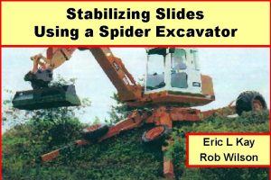
 Kaiser spider excavator
 Militarism definition
 Kaiser mfa northern california
 Kaiser permanente santa cruz
 Kaiser permanente spokane walk in clinic
 Why did the kaiser abdicate?
 Kaiser raman spectroscopy
 Kaiser application status
 Kaiser permanente value compass
 Did kaiser wilhelm have a withered arm
 Pirámide de kaiser niveles
 Kaiser stockton
 What is shell.shock
 Apply for kaiser
 Noel kaiser
 Woher nahmen könige und kaiser ihre macht
 Dr kaiser tamás
 Kaiser permanente springfield
