SKULL Dr Nivin Sharaf MD LMCC Objectives Brain

SKULL Dr. Nivin Sharaf MD LMCC

Objectives • Brain storm basic functions of the human skull • To identify basic anatomical features of the skull • To be able to recognize different bony landmarks of the skull • To Identify outer bony features of bones of the skull • Identify sutures, pterion, and base of the skull “outer view” • Understand importance of different bony foramina of the skull” details TB discussed in CNS”


Parts SKULL The skull has 22 bones, excluding the ossicles of the ear. Except for the mandible, which forms the lower jaw, the bones of the skull are attached to each other by sutures, are immobile, and form the cranium. • The cranium can be subdivided into: an upper part (the calvaria), which surrounds the cranial cavity containing the brain; • a lower anterior part-the facial skeleton (viscerocranium). The bones forming the calvaria are the paired temporal and parietal bones, and the unpaired frontal, sphenoid, ethmoid, and occipital bones. The bones forming the facial skeleton are the paired nasal bones, palatine bones, lacrimal bones, zygomatic bones, maxillae, inferior nasal conchae, and the unpaired vomer. The mandible is not part of the cranium nor part of the facial skeleton.

Neck The neck extends from the head above to the shoulders and thorax below. Its superior boundary is along the inferior margins of the mandible and bone features on the posterior aspect of the skull. The posterior neck is higher than the anterior neck to connect cervical viscera with the posterior openings of the nasal and oral cavities.
 Facial Skeleton consists of 14 irregular bones: Lacrimal 2 Nasal 2 Maxillae")
Regions(Anterior View) Facial Skeleton consists of 14 irregular bones: Lacrimal 2 Nasal 2 Maxillae 2 Zygomatic 2 Palatine 2 Inferior conchae 2 Mandible 1 Vomer 1

Regions Cont.
")
Regions (Extended neck)

Regions Lateral View Cont.

http: //www. gwc. maricopa. edu/class/bio 201/skull/Skull. Slideshow 1/index_PP 1. htm 8 bones form the adult’s Neurocranium Frontal Parietal 2 Temporal 2 Occipital 1 Sphenoid 1 Ethmoid 1 1 Glabella Metopic suture

Posterior View &Sutures

Superior View &Sutures Cont.

Sutures Cont.

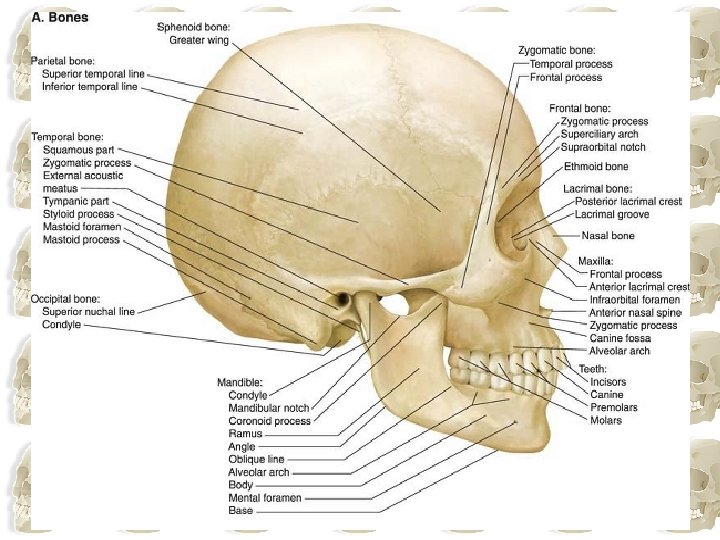
Skull: Simple Lateral View

Skull Lateral Anatomical View

Observe The Pterion: Intersects the course of the anterior division of the middle meningeal artery

Pterion. . How to identify surface anatomy?

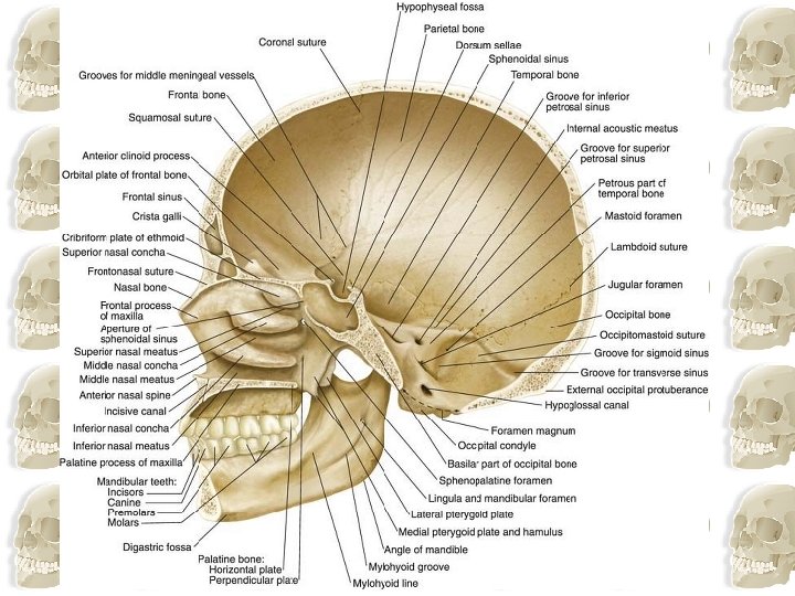
Inferior View

FYI!
 I. Olfactory Sphenoid (optic) II. Optic III. Oculomotor IV.")
Inferior View Foramina Ethmoid (olfactory) I. Olfactory Sphenoid (optic) II. Optic III. Oculomotor IV. Trochlear VI. Abducens Temporal (otic) VIII. Acoustic/Auditory/ Vestibulocochlear Face/Jaws V. Trigeminal VII. Facial Throat (rest of body) IX Glossopharyngeal X. Vagus XI. Spinal Accessory XII. Hypoglosal

The Cranial Cavity

Sphenoid bone

FYI!!
 FYI!!")
Cont. Important openings &structures passing through them( Base of skull) FYI!!

Mandible

Mandible • The final bony structure visible in a lateral view of the skull is the mandible. • Inferiorly in the anterior part of this view, it consists of the anterior body of mandible, a posterior ramus of mandible, and the angle of mandible where the inferior margin of the mandible meets the posterior margin of the ramus • The teeth are in the alveolar part of mandible of the body and the mental protuberance is visible in this view. • The mental foramen is on the lateral surface of the body and on the superior part of the ramus a condylar and coronoid process extend upwards. • The condylar process is involved in articulation of the mandible with the temporal bone and the coronoid process is the point of attachment for the temporalis muscle.

References • Clinical Anatomy by Region “Vishal” 6 th edition • Clinically oriented anatomy “Keith Moore” Fourth edition • 2009 Lippincott Williams & Wilkins • Gray’s Anatomy for students • http: //www. gwc. maricopa. edu/class/bio 201/s kull/skulltt. htm

- Slides: 30