SkinSoft Tissue Infections Emily Blodget MD MPH Assistant

Skin/Soft Tissue Infections Emily Blodget, MD, MPH Assistant Professor of Medicine Division of Infectious Diseases

Cellulitis �Skin infection that result from bacterial entry via breaches in skin barrier �Toe web space bacteria �Fungal foot infections �Pressure ulcers �Venous leg ulcers

Epidemiology � 200 cases per 100, 000 patient-years �Most frequently among middle-aged and older adults �Erysipelas – young children and older adults �Incidence is increasing – 10% of infectious disease related US hospitalizations from 1998 -2006 �Number of US hospitalizations increasing by 73% from 12 per 10000 in 1997 to 21 per 10000 in 2011

Predisposing Factors �Disruption of skin barrier as a result of trauma �Inflammation (eczema, radiation therapy) �Preexisting skin infection (impetigo or tinea pedis) �Varicella �Edema �Lymphatic obstruction following surgical procedures �Age �Obesity

Histologic Features �Nonspecific �Dermal edema �Lymphatic dilation �Diffuse, heavy neutrophil infiltration around blood vessels

Cultures �Usually negative �If positive the burden of bacteria is low �Small number of bacteria responsible
 �Staph aureus �Gram-negative aerobic")
Microbiology �Beta-hemolytic streptococci (group A, B, C, G and F) �Staph aureus �Gram-negative aerobic bacilli – minority of cases �Study of 179 hospitalized patients – beta-hemolytic streptococci – accounted for 73% of cases (blood culture or serologic testing for anti-streptolysin-O and anti-DNAse-B antibodies) �Lack of identifiable etiology in 27%
 �Systematic review – 808 adult and pediatric cellulitis patients undergoing needle aspiration")
Microbiology (cont’d) �Systematic review – 808 adult and pediatric cellulitis patients undergoing needle aspiration or punch biopsy – 16% had cultures that established a diagnosis �Positive culture results – 51% S. aureus and 27% were S. pyogenes �Purulent cellulitis not excluded from the systemic review but abscess was excluded

Microbiology �Blood cultures identified bacteria in only 7. 9% of 1578 patients in a systematic review � 19% - S. pyogenes � 38% other β-hemolytic strep � 14% S. aureus � 28% gram negative organisms �In cases of non-purulent and uncomplicated cellulitis – addition of anti-MRSA did not improve outcome

Microbiology �Study of 422 patients presented with purulent skin and soft tissue infections- 11 ED throughout the US �Skin surface swab cultures revealed MRSA in 59% patients , MSSA in 17% and β hemolytic in 2. 6% �Consider MRSA in purulent infections in high risk populations – athletes, children, military recruits, residents of long term care facilities and IV drug users

• Streptococcus- spreads widely along tissue planes. No pus should be found. If there is any pus, there is more than strep there! • Staph aureus/MRSA – produces localized pus and abscesses with small area of surrounding erythema, rather than diffuse inflammation.

Staph abscesses

Cellulitis in special circumstances �Pasteurella multocida and Capnocytophaga canimorsus �Aeromonas hydrophila and Vibrio vulnificus �Streptococcus iniae �Clostridium species �Pseudomonas aeruginosa �Group B streptococcus

Cllinical Manifestations �Skin erythema �Edema �Warmth �Erysipelas involves the upper dermis and superficial lymphatics �Cellulitis – deeper dermis and subcutaneous fat �Erysipelas lesions – raised above the level of surrounding skin and clear line of demarcation
 �Dilated and edematous skin lymphatics �Bulla formation �lymphangitis")
Clinical Manifestations (cont’d) �Dilated and edematous skin lymphatics �Bulla formation �lymphangitis

�Lower extremities – most common site of involvement for cellulitis �Usually unilateral �Fever is variable �Periorbital cellulitis �Abdominal wall cellulitis �Lymphangitis and inflammation of regional lymph nodes �Necrotizing fasciitis

Erysipelas

Diagnosis �Clinical manifestations �Blood culture, needle aspiration and biopsy – usually not necessary in mild infection �Blood culture – positive in less than 5% of cases �Culture results from needle aspiration can vary - <5% - 40% �Biopsy – culture yields a pathogen 20 -30%

Laboratory findings �WBC elevations – 34 -50% �ESR – 59 -91% �CRP – 77 -97% �Necrotizing fasciitis – serum lactate of 2 – sensitivity of 100%, specificity of 76% (Murphy et. al) �Adjunct tools �Radiographic imaging- ppv of 76% and npv of 100% � 36% necrotizing fasciitis included gas
 �Cultures of blood, pus or bullae – warranted in systemic toxicity, extensive")
Diagnosis (cont’d) �Cultures of blood, pus or bullae – warranted in systemic toxicity, extensive skin involvement, underlying comorbidities, special exposures, recurrent or persistent cellulitis �Cultures of swabs from intact skin – not warranted – commonly polymicrobial or reflect colonization �Radiographic imaging – useful for excluding occult abscess and distinguishing cellulitis from osteomyelitis, necrotizing fasciitis (US or MRI) �Risk of DVT in patients with cellulitis – low (incidence of 3. 1%)

Rapid progressive erythema �Necrotizing fasciitis �Toxic shock syndrome �Gas gangrene
")
Differential �Skin abscess �Deep venous thrombosis �Stasis dermatitis – most often mimics cellulitis (unilateral) �Erythema migrans �Herpes zoster �Septic arthritis �Septic bursitis �Osteomyelitis

Non-infectious causes �Contact dermatitis �Acute gout �Drug reaction �Vasculitis �Insect bite �Panniculitis

Treatment �Non-antibiotic- elevation of affected areas and treatment of underlying conditions �Hydrate skin to avoid dryness and cracking
 �Cochrane review of 25 rcts could not give treatment recommendations as no")
Treatment (cont’d) �Cochrane review of 25 rcts could not give treatment recommendations as no 2 studies used the same treatment regimen �Cellulitis management in Canada – ED had 25 different initial treatment regimens and 40 different antibiotic regimens when discharged
")
�Nonpurulent without systemic signs of infection – antistreptococcal antimicrobial agents (cephalexin, dicloxacillin, amoxicillin/clavulanate, clindamycin) �Multicenter retrospective cohort study – outpatients treated for uncomplicated cellulitis – no statistically different failure rates when comparing oral β lactams with non-β-lactam group (bactrim, tetracyclines, clindamycin) – increased discontinuation rate of nonβ-lactam group

Treatment �Multicenter, double blind rct compared cephalexin to cephalexin + bactrim for non-purulent, uncomplicated cellulitis – no benefit to addition of antibiotics against MRSA �Cure rate of 82 v. 85% �Systemic signs of infection – predicts failure of empiric outpatient antibiotics �Moderate cellulitis – cellulitis with any 1 criterion for SIRS � 2 or more SIRS criteria – consider iv regimen (cefazolin, ceftriaxone, pen G or clindamycin)

Severe Cellulitis �Vancomycin �Used in cases associated with penetrating trauma �Evidence of MRSA infection or colonization elsewhere �Active IV drug use �Linezolid – alternative to vancomycin
 �Severe immunocompromise �SIRS and hypotension �Rapid progression �Empiric vancomycin and zosyn")
Severe cellulitis (cont’d) �Severe immunocompromise �SIRS and hypotension �Rapid progression �Empiric vancomycin and zosyn �Signs of shock add clindamycin for toxic shock syndrome

Purulent cellulitis �Purulent cellulitis with 1 criterion for SIRS – initially treated with same antibiotic for mild disease with suspicion for MRSA (bactrim, doxycycline, minocycline) � 2 or more criteria for SIRS- consider IV therapy (oxacillin, nafcillin or cefazolin) if MSSA suspected or vancomycin, clindamycin or linezolid if MRSA suspected

Newer agents �Dalbavancin – administered once weekly �Oritavancin – single dose as effective as twice-daily iv vancomycin administered for 7 -10 days �Tedizolid �Telavancin

Antimicrobial management �Mild cellulitis – treated with oral antibiotics �Parenteral antibiotics – signs of systemic toxicity, rapid progression of erythema, immunocompromise, proximity to an indwelling device (prosthetic joint or a vascular graft) �Consider parenteral therapy for patients with persistence or progression of symptoms despite 48 -72 hours of appropriate oral therapy

Duration of therapy �Based on clinical response �Outpatient regimen – 5 -10 days �Immunocompromised may require 7 -14 days �Clinical reassessment 24 -48 hours after treatment initiation for improvement in pain, redness, swelling or warmth

Recurrent Cellulitis � 22 -49% patients with cellulitis report at least 1 previous episode �Occur in approximately 14% cellulitis cases within 1 year and 45% cases within 3 years �Occur at the same location �Identification and treatment of underlying conditions

Prophylactic antibiotics �Considered for patients with 3 -4 episodes of cellulitis per year �Oral penicillin 250 mg or 1 g twice daily �Dicloxacillin 500 mg twice daily �Clindamycin 150 mg daily � 2013 double blind rct of 274 patients with 2 or more episodes of cellulitis – 250 mg twice daily of penicillin vs. placebo for 12 months �Significantly reduced risk of recurrent leg cellulitis �Effect diminished when penicillin discontinued �Prophylaxis failure (obese, pre-existing edema)

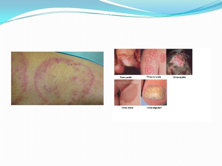
Fungal skin infections �Dermatophyte infections – most common fungal infections on skin, nail and hair �Filamentous fungi in genera Tricophyton, Microsporum and Epidermophyton �Major subtypes �Tinea corporis �Tinea pedis �Tinea cruris �Tinea capitum �Tinea unguium

Tinea infection �Typically superficial and only involve the epidermis �KOH preparation is the most rapid method to confirm the diagnosis �Topical or systemic antifungal therapy – azoles, terbinafine and griseofulvin – extensive or refractory cutaneous infections �Corticosteroid therapy – not necessary to achieve cure and may cause skin atrophy

Intertrigo �Skin folds – moist erythema, malodor, weeping, pruritus and tenderness �Initiation factor – friction and moisture associated with lack of air circulation in deep skin folds �Candidal infection may initiate or aggravate intertrigo �Fungal infection or colonization of the skin – contributes through initiation of innate or acquired immunity-mediated inflammatory cascades

Treatment �Minimizing moisture and friction �Daily cleansing of intertriginous skin with a mild cleanser followed by drying of affected area with a hair dryer on a cool setting �Aeration of affected area when feasible �Daily application of drying powders �Use of absorbent material or clothing, such as cotton or merino wool, to separate skin in folds �Application of barrier creams in areas that may come in contact with urine or feces �Treatment of hyperhidrosis in the affected area �Weight loss in overweight or obese patients �Appropriate treatment of coexisting diabetes mellitus
 �Topical azole antifungal �ketoconazole � clotrimazole �miconazole, � econazole presence of candidal")
Treatment (cont’d) �Topical azole antifungal �ketoconazole � clotrimazole �miconazole, � econazole presence of candidal infection and the additional antibacterial and anti-inflammatory effects of these drugs Topical corticosteroid if there is marked pruritus (2. 5% hydrocortisone cream)

- Slides: 42