Skin Tumors Dr Saleh Abualhaj Plastic Surgeon Skin

Skin Tumors Dr. Saleh Abualhaj Plastic Surgeon

Skin embryology A. Epidermis: Ectoderm B. Dermis: Mesoderm C. Other cells 1. Melanocytes: Neural crest 2. Merkel cells: Neural cells 3. Langerhans cells: Mesenchymal Neural cell origin tumor : Usually have poor prognosis , aggressive , mets

Skin histology A. Epidermis 1. Keratinocytes origin of SCC a. Primary cell in epidermis b. Start in basal layer (stratum germinativum or basale) and make their way carcinoma to surface becoming a dead cornified layer (stratum corneum). 2. Melanocytes a. Found in basal layer b. Protect against ultraviolet (UV) radiation 3. Merkel cells: Mechanoreceptorsa 4. Langerhans cells: Antigen-presenting cells in stratum spinosum origin of basal cell

B. Dermis 1. Cell types: Fibroblast, macrophage, and mast cell 2. Papillary dermis Dermis layers important in classification of tumors esp. in case of malignant melanoma a. Similar thickness to epidermis b. High content of type III collagen, less type I c. Site of collagenase activity. d. Intertwines with the rete ridges of the epidermis. e. Contains terminal networks of Meissner corpuscles and capillaries. 3. Reticular dermis a. Majority of the dermal layer b. Mostly type I collagen bundles with elastic fibers between c. Contains roots of the hair, sebaceous glands, sweat glands, receptors, nails, and blood vessels

Skin malignancies A. Generally grouped into three types ( listed from most common to least ) B. Basal cell carcinoma (BCC) > (most common and better prognosis ) C. Squamous cell carcinoma (SCC) D. Melanoma > ( worse prognosis ) 1. The ratio of BCC to SCC to melanoma is ≈40: 1 ( BCC is the most common the SCC then melanoma )
 Incidence 1. BCC is the most common skin cancer ,")
Basal Cell Carcinoma (BCC) Incidence 1. BCC is the most common skin cancer , accounting for ≈80% of all skin cancers. 2. Roughly 2. 8 million new cases per year in the United States.

Risk factors of BCC ( if there is a risk factor in the Hx think of malignancy ) 1. Sun exposure (increased with lower latitudes, high altitude) : 36% of BCCs originate from the area of previously diagnosed actinic keratosis (AKs), but have distinct cells of origin. + occupation : ( degree of exposure to sun ) 2. Advancing age ( risk increase with age ) 3. Fair complexion 4. Long-term exposure to psoralens and UVA therapy (i. e. , PUVA therapy forpsoriasis) 5. Immunosuppression, most commonly seen in transplant patients 6. Nevus sebaceus of Jadassohn, a superficial skin lesion typically in the head and neck regions, presents as an irregular, raised, yellow to pink, non–hairbearing raised mass. They are usually present at birth or develop in early childhood, and approximately 15% undergo malignant transformation to BCC. 7. Arsenic exposure
 mandibular cyst ,")
Syndromes associated with BCC: a. Basal cell nevus syndrome (Gorlin’s syndrome) mandibular cyst , palm pits , multiple BCC seen early in childhood with malignant b. Xeroderma pigmentosum (XP): Patients have increased incidence of BCC, SCC, and malignant melanoma ( increase RISK OF ALL SKIN CA ) + pathophysiology is important c. Albinism

BCC disease biology and characteristics A. Basal keratinocytes are the cell of origin, residing in the basal layer of the Epidermis at the dermoepidermal junction. B. No universal clinical precursor lesion C. BCC is most common in areas with high concentrations of pilosebaceous follicles and thus >90% are found on the head and neck. D. Metastasis is rare—termed “barely a cancer” by some researchers E. Morbidity is caused by invasion of the tumor into underlying structures, including the sinuses, orbit, and brain. Typically, only a problem if neglected for many years.
 1. Nodular BCC a.")
Types of BCC ( definite dx is by histopathology ) 1. Nodular BCC a. The *most common type, usually presenting as a single lesion consisting of pearly papules with telangiectasias, pruritus, and occasional bleeding. b. Lesion breakdown over time leads to nodulo-ulcerative BCC (“Rodent ulcer”). Lesions usually on head and neck. 2. Superficial spreading BCC ( some time U cant differentiate it from scare) a. Slow-growing, erythematous, with minimal induration, and located primarily on the trunk. b. It is easily confused with other scaly, eczematous dermatoses. c. The lesions are shallow with a characteristic horizontal growth pattern and often present in multiples.
 BCC a. Flat, often yellowish or hypopigmented, sometimes resembling scars")
3. Morpheaform (sclerosing, fibrosing) BCC a. Flat, often yellowish or hypopigmented, sometimes resembling scars or normal skin. b. The true extent of the lesion is usually greater than the clinical appearance. (Bad prognosis) c. There is a high incidence of recurrence or incomplete excision due to “fingerlike” extensions. d. Margins of 1 cm or Mohs extirpation is warranted. 4. Pigmented BCC: Similar to nodular BCC; easily confused with melanoma due to : its deep pigmentation and nodularity 5. Adnexal BCC a. Uncommon and found in older individuals. b. Tumors arise from sweat glands, and although they exhibit slow growth, they are locally invasive, with a high incidence of local recurrence.

# If there is a high suspicion of skin CA take biopsy ( excisional or incisional ) according to the size of the lesion histopathology Treatment of BCC A. Standard surgical techniques: ≈95% cure rate 1. Wide local excision of BCC: 3 - to 5 -mm margins for nonaggressive types and 7 -mm up to 1 cm margins for morpheaform type !!. Safety margins are important� *BCC No LN Metastasis so it just require local excision of the tumor (full thickness of the skin ) * No need for CT or tumor staging in BCC a. Frozen sections may be used to confirm negative margins intraoperatively. False negatives are common. 2. Mohs surgery: Sequential horizontal excision with immediate frozen section testing by dedicated Mohs dermatopathologist a. *Indications include morpheaform BCC and/or lesions in aesthetically Sensitive areas (nose, eyelid, lip, etc. ) B. There is other method of treatment as radiological or curettage but not impo.

Squamous Cell Carcinoma Incidence Second most common skin cancer after BCC.

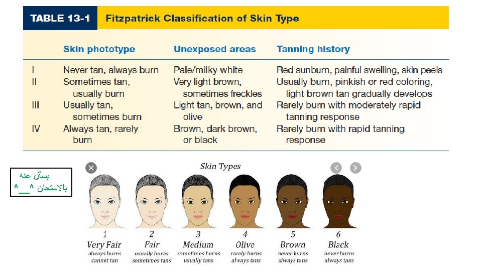
Risk factors : 1. UV radiation: Sun exposure and tanning booth use; PUVA therapy for psoriasis 2. Chemical exposure, including some pesticides, organic hydrocarbons such as coal tar, fuel oil, paraffin oil, and arsenic (in welding materials) 3. Viral infection: Some types of human papillomavirus (HPV) ; herpes simplex virus very impo in head and neck tumor 4. Radiation: Long latency between exposure and disease. 5. Marjolin’s ulcer: SCC arising in a chronic wound (i. e. , chronic burn scars and pressure sores) secondary to genetic changes caused by chronic inflammation. - Any non healing ulcer you should think of CA - Bad prognosis 6. Impaired immunity: That is, immunosuppression for transplants and AIDS. Ratio of SCC to BCC in these patients is 2: 1. 7. Fitzpatrick skin type

SCC disease biology and characteristics *Precursor lesions ( premalignant , 10 % risk at least to be malignant ) 1. Actinic keratoses (AKs, or solar keratoses) a. Erythematous macules and papules with coarse, adherent scale b. Histologically resembles SCC in situ (pre-malignant c. AK is considered a precursor lesion; up to 5% progress to SCC; in turn, 65% of all SCC arise from sites of Aks 2. Bowen’s disease (SCC in situ) a. Exhibits full-thickness cytologic atypia of the keratinocytes b. Erythroplasia of Queyrat is SCC in situ of the glans penis.

3. Leukoplakia a. Presents as a white patch on oral or other mucosa. b. Malignant transformation occurs in 15%. 4. Keratoacanthoma a. Benign skin tumor that is composed of squamous cells and keratin; may clinically resemble SCC. b. Etiology is unknown but thought to originate from hair follicles. c. Typically has a rapid 6 -week growth phase followed by involution over the next 6 months. However, can progress to SCC in 5% to 10% of cases. d. Excision is the treatment of choice; may be difficult to differentiate from SCC histologically.

Types of SCC 1. Verrucous SCC: Slow-growing, exophytic , fungated , and less likely to metastasize. 2. Ulcerative SCC: Grows rapidly and is locally invasive. a. Ulcerative SCC has very aggressive growth characteristics, raised borders, and central ulceration. b. <50% 5 -year survival if spread to lymph nodes in the head and neck. 3. Majorlin’s ulcer a. Arise from chronic wounds (burn, pressure ulcer, fistula, osteomyelitis tracks) b. Commonly metastasize to lymph nodes.

> In incisional biopsy take tissue from 4 quadrant SCC treatment options A. Standard surgical techniques: 90% to 95% cure rates; similar to BCC options 1. Wide local excision of SCC: 5 - to 10 -mm margins are usually sufficient. ( mm not Cm �) Frozen sections may be used to confirm negative margins intraoperatively. a. If <2 cm, low grade and extends to dermis, 4 -mm margin b. If >2 cm, grade 2 to 4 , high risk or extension into fat, 6 -mm margin - Needs staging by CT ! ( Risk of metastasis ) - May need LN dissection if involved and sentinel LN biopsy ! 2. Mohs surgery: Sequential horizontal excision with frozen section testing. Highest cure rate for SCC: 94% to 99%. - Indications : : include recurrent, high-risk SCC, and/or lesions in aesthetically sensitive areas (nose, eyelid, lip, etc. )
 nodes. 2. FNA: Confirm spread of")
Regional lymphadenectomy 1. Indicated for clinically positive (palpable) nodes. 2. FNA: Confirm spread of SCC to palpable lymph node. 3. ELND: Indicated for a tumor extending down to parotid capsule or a large lesion contiguous with a draining nodal basin. 4. SLN biopsy: Considered for high-risk SCC without palpable nodes controversial). D. Adjuvant radiation therapy: Used postexcision for high-risk cutaneous SCC.

Melanoma : least common , , most aggressive Epidemiology 1. Incidence is increasing, faster than any other cancer in Western world 3. Lifetime risk in general population is 2% for children born today. 4. Less than 3% of all skin cancers, but cause of 75% of skin cancer-related deaths. 5. Prognosis of metastatic disease has changed little in past 40 years (unlike many other cancers). Very aggressive tumor
 , freckling, light")
Risk factors 1. Phenotypic include fair skin (Fitzpatrick I and II) , freckling, light eye color, and light hair color (stronger risk factor than eye color). Darker skin is protective against melanoma. 2. Geographic: High altitudes, lower latitudes have increased UV exposure, and therefore increased risk. 3. Gender: Females have lower risk and better prognosis ; however, gender-based differences in risk are lessenings. Lower extremity is the most common site in (females) ; males more commonly have lesions on the head and trunk.

4. Race: Incidence is lower , but prognosis is worse for African-Americans , (Why ? ! ) due to delayed diagnosis and/or worse disease subtype. 5. Affluence: Unlike most cancer types, higher socioeconomic status correlates with higher risk. 6. History of UV radiation exposure (both UVA and UVB): Evidence for direct causality is less clear than for other skin cancer types. A history of blistering sunburns, particularly in early life, correlates to increased risk of some melanoma types. 7. Previous melanoma is a strong predictive factor and confers a 3% to 5% chance of developing a second melanoma.
 i. Heterogeneous group of syndromes; due mutations in various DNA")
c. Xeroderma pigmentosum (XP) i. Heterogeneous group of syndromes; due mutations in various DNA repair genes. ii. DNA damage by UV leads to early death secondary to metastatic spread of skin tumors. iii. Typically presents in early childhood with multiple BCCs; SCCs and melanomas typically cause death. iv. Restriction from sunlight exposure is mandatory, with aggressive surveillance/ treatment of skin lesions. Bad prognosis

Classification of melanoma types 1. Superficial spreading melanoma a. *Most common type, ≈70% cases b. Intermediate in malignant potency ( good prognosis because it has > a horizontal spread ) c. Most likely to arise from a preexisting nevus d. Affects both genders equally e. Median age at diagnosis is 50 years f. Upper back in men and lower legs in women are most common sites g. Irregular, asymmetric borders with color variegation h. Radial growth phase early, vertical growth phase late

2. Nodular melanoma a. Second most common: 15% to 30% cases b. *Most aggressive type (worse prognosis ) c. Typically do not arise from preexisting nevi d. Men are affected twice as frequently as women e. Median age at diagnosis is 50 years f. No clear association with sunlight exposure g. Typically bluish-black, with uniform, smooth borders h. 5% are amelanotic—associated with a poorer prognosis because of delayed diagnosis i. Vertical growth phase is a hallmark feature ( cause of poor prognosis ) ; no radial growth
 a. 10% to 15% of cutaneous melanomas b. *Least")
3. Lentigo maligna melanoma (LMM) a. 10% to 15% of cutaneous melanomas b. *Least aggressive type c. Most clearly associated with sunlight/UV exposure d. Head, neck, and arms of elderly (sun-exposed areas) typically affected e. Women are affected more frequently than men f. The median age at diagnosis is 70 years g. Usually greater than 3 cm in diameter; irregular, asymmetric with color variegation, areas of regression may appear hypopigmented. h. Precursor lesion is lentigo maligna or Hutchinson freckle (histologically equivalent to melanoma in situ, or AJMH): radial growth phase only. Transition to vertical growth phase marks development of LMM. i. Malignant degeneration is characterized by nodular development.

4. Acral lentiginous melanoma a. 2% to 8% of melanomas in Caucasians, 35% to 60% of melanomas in African-Americans, Hispanics, and Asians b. Presents in palms, soles, and beneath nail plate (subungual). Must be distinguished from melanonychia, a benign, linear, pigmented streak in the nail, common in African and Asian populations. Due to the risk of melanoma, biopsy of suspect lesions should be performed. c. Median age at diagnosis is ≈60 years d. Irregular pigmentation, large size (>3 cm) common e. Most common site is : : great toe or thumb. f. Long radial growth phase, transition to vertical growth phase occurs with high risk of metastasis. Bad prognosis # The most important prognostic factor in malignant melanoma is depth = vertical extension.

Noncutaneous melanoma 1. Mucosal melanoma a. Mucosal melanomas represent <2% of melanomas, most commonly presenting within the genital tract, anorectal region, and head and neck mucosal surfaces. b. Difficult to detect; typically advanced at the time of diagnosis with poor prognosis. c. Radical excision is of questionable benefit. 2. Ocular melanoma a. Represent 2% to 5% of melanomas (most common noncutaneous melanoma) b. b. Interference with vision leads to earlier diagnosis. c. Melanomas of iris are similar to cutaneous melanomas in genetics/behavior; melanomas of the posterior uvea act more like mucosal melanomas and have a worse prognosis. d. The eye has no lymphatic drainage; therefore, no nodal metastasis is seen e. The liver is the main site of metastatic disease f. Treatment is by enucleation only
 1. Represent 3% of melanomas")
Melanoma with an unknown primary ( unknown origin ) 1. Represent 3% of melanomas 2. Diagnosis is by exclusion 3. Nodal metastases are the most common presentation ( axillary , inguinal ) 4. Prognosis is similar to metastatic melanomas with a known primary. bad prognosis Only dissection of LN. # WHY this happen ? ! As a theory : that metastatic tumor suppress the primary tumor !

Diagnosis and staging of melanoma A. Physical examination is only 60% to 80% sensitive for diagnosing melanoma. Full-body photography to monitor atypical nevi may increase sensitivity. B. Common clinical features of melanoma lesions (nevi) : (ABCDE) 1. Asymmetry 2. Border irregularity & Bleeding 3. Color variation 4. Diameter >6 mm 5. Enlarging/evolving lesion

Diagnosis of primary melanoma is made by histologic analysis of * full-thickness biopsy specimens 1. Excisional biopsy is preferred for lesions <1. 5 cm in diameter. If possible, excise lesion with 1 - to 2 -mm margins. 2. Incisional biopsy is appropriate when suspicion is low, the lesion is large (>1. 5 cm) or is located in a potentially disfiguring area (face, hands, and feet), or when it is impractical to perform complete excision. Incisional biopsy does not increase risk of metastasis or affect patient survival. 3. Permanent sectioning is used to determine tumor thickness 4. Avoid shave biopsies, since they forfeit the ability to stage the lesion based on thickness

# Very important In malignant melanoma margin in *cm

Treatment 6. Wide local excision for tissue diagnosis can decrease the efficacy of future lymphatic mapping because of disruption of local lymphatics. Biopsy incisions should result in scars parallel to lymphatic drainage. according to depth ( in more than. 75 mm depth we need to do sentinel LN biopsy. ) 7. Orientation of biopsy incisions should also take definitive surgical therapy into consideration. a. Extremity biopsies should use longitudinal incisions. b. Transverse incisions are sometimes preferable for preventing contractures over joints. c. Head and neck incisions should be placed within relaxed skin tension lines, keeping facial aesthetic units in mind.
 Tumor thickness ( BY mm or histological layer ) 2)")
Major prognostic factors: 1) Tumor thickness ( BY mm or histological layer ) 2) Nodal status , and 3) Metastases—TNM LN Mets occur in stage 3 1. Breslow thickness is reported in millimeters; thus, it is more accurate and reproducible than Clark level and is a better prognostic indicator. 2. Clark level is based on invasion through the histologic layers of the skin; more subjective (SO) According to this grading we determine the safety margin and LN biopsy. - We do sentinel LN biopsy if the LNs are clinically negative , if +ve we don’t do it ( dissection immediately).

melanoma treatment A. Definitive management of melanoma 1. Wide local excision is the treatment of choice. 2. Recommended surgical margins depend on tumor thickness 3. Subungual melanoma requires amputation proximal to the DIPJ for fingers and proximal to IP joint for the thumb.

# In melanoma and SCC if LN is +ve we staging of LN mets by chest /abdomen CT scan , if the pt has symptoms Headache brain ct Elevated liver enzyme liver u/s Surveillance and treatment of melanoma recurrence 1. Asymptomatic patients should be seen every 3 to 4 months for 2 years, then every 6 months for 3 years, and then annually. The most accurate way to detect metastatic disease is to take a thorough history. 2. Chest X-ray and liver function tests (LDH and alkaline phosphatase) are usually sufficient; more extensive workups including CT scans have not altered outcomes. 3. Local recurrences typically occur within 5 cm of the original lesion, usually within 3 to 5 years after primary excision; most often this represents incomplete excision of the primary tumor. 4. The most common sites of recurrence are the skin, subcutaneous tissues, distant lymph nodes, then other sites (lung, liver, brain, bone, GI tract). 5. Re-excision is the primary treatment for local, small, isolated lesions 6. Surgery is effective for palliation in patients with isolated recurrences in skin, CNS, lung, or GI tract.

• BCC • Wide local excision only • No need to post op chemo or radiotherapy • SCC • • Wide local excision Staging More than T 3 LN biopsy Post op radiotherapy , ( post op chemo isn’t effective ) • Malignant Melanoma • Biopsy according to depth wide local excision • Post op chemo and radiotherapy
- Slides: 39