SKIN SUPERFICIAL DEEP FASCIA Dr Mukesh Singla SKIN


: • Single layer of cuboidal")


: • Non-pigmented granular dendrocytes.")


 • Absent from lips, glans & nail bed. • Mode")


 • Langer lines of skin tension, or sometimes called cleavage lines")


- Slides: 72
SKIN, SUPERFICIAL & DEEP FASCIA Dr Mukesh Singla
SKIN
DEFINITION • • • GENERAL COVERING OF THE EXTERNAL SURFACE OF THE BODY FORMS 15% OF THE TOTAL BODY WEIGHT THICKNESS-1. 5 to 5. 0 mm LARGEST ORGAN OF THE BODY
INTEGUMENTARY SYSTEM
Some Facts about Skin • Surface area: 1. 5 -2. 0 sq meters • Growth rate of nail: 0. 1 mm per day • Growth rate of hair: 1. 5 -2. 2 mm per week • Life span of hair: Eyelashes, axilla- 4 months Scalp – 4 years
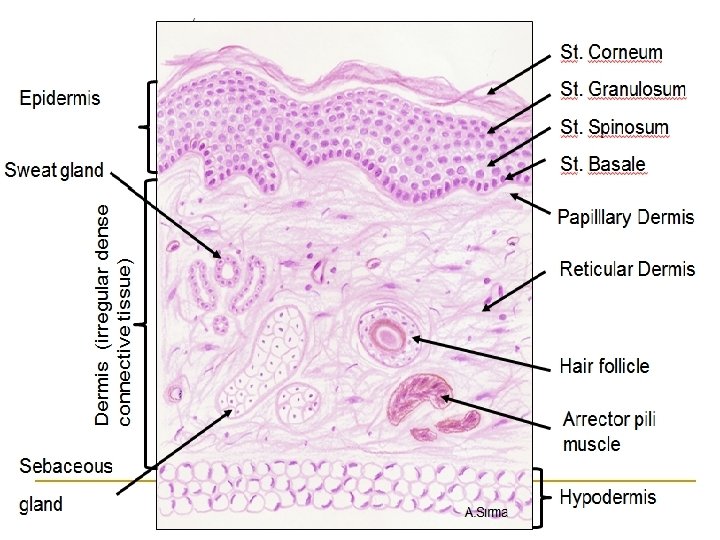
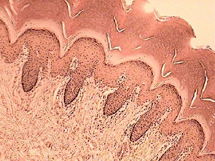
STRUCTURE OF SKIN • TWO DISTINCT LAYERS * EPIDERMIS * DERMIS • EPIDERMIS : SUPERFICIAL AND AVASCULAR • DERMIS : DEEP AND VASCULAR
TERMS USED FOR MOVEMENTS
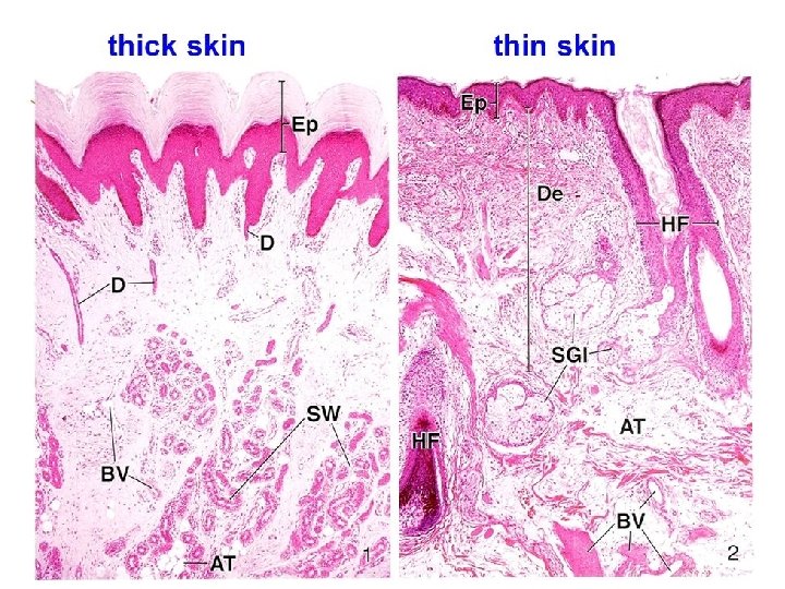
TYPES OF SKIN • THICK SKIN - EPIDERMIS VERY THICK USUALLY HAIRLESS ON PALMS OF HAND & SOLES OF FEET • THIN SKIN – COVERS GREATER PART OF BODY & IS USUALLY HAIRY • EXCEPTION – SCALP : THICK AND HAIRY
Layers Of Skin Epidermis: • Composed of keratinized stratified squamous epithelium. Dermis: • Papillary region composed of loose connective tissue. • Reticular region composed of dense irregular connective tissue. Hypodermis: • Composed of areolar tissue with abundant adipocytes
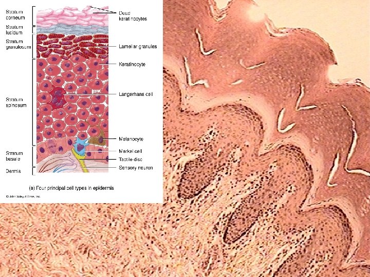
Cells • Keratinocytes • Melanocytes-pigment forming cells from neural crest cells • Merkel cells-sensory mechanoreceptors • Langerhans cells-antigen presenting cells from bone marrow • Free nerve endings
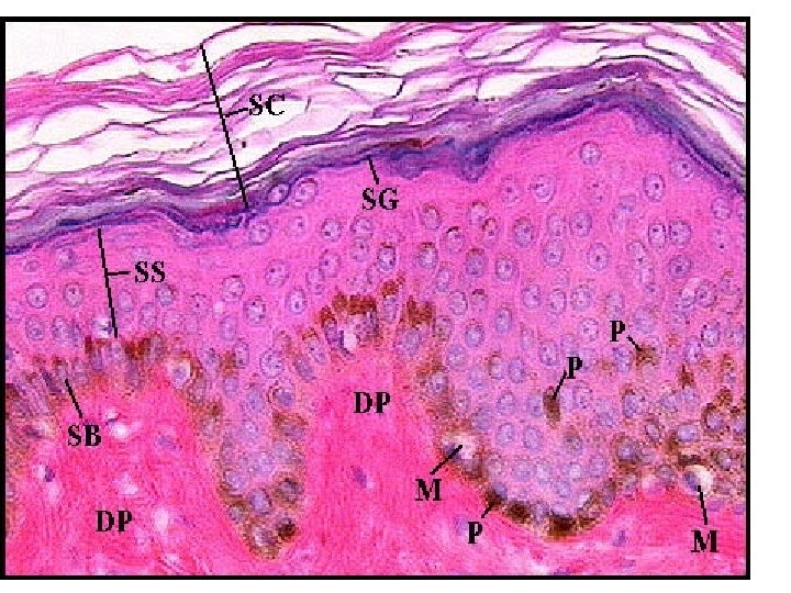
LAYERS OF THE EPIDERMIS Stratum Basale (Germinal/ Malpighian layer): • Single layer of cuboidal cells resting on basement membrane. • High mitotic activity. Stratum Spinosum: • Several layers of polygonal cells. • Cells are held together by desmosomes. Stratum Granulosum: • 3 -5 layers of flattened polygonal cells. • Cells contain keratohyaline granules.
contd…. Stratum Lucidum: • Seen only in non-hairy or thick skin. • Cells are flattened, translucent, eosinophilic with indistinct boundaries & nucleus. • Contains a product of keratohyaline i. e. eleidin. Stratum Corneum: • Composed of structureless dehydrated dead cells. • Flattened & scale-like. • Filled with keratin. • Superficial layer is continuosly sloughed off.
Stratum Basale
Stratum Spinosum
Stratum Granulosum
Stratum Lucidum
Stratum Corneum
SPECIALIZED CELLS OF THE EPIDERMIS Keratinocytes: • Most common cells of the epidermis. • Provides protection and waterproofing sealant. Melanocytes: • Rounded cells with dendrite-like branches. • Present in Stratum basale. • Produces melanin pigment responsible for the colour of skin. • Melanin is a brown/black pigment that absorbs UV-light.
SPECIALIZED CELLS OF THE EPIDERMIS Langerhans Cells (antigen presenting cells): • Non-pigmented granular dendrocytes. • Present in Stratum spinosum. • Nucleus is indented at many places & cytoplasm contains rod-shaped granules. • They participate in immune responses against bacteria and viruses. Merkel Cells: • Found in Stratum basale. • Sensory cells innervated by sensory nerves. • Abundant in fingertips, oral mucosa & hair follicles. • Function as mechanoreceptors.
PIGMENTATION OF SKIN The colour of skin depends upon following factors: • Carotene: yellow-orange pigment (precursor of vitamin A) found in stratum corneum & dermis. • Melanin: produced in epidermis by melanocytes gives black colour to the skin. • Hemoglobin (in blood vessels of dermis): gives pink colour to the skin.
LAYERS OF THE DERMIS • Papillary layer: -Narrow band of loose connective tissue. -In contact with basement membrane of stratum basale. -Dermal papillae (finger- like processes) - provide mechanical anchorage and supply nerves and blood vessels • Reticular layer: -Dense irregular connective tissue. -Thick elastic fibres. -may be involved in development of skin lines Dermal 4 papilla 3 1 2
TYPES OF SKIN Thin Skin Thick Skin Layers of epidermis St. corneum & spinosum are thin while thick while lucidum is absent. present. Thickness of epidermis 0. 10 -0. 15 mm 0. 6 -4. 5 mm Epidermal ridges Absent Present (well developed dermal paplillae) Hair follicles, arrector pili Present muscle & sebaceous gland Absent Sweat glands Few Many Sensory receptors Less More Distribution Covers all parts of body Present in palms, palmar except palms & soles surface of digits & soles
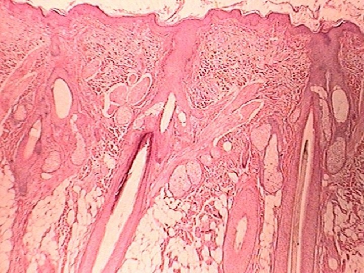
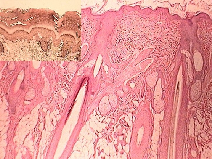
APPENDAGES OF THE SKIN HAIRS: • Keratinized filaments derived from invagination of the basal layer of epidermis into the dermis. • Parts- a) Root: enclosed by hair follicle. b) Shaft: projects above the surface. • Hair follicle: tubular invagination, partly epidermal and partly dermal in origin.
contd…. . Structure of shaft and root: • Medulla • Cortex • Cuticle Hair follicle: • Tubular invagination of epidermis & dermis in which hair root resides. • Layers: 3 (inner root sheath, outer root sheath, connective tissue sheath).
contd…. . • Hair bulb: lower expanded end of hair follicle. • Hair papilla: the indentation at the base of hair bulb by part of the dermis.
contd…. . Arrector Pilorum Muscle: • Smooth muscle innervated by sympathetic nerves. • Extends from papillary layer of dermis to the connective tissue sheath of a hair follicle. • Contraction of muscle presses the sebaceous gland which squeezes out sebum. • Formation of “goose flesh”.
APPENDAGES OF THE SKIN NAILS: • Hardened keratin plates on the dorsal surface of the tips of fingers & toes. • Parts: a) Root b) Free border c) Body • Nail bed: tissue on which the nail rests. Made up of stratum basale & spinosum.
APPENDAGES OF THE SKIN SEBACEOUS GLANDS: • Distributed all over the dermis of the skin, except for the palms & soles. • Abundant in the scalp, face, around the apertures of the ear, nose, mouth & anus.
APPENDAGES OF THE SKIN SEBACEOUS GLANDS: • Holocrine in nature. • Number of alveoli connected to broad duct that opens into hair follicle. • Produces an oily secretion called sebum.
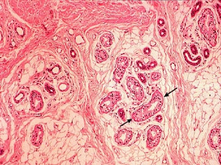
SWEAT GLANDS (SUDORIFEROUS GLANDS) • Absent from lips, glans & nail bed. • Mode of secretion: merocrine • Simple tubular gland • 2 parts: (a) Secretory portion (b) Excretory duct Secretory portion: • Twisted coil like structure with simple cuboidal epithelium. • 3 types of cells: clear cells, dark cells, myoepithelial cells. Excretory duct: • Long & extends from secretory portion to surface of epidermis.
contd…. 2 types: Eccrine: • Most numerous in the soles & palms. • Produces thin watery secretion. Apocrine: • Confined to axilla, eyelids (Moll’s glands), nipple & areola of breast, perianal region, and the external genitalia. • Produces thick odourous secretion. • Ceruminous glands & lactating mammary glands are modified apocrine sweat glands.
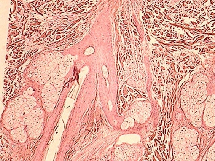
Sweat Gland
SURFACE IRREGULARITIES • FLEXURE LINES - Permanent lines along which the skin folds during habitual movements of joints • CLEAVAGE LINES - According to arrangement of fibres in deep fascia, horizontal in trunk, in old age fibres atrophy and skin wrinkles
PAPILLARY RIDGES • Palms and soles and digits • Form narrow ridges separated by fine parallel grooves, • corrospond to dermal papillae • Study is called dermatoglypics • Pattern of finger prints- loops , whorls and arches and composite
Langer's lines(Cleavage lines) • Langer lines of skin tension, or sometimes called cleavage lines • Correspond to the natural orientation of collagen fibers in the dermis, • Are generally parallel to the orientation of the underlying muscle fibers. • Langer's lines have relevance to forensic science and the development of surgical techniques
Langer's lines • Langer lines of skin tension, or sometimes called cleavage lines • Correspond to the natural orientation of collagen fibers in the dermis, • Are generally parallel to the orientation of the underlying muscle fibers. • Langer's lines have relevance to forensic science and the development of surgical techniques
Applications of Langer Lines • Incisions made parallel to Langer's lines may heal better and produce less scarring than those that cut across. Conversely, incisions perpendicular to Langer's lines have a tendency to pucker and remain obvious, although sometimes this is unavoidable • In old age, elastic fibres atrophy and skin becomes wrinkled
Linea gravidarum • Rupture of fibre bundles of dermis due to excessive stretching result in prominent white lines. • Seen in anterior abdominal wall in pregnancy.
Rule of Nine: To estimate the extent of damaged skin in burn injuries. • First degree burn- only epidermis. • Second degree burnepidermis + upper region of dermis. • Third degree burnentire thickness of skin.
Dermatomes • The strip of skin supplied by a single spinal nerve is called dermatome.
FUNCTIONS OF SKIN Protective shield for the body Barrier to water Thermoregulation Important sense organ (pain, touch, temperature & pressure) • Absorption of ultraviolet radiation from sun for the production of vitamin D • •
APPLIED ANATOMY § Skin is pale in anemia, yellow in jaundice and blue in cyanosis § Skin incisions should be made parallel to lines of cleavage to have the smallest scar § Sebaceous cyst is common in scalp due to obstruction to sebaceous duct
SUPERFICIAL FASCIA
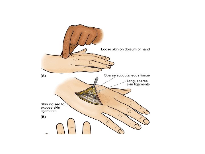
SUPERFICIAL FASCIA § DEFINITION- General coating of the body beneath the skin made up of loose areolar tissue and fat § ABUNDANCE OF FAT- Front of thigh and anterior abdominal wall § ABSENCE OF FAT- Eyelids & external ear § In females there is more fat and it is evenly distributed
TERMS USED FOR MOVEMENTS
IMPORTANT FEATURES § Most distinct in lower part of anterior abdominal wall & limbs § Very thin on dorsal aspect of hands & feet, sides of neck, face. § Very dense in scalp, palms and soles.
IMPORTANT FEATURES § IT CONTAINS 1. Cutaneous nerves & vessels 2. Groups of lymph nodes 3. Subcutaneous muscle in neck
FUNCTIONS • Helps in movements of skin • Allows for the passage of the vessels & nerves to the skin • Conserves body heat as fat is a bad conductor of heat • Fat fills hollow spaces like axilla & orbits
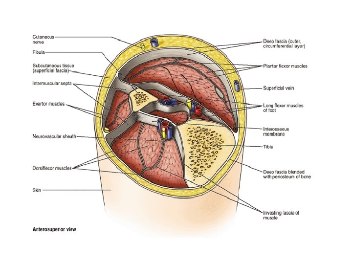
DEEP FASCIA • Deep fascia is a fibrous sheet which covers the body beneath the superficial fascia. • It is devoid of fat, and is usually inelastic and tough
DISTRIBUTION OF DEEP FASCIA • Best seen in limbs where it forms tough and tight sleeves • Well defined in the neck where it • forms a collar • Not well formed on the trunk and face • Blends with periosteum of a subcutaneous bone
MODIFICATIONS OF DEEP FASCIA • Inter muscular septa which divides limbs into compartments • Thickenings form retinacula around joints like wrist and ankle • Forms sheath around large arteries • In palms & soles form aponeurosis • Form investing layer of fascia in region of neck giving it shape.
INTERMUSCULAR SEPTA
RETINACULA
INVESTING LAYER OF FASCIA
FUNCTIONS • Keeps underlying structures in position • Provides extra surface for muscle attachment • Helps in venous return • Helps muscles in action by applying tension and pressure • Retinacula keep tendons in position
MCQ Q 1. Which layer is present only in thick skin: a. b. c. d. Stratum basale Stratum spinosum Stratum granulosum Stratum lucidum
MCQ Q 2. The characteristic feature of reticular layer of dermis is: a. b. c. d. High mitotic activity Contains keratin granules Dense irregular connective tissue Finger like processes
MCQ Q 3. Secretion of sebaceous glands is aided by contraction of: a. b. c. d. Arrector pilorum muscle Myoepithelial cells Papillary layer of dermis Reticular layer of dermis
MCQ Q 4. Langerhans cells are present in: a. b. c. d. Stratum basale Stratum spinosum Stratum granulosum Stratum lucidum
MCQ Q 5. The sensory cells of epidermis are: a. b. c. d. Melanocytes Keratinocytes Langerhans cells Merkel cells