SKIN N O R M A L Skin

 Skin, epidermis,")


 TERMS • • • • macule patch papule nodule plaque vesicle bulla")

 TERMS • • • • hyperkeratosis parakeratosis hypergranulosis acanthosis papillomatosis acantholysis spongiosis")


 MELASMA LENTIGO NEVUS “DYSPLASTIC” NEVUS MALIGNANT MELANOMA")


")


 KERATOSIS, i. e. precursor to SCC • SQUAMOUS CELL CARCINOMA,")
")
 • DERMATOFIBROSARCOMA PROTUBERANS (DFP) • MALIGNANT FIBROUS")
 • Mycosis Fungoides (T-Cell cutaneous lymphoma) • Mastocytosis")
 –ECZEMA –ERYTHEMA MULTIFORME • CHRONIC –PSORIASIS")

 • A myriad of ACUTE inflammatory disorders, with allergic,")


 • • BULLOUS PEMPHIGOID DERMATITIS HERPETIFORMIS EPIDERMOLYSIS BULLOSA PORPHYRIA •")

 • ERYTHEMA INDURATUM")
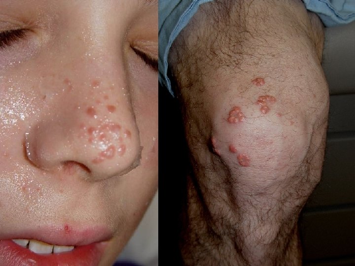
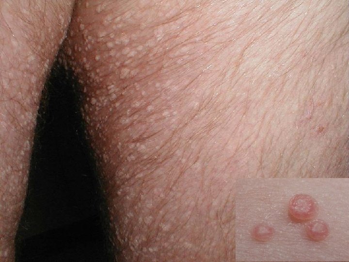
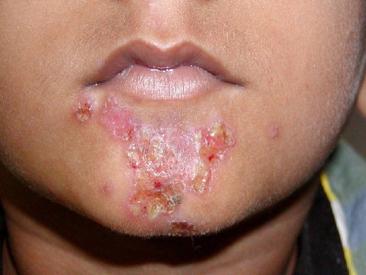
 MULLUSCUM CONTAGIOSUM, viral IMPETIGO, bacterial, staph strep")

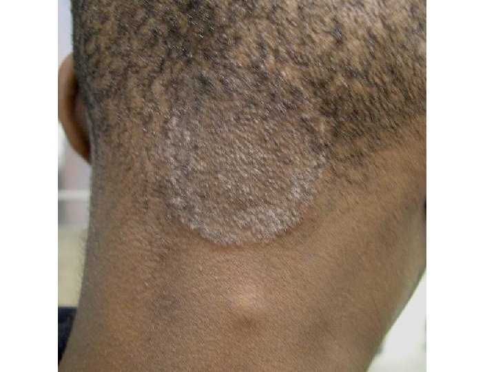
 • …Barbae • …Corporis (Ringworm) • …Cruris (Jock itch)")


- Slides: 115
SKIN
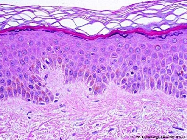
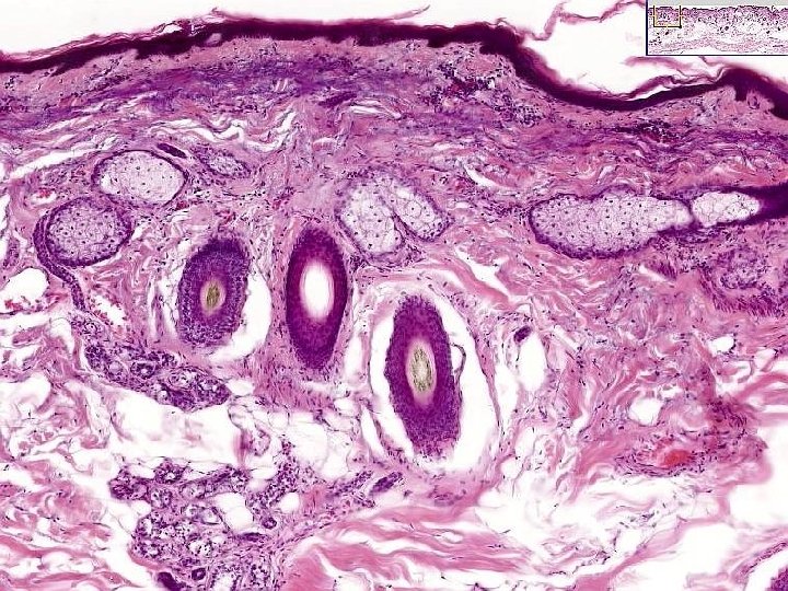
N O R M A L Skin, epidermis, keratinocytes, stratum basale (germinativum) Skin, epidermis, keratinocytes, stratum spinosum (prickle cells) Skin, epidermis, keratinocytes, stratum granulosum Skin, epidermis, keratinocytes, stratum lucidum Skin, epidermis, keratinocytes, stratum corneum, thin skin Skin, epidermis, keratinocytes, stratum corneum, thick skin Skin, epidermis, melanocytes Skin, epidermis, Langerhans cells Skin, epidermis, Merkel cells Skin, epidermis, appendage(s) Skin, epidermis, appendage, hair follicle, shaft Skin, epidermis, appendage, hair follicle, sebaceous gland Skin, epidermis, appendage, sweat gland, eccrine Skin, epidermis, appendage, sweat gland, apocrine Skin, basement membrane Skin, dermis, papillary Skin, dermis, reticular Skin, hypodermis (sub-cutis, pannus)
A B N O R M A L Macroscopic, macule Macroscopic, patch Macroscopic, papule Macroscopic, nodule Macroscopic, plaque Macroscopic, vesicle Macroscopic, bulla Macroscopic, blister Macroscopic, pustule Macroscopic, wheal Macroscopic, scale Macroscopic, lichenification Macroscopic, excoriation Macroscopic, onycholysis microscopic, hyperkeratosis microscopic, parakeratosis microscopic, hypergranulosis microscopic, acanthosis microscopic, papillomatosis microscopic, acantholysis microscopic, spongiosis microscopic, hydropic swelling (ballooning) microscopic, exocytosis microscopic, erosion microscopic, ulceration microscopic, vacuolization microscopic, lentiginous
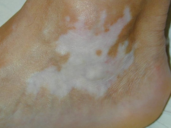
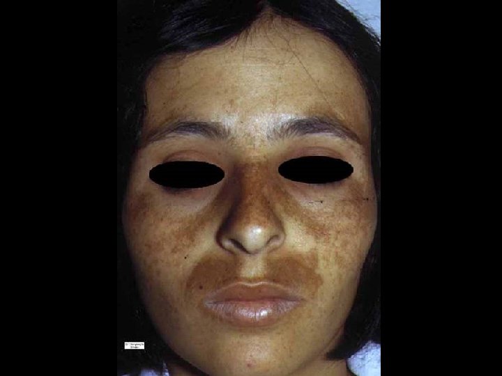
A B N O R M A L Pigmentation disorders, vitiligo Pigmentation disorders, freckle (ephelis) Pigmentation disorders, melasma Pigmentation disorders, lentigo Pigmentation disorders, nevus, melanocytic Pigmentation disorders, nevus, dysplastic Pigmentation disorders, malignant melanoma Epidermal neoplasms, benign, seborrheic keratosis Epidermal neoplasms, benign, acanthosis nigricans Epidermal neoplasms, benign, fibroepithelial polyp (skin tag) Epidermal neoplasms, benign, epithelial inclusion cyst (wen) Epidermal neoplasms, benign, appendage tumors Epidermal neoplasms, benign, keratoacanthoma Epidermal neoplasms, malignant, actinic keratosis Epidermal neoplasms, malignant, squamous cell carcinoma (SCC) Epidermal neoplasms, malignant, basal cell carcinoma (BCC) Epidermal neoplasms, malignant, Merkel cell tumor Dermal neoplasms, fibrous histiocytoma (dermatofibroma) Dermal neoplasms, dermatofibrosarcoma protuberans Dermal neoplasms, xanthomas Dermal neoplasms, vascular tumors Tumors of cellular “immigrants”, Langerhans cells Tumors of cellular “immigrants”, t- cell lymphomas (Mycosis Fungoides) Tumors of cellular “immigrants”, mast cells
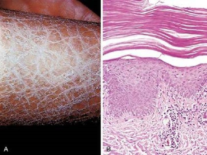
A B N O R M A L Epidermis, maturation disorder, ichthyosis Epidermis/Dermis, inflammation, acute, urticaria Epidermis/Dermis, inflammation, acute, eczema Epidermis/Dermis, inflammation, acute, erythema multiforme Epidermis/Dermis, inflammation, chronic, psoriasis Epidermis/Dermis, inflammation, chronic, seborrheic dermatitis Epidermis/Dermis, inflammation, chronic, lichen planus Epidermis/Dermis, inflammation, chronic, lupus erythematosus Epidermis/Dermis, infection/infestation, (verrucae) Epidermis/Dermis, infection/infestation, molluscum contagiosum Epidermis/Dermis, infection/infestation, impetigo Epidermis/Dermis, infection/infestation, fungus Epidermis/Dermis, infection/infestation, arthropods, bites Epidermis/Dermis, infection/infestation, arthropods, stings Epidermis/Dermis, infection/infestation, arthropods, infestations Epidermis/Dermis, bullae (blisters) Epidermis/Dermis, bullae, pemphigus Epidermis/Dermis, bullae, bullous pemphigoid Epidermis/Dermis, bullae, dermatitis herpetiformis Epidermis/Dermis, bullae, epidermolysis bullosa Epidermis/Dermis, bullae, porphyria Epidermis/Dermis, adnexae (appendages), acne vulgaris Hypodermis (pannus), inflammation (panniculitis) Hypodermis (pannus), inflammation, erythema nodosum Hypodermis (pannus), inflammation, erythema induratum
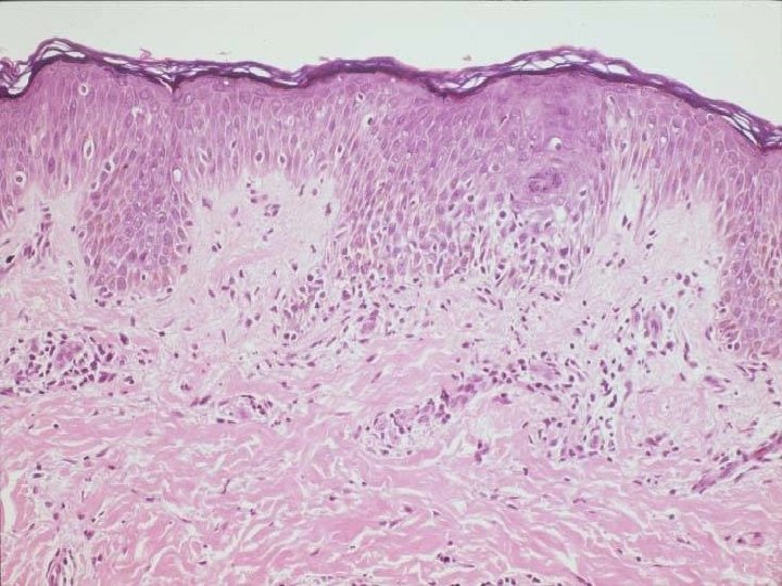
NORMAL SKIN
NORMAL SKIN, with labels
MACRO-scopic (clinical) TERMS • • • • macule patch papule nodule plaque vesicle bulla blister pustule wheal scale lichenification excoriation onycholysis
MACROSCOPIC TERMS Macule: Circumscribed lesion of <5 mm in diameter characterized by flatness and usually discolored (often red) Patch: Circumscribed lesion of >5 mm in diameter characterized by flatness and usually discolored (often red) Papule: Elevated dome-shaped or flat-topped lesion <5 mm across. Nodule: Elevated lesion with spherical contour >5 mm across. Plaque: Elevated flat-topped lesion, usually >5 mm across (may be caused by coalescent papules). Vesicle: Fluid-filled raised lesion <5 mm across. Bulla: Fluid-filled raised lesion >5 mm across. Blister: Common term used for vesicle or bulla. Pustule: Discrete, pus-filled, raised lesion. Wheal: Itchy, transient, elevated lesion with variable blanching and erythema formed as the result of dermal edema. Scale: Dry, horny, plate-like excrescence; usually the result of imperfect cornification (i. e. , keratinization). Lichenification: Thickened and rough skin characterized by prominent skin markings; usually the result of repeated rubbing in susceptible persons. Excoriation: Traumatic lesion characterized by breakage of the epidermis, causing a raw linear area (i. e. , a deep scratch) Onycholysis: Separation of nail plate from nail bed.
micro-scopic (histologic) TERMS • • • • hyperkeratosis parakeratosis hypergranulosis acanthosis papillomatosis acantholysis spongiosis hydropic swelling (ballooning) exocytosis erosion ulceration vacuolization lentiginous
MICROSCOPIC TERMS Hyperkeratosis: Thickening of the stratum corneum, often associated with a qualitative abnormality of the keratin. Parakeratosis: Modes of keratinization characterized by the retention of the nuclei in the stratum corneum. On mucous membranes, parakeratosis is normal. Hypergranulosis: Hyperplasia of the stratum granulosum, often due to intense rubbing. Acanthosis: Diffuse epidermal hyperplasia. Papillomatosis: Surface elevation caused by hyperplasia and enlargement of contiguous dermal papillae. Dyskeratosis: Abnormal keratinization occurring prematurely within individual cells or groups of cells below the stratum granulosum. Generally the same as DYSPLASIA. Acantholysis: Loss of intercellular connections resulting in loss of cohesion between keratinocytes. Spongiosis: Intercellular edema of the epidermis. Hydropic swelling (ballooning): Intracellular edema of keratinocytes. Exocytosis: Infiltration of the epidermis by inflammatory or circulating blood cells. Erosion: Discontinuity of the skin exhibiting incomplete loss of the epidermis. Ulceration: Discontinuity of the skin exhibiting complete loss of the epidermis and often of portions of the dermis and even subcutaneous fat. Vacuolization: Formation of vacuoles within or adjacent to cells; often refers to basal cell-basement membrane zone area. Lentiginous: Referring to a linear pattern of melanocyte proliferation within the epidermal basal cell layer. Lentiginous melanocytic hyperplasia can occur as a reactive change or as part of a neoplasm of melanocytes.
SKIN PATHOLOGY • DEGENERATION • INFLAMMATION, i. e. , DERMATOSES • NEOPLASMS: Epidermis, Dermis, Benign, Malignant
SKIN PATHOLOGY • Pigmentation • Epidermal tumors, benign • Epidermal tumors premalignant • Epidermal tumors, malignant • Dermal tumors • “Immigrant” tumors • Maturation disorders • • Dermatoses, acute Dermatoses, chronic Blisters (Bullae) Appendage (adnexal) disorders • Panniculitis • Infection/Infestation
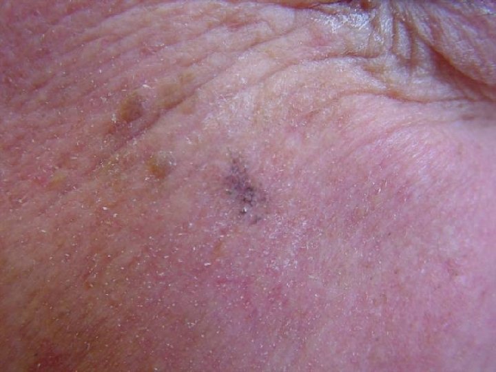
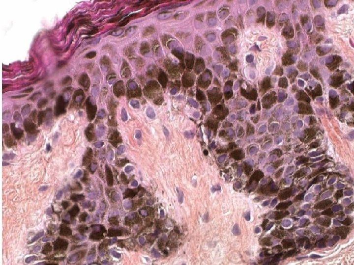
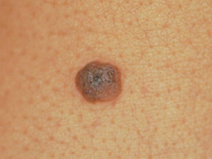
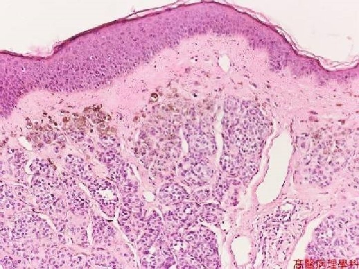
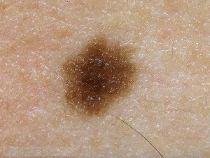
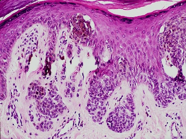
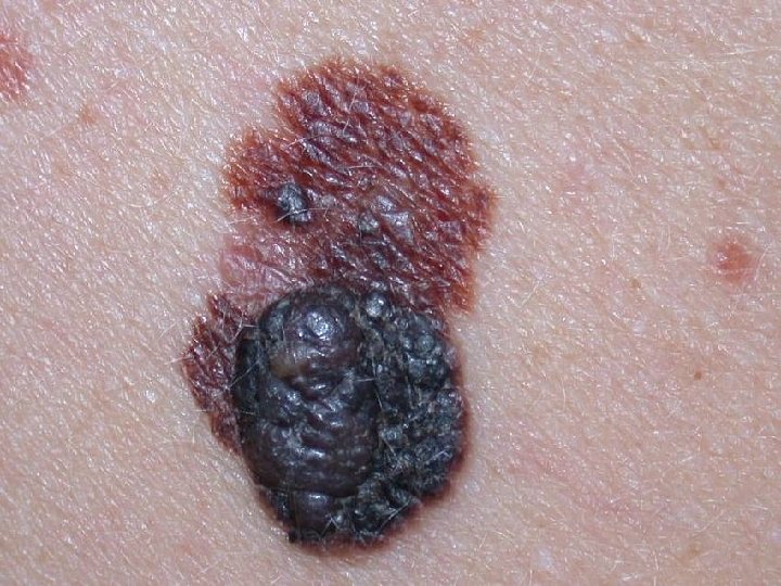
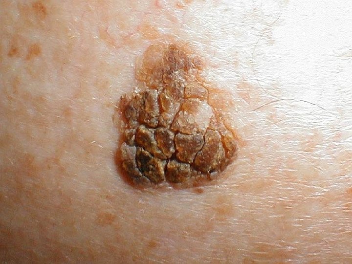
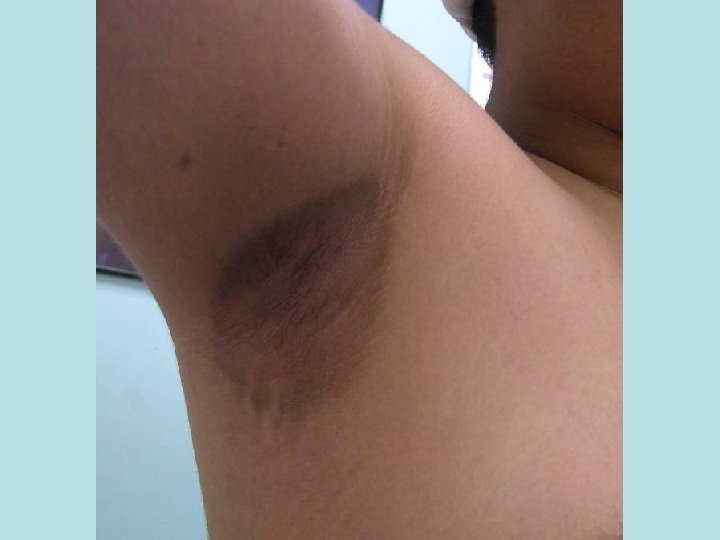
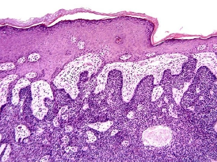
PIGMENTATION DISORDERS • • VITILIGO FRECKLE (EPHELIS) MELASMA LENTIGO NEVUS “DYSPLASTIC” NEVUS MALIGNANT MELANOMA
NEVI • Many, many adjectives and classifications. • The MAIN things to differentiate from melanomas • Junctional (more pigmented, more closely associated with melanoma) • Intradermal • Compound (both)
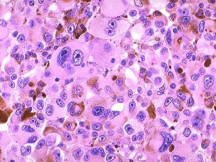
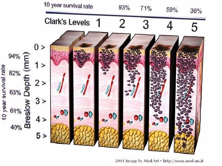
MALIGNANT MELANOMA • Incidence rising, VERY much • Related to SUN like ALL skin cancers are • The only primary skin cancer that can kill you (except for the RARE Merkel cell tumor) • QUICKLY METASTASIZES • Has both VERTICAL and HORIZONTAL growth phase but prognosis is 100% related to the VERTICAL, (BRESLOW staging, TNM too) • DIFFICULT to differentiate from NEVUS clinically and often microscopically
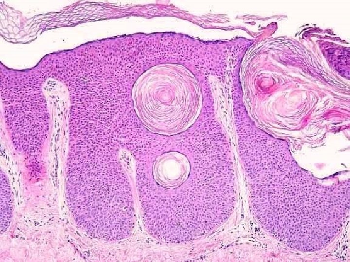
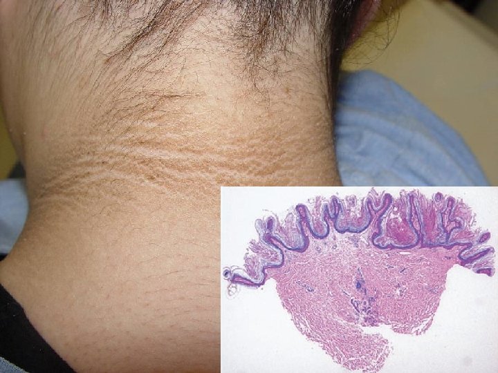
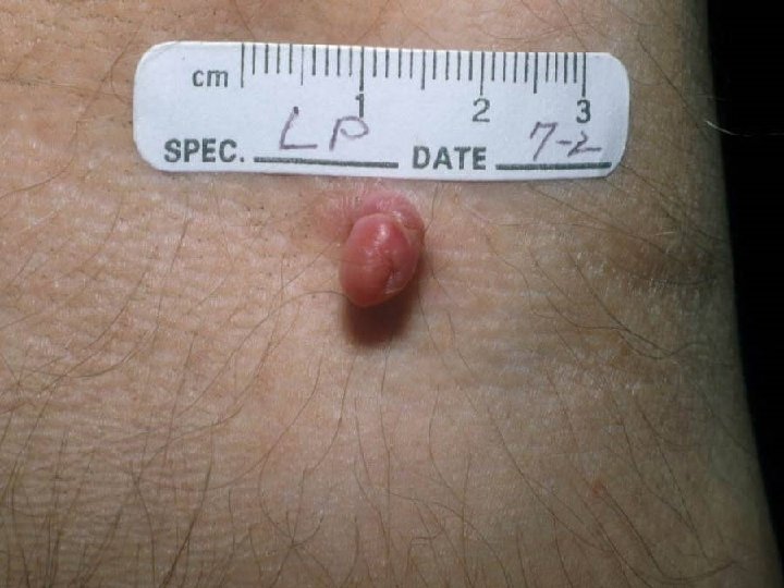
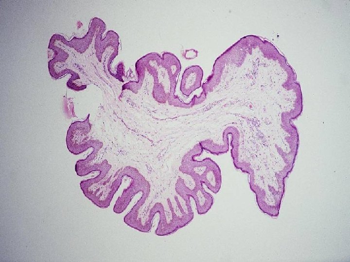
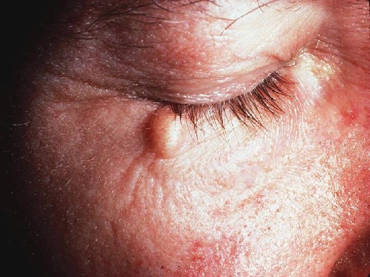
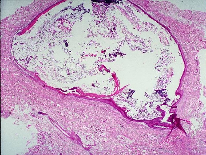
BENIGN Epidermal Tumors • • • Seborrheic Keratosis Acanthosis Nigricans Fibroepithelial Polyp (skin tag) Epidermal (inclusion) Cyst Adnexal tumors : Eccrine, Apocrine • Keratoacanthoma
ADNEXAL TUMORS • HAIR FOLLICLES • SEBACEOUS GLANDS • SWEAT GLANDS –ECCRINE –APOCRINE
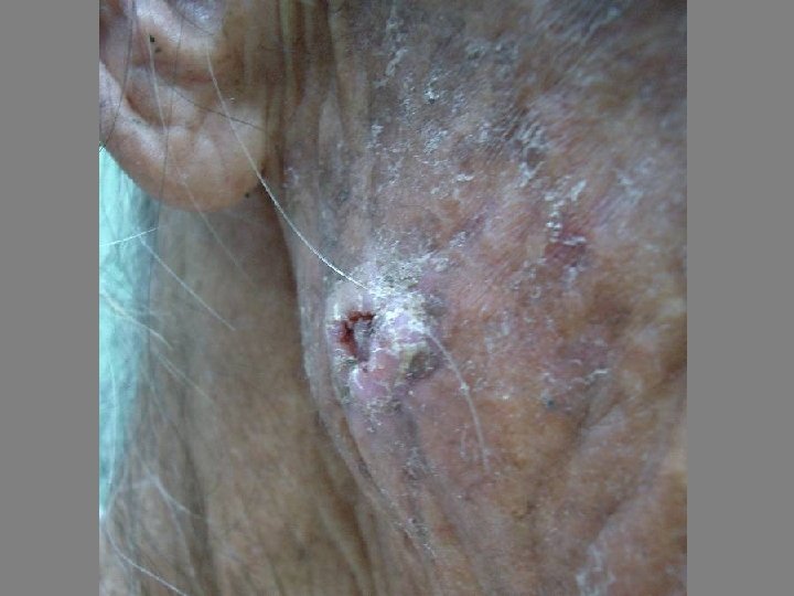
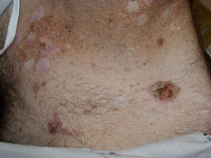
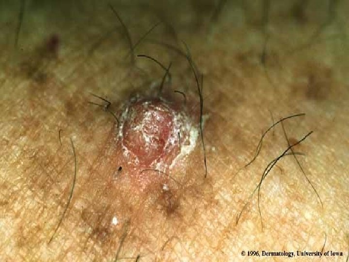
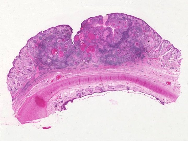
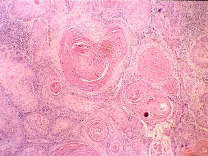
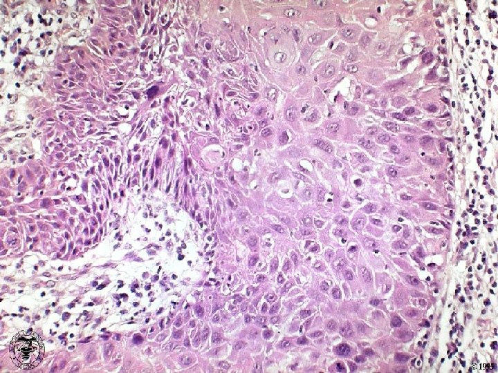
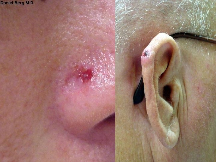
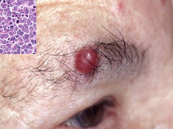
PREMALIGNANT/MALIGNANT • ACTINIC (Solar) KERATOSIS, i. e. precursor to SCC • SQUAMOUS CELL CARCINOMA, squamous “pearls”, intercellular bridges • BASAL CELL CARCINOMA, by far, MOST COMMON, BLUE palisading nests • MERKEL CELL CARCINOMA (TUMOR), VERY MALIGNANT AND LETHAL, LOOK LIKE SMALL CELL CA. OF LUNG
GENERAL COMMENTS • BOTH SCC and BCC related to SUN (i. e. , radiation) exposure. (as is MM also) • SCC also related to As, carcinogens, chaw, betel nut, HPV, familial, etc. • BOTH SCC and BCC can do local damage but very rarely metastasize or kill. • MERKEL CELL tumors metastasize early and extensively, like melanomas.
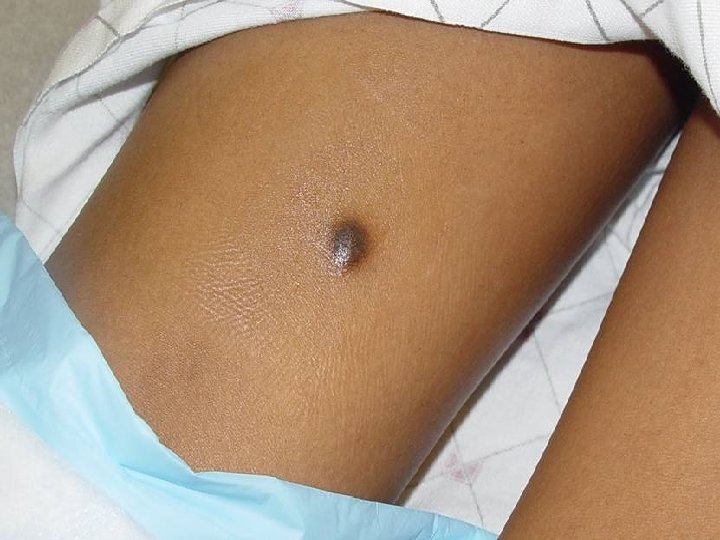
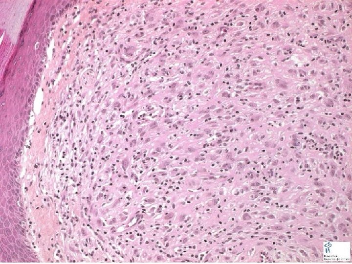
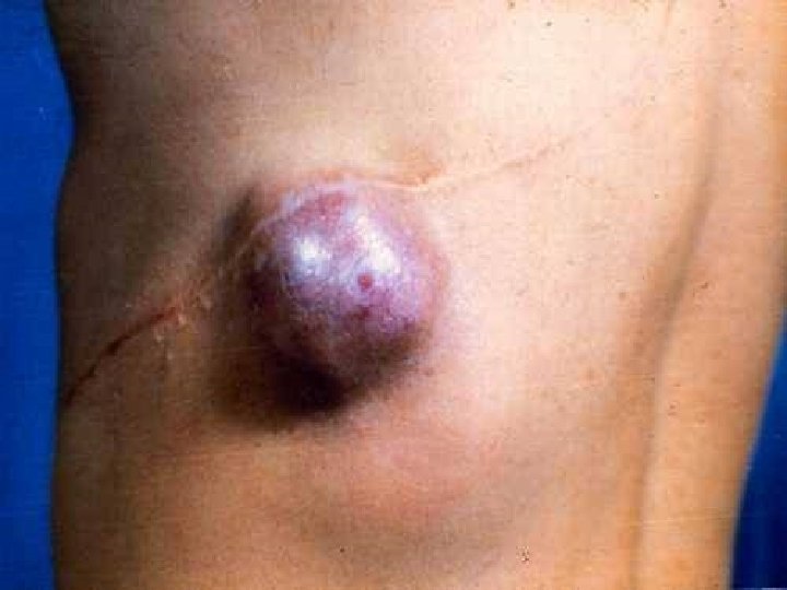
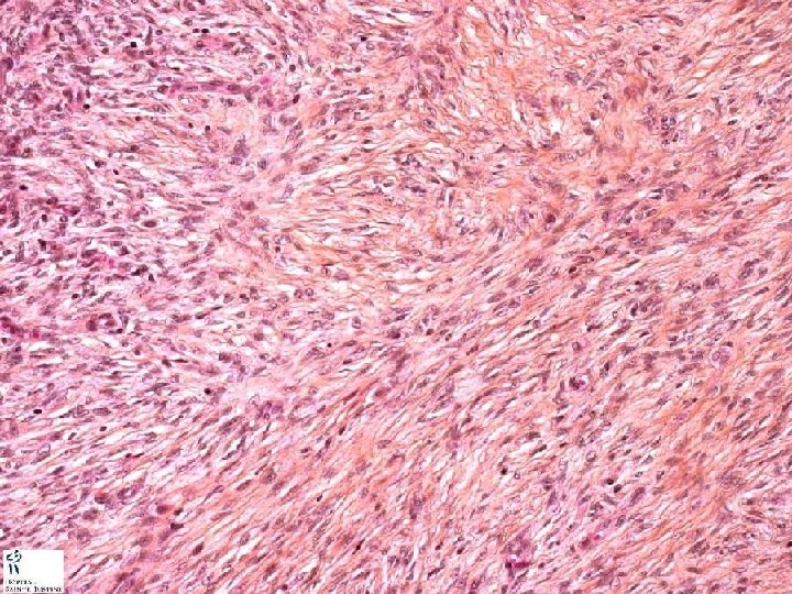
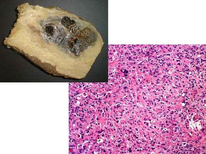
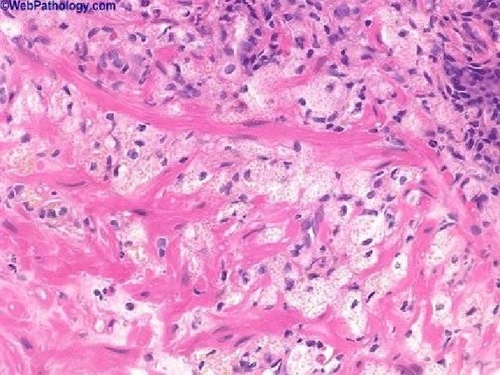
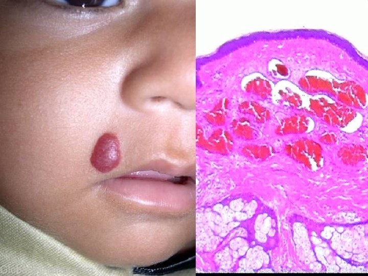
DERMIS TUMORS • DERMATOFIBROMA (BENIGN FIBROUS HISTIOCYTOMA) • DERMATOFIBROSARCOMA PROTUBERANS (DFP) • MALIGNANT FIBROUS HISTIOCYTOMA (MFH) • XANTHOMA • VASCULAR TUMORS of various types
Cellular “Immigrants” • Langerhans cells (Histiocytosis) • Mycosis Fungoides (T-Cell cutaneous lymphoma) • Mastocytosis (mast cell tumors)
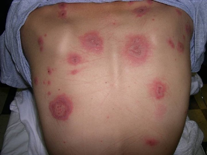
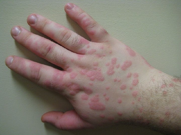
DERMATOSES • ACUTE –URTICARIA (i. e. , “HIVES”) –ECZEMA –ERYTHEMA MULTIFORME • CHRONIC –PSORIASIS – SEBORRHEIC DERMATITIS – LICHEN PLANUS – LUPUS ERTHYMATOSUS
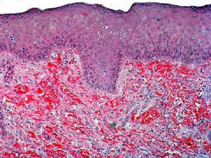
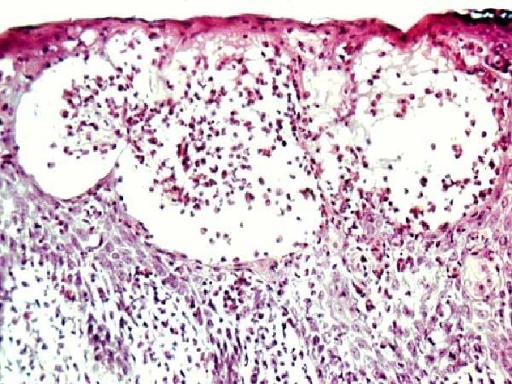
URTICARIA • DERMAL EDEMA • DILATATION of VASCULAR SPACES • EARLY PERIVASCULAR CUFFING OF INFLAMMATORY CELLS
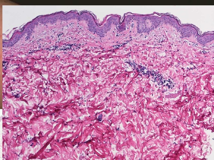
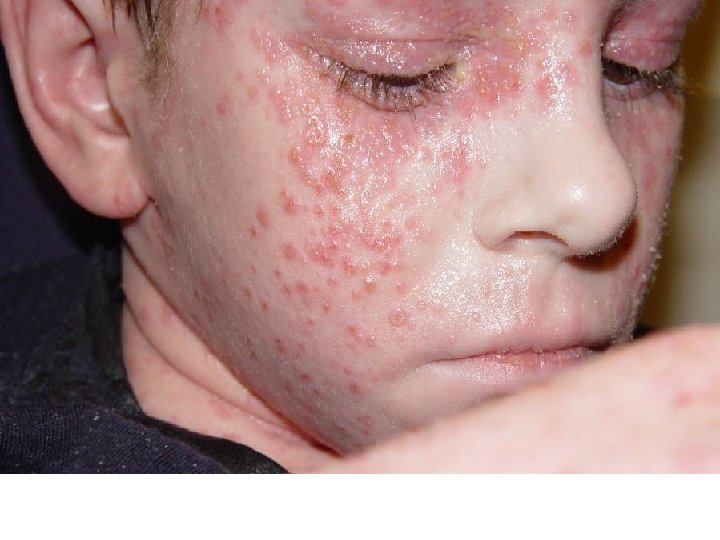
ECZEMA (aka, acute eczematous dermatitis) • A myriad of ACUTE inflammatory disorders, with allergic, drug related, sun related etiologies • The common histologic feature is SPONGIOSIS
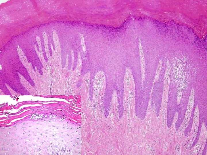
PSORIASIS • 1 -2% of USA • Elbows, Knees • Parakeratosis, generalized epidermal hyperplasia, elongation of the rete pegs, extensive chronic inflammatory cell infiltrates, “MUNRO” intraepidermal microabscesses
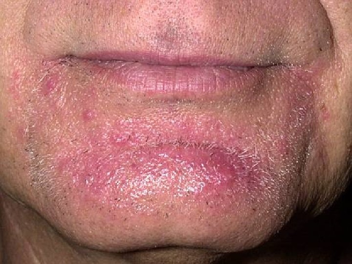
SEBORRHEIC DERMATITIS LICHEN IN HIV PLANUS LUPUS
STASIS DERMATITIS
STASIS DERMATITIS
STASIS DERMATITIS
STASIS DERMATITIS
STASIS DERMATITIS
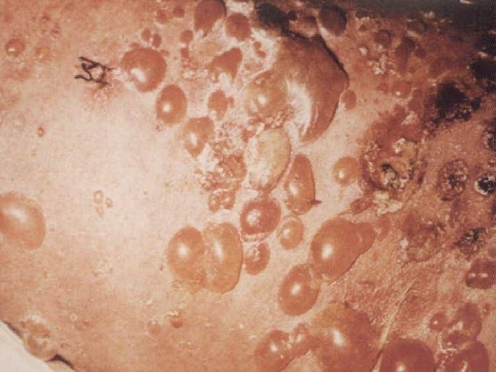
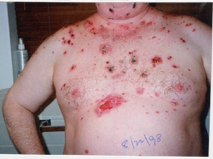
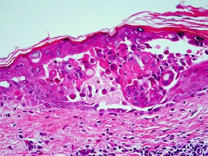
BULLOUS DISEASES • PEMPHIGUS(VULGARIS) • • BULLOUS PEMPHIGOID DERMATITIS HERPETIFORMIS EPIDERMOLYSIS BULLOSA PORPHYRIA • “ACANTHOLYSIS” is the common unifying finding, as is basement membrane immunoglobulins
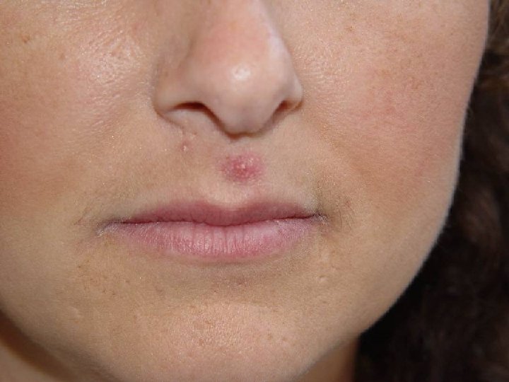
ACNE VULGARIS • Bread and Butter of dermatology practice • Sebaceous duct blockage with secondary inflammation is main feature • bacterial lipases of Propionibacterium acnes break down sebaceous oils, and the resulting fatty acids acts as irritants
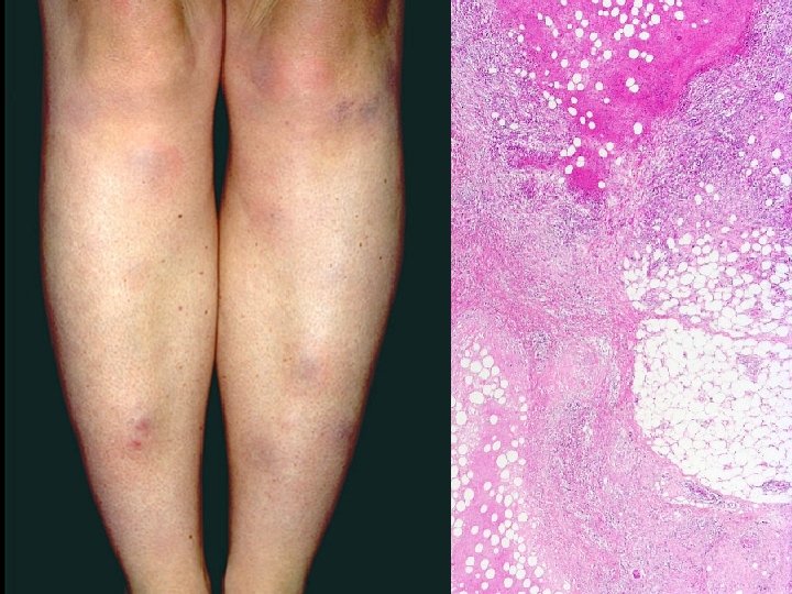
PANNICULITIS • ERYTHEMA NODOSUM, (red nodules on legs) • ERYTHEMA INDURATUM
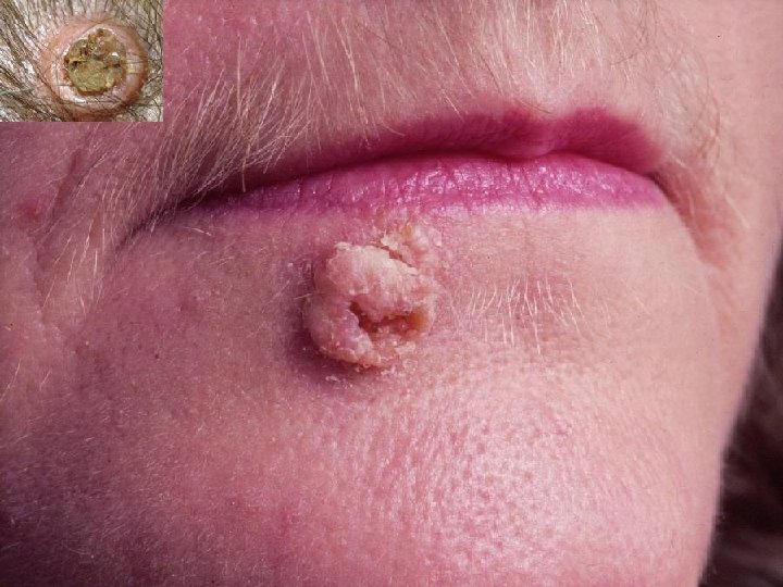
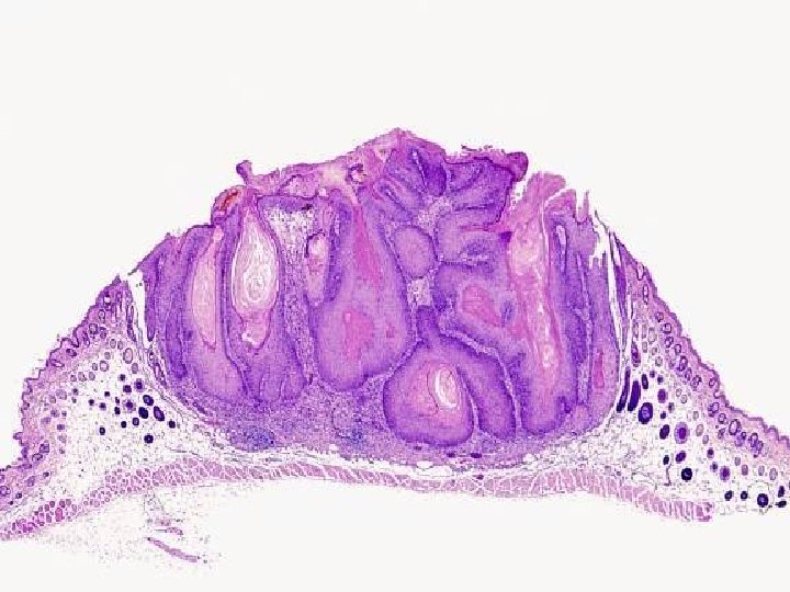
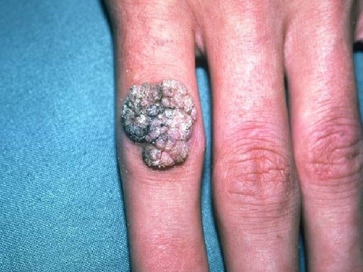
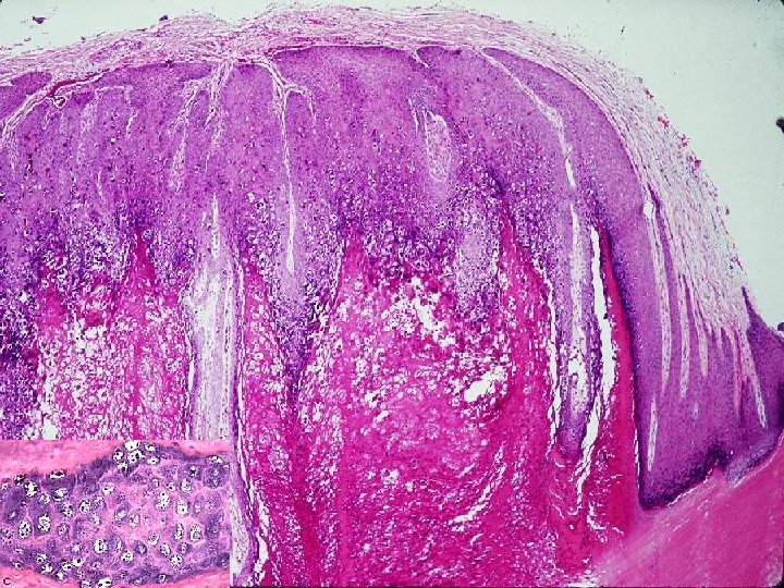
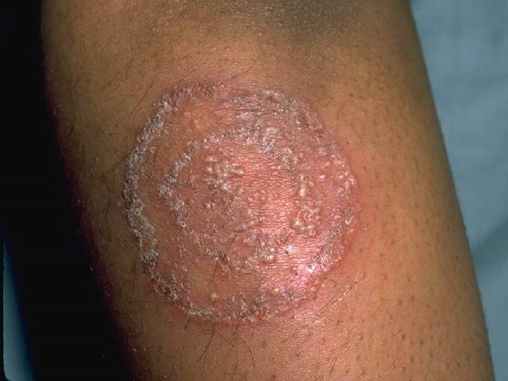
INFECTION/INFESTATION • • • VERRUCAE, viral (HPV) MULLUSCUM CONTAGIOSUM, viral IMPETIGO, bacterial, staph strep FUNGI ARTHROPODS
RED PURPLE BLUE
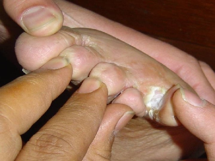
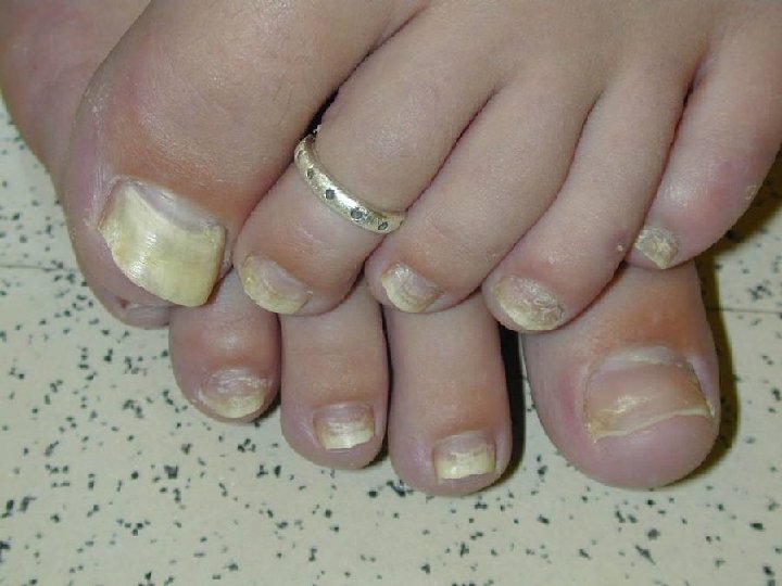
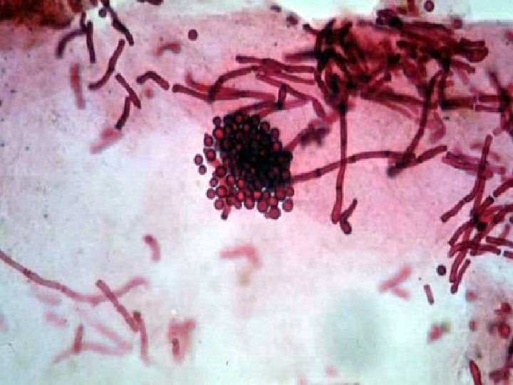
TINEAS… • …Capitis (Scalp ringworm) • …Barbae • …Corporis (Ringworm) • …Cruris (Jock itch) • …Pedis (Athlete’s foot) • Onychomycosis (nail)
TINEAS • Trichophyton species • Microsporum species • Epidermophyton species
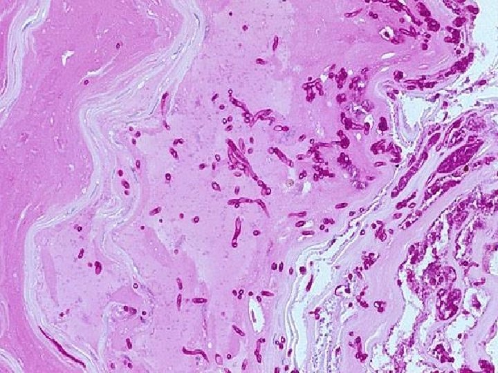
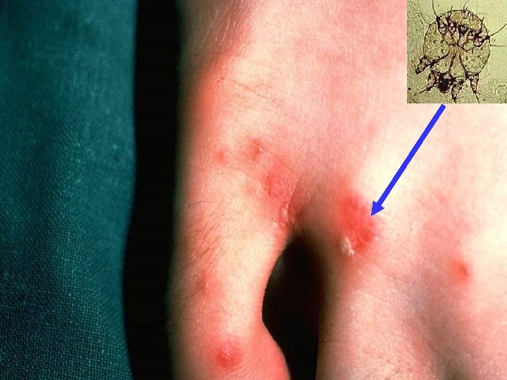
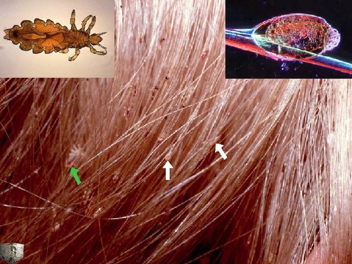
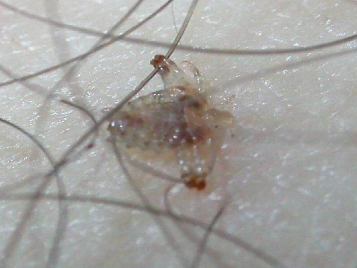
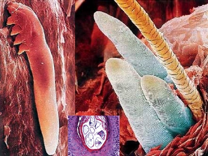
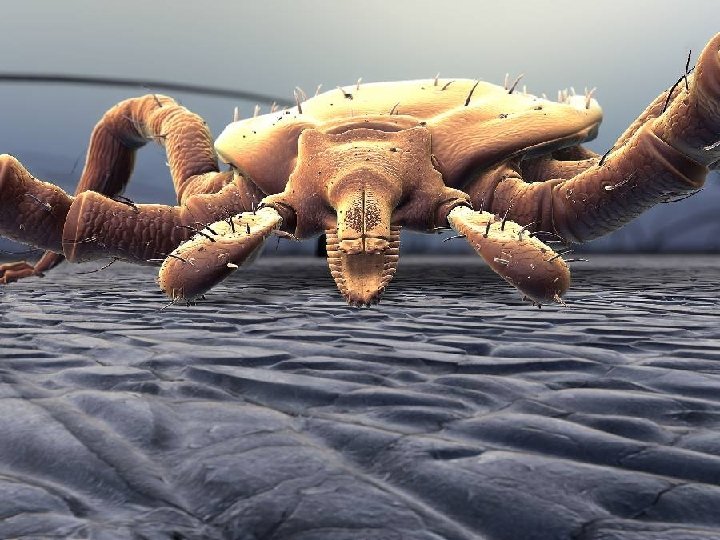
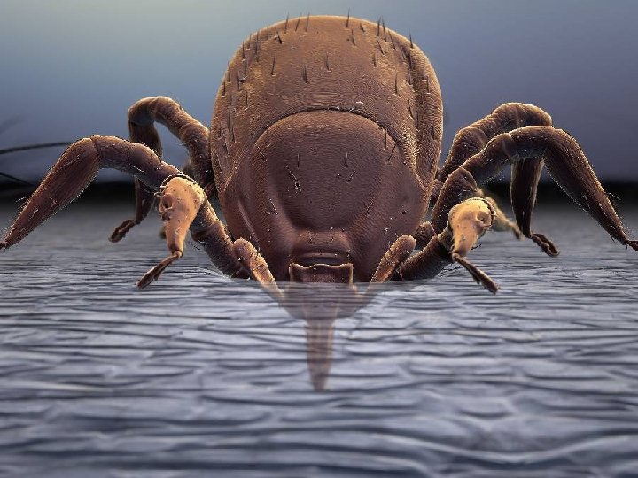
ARTHROPODS • Bites • Stings • INFESTATIONS
ARTHROPODS • Scabies • Pediculosis • Demodex • Ticks, Mites