Skin Examination Pharmacy Practice 742 Physical Assessment The


 or")


- Slides: 47
Skin Examination Pharmacy Practice 742: Physical Assessment
The Skin: History n Three important aspects to seek out: n n n symptoms attributed to the skin lesion chronology of appearance, change, and disappearance of the lesions conditions of exposure, injury, or medication that may have induced or altered the disease
The Skin: History n Original lesion n n n exact site duration appearance distribution progression Symptoms n local n n n pruritis pain burning n Setting & Timing of Attacks n n n occupation topical agents drug history season of year environment
Skin: Physical Examination n Three categories of observation should be made in sequence: n n n First, anatomic distribution of the lesion Second, configuration of groups of lesions Third, the morphology of the individual lesions
Skin: Physical Examination n Inspection n n natural lighting preferred, need complete exposure of all skin surfaces. remember to scan nails, hair, mucous membranes n Location and Distribution n exact, measure, symmetry?
Skin: Physical Examination n Inspection n Color: n n n variation common, even within same person. Melanin n maybe diffuse or localized n increased: Addison’s Disease, hyperthyroidism, pregnancy, sunlight exposure n decrease: albinism and vitiligo Erythema n appearance of increased amounts of oxygenated blood in dermal vasculature
Skin: Physical Examination n Inspection n Color: n n n Cyanosis n blue tint from venous blood (deoxygenated hemoglobin) seen associated with congestive heart failure, pneumonia Extravasation of blood products n ecchymosis, petechiae Pallor n decrease hemoglobin in vessels close to skin secondary to anemia, shock
Skin: Physical Examination n Inspection n Color: n n Depositions of abnormal pigments n Jaundice from bilirubin n Carotenemia from carotene (diabetes, excess ingestion of yellow vegetables (carrots) n Gray from heavy metals (Au-gold, Ag-silver, Bi-bismuth) n Blue-gray from amiodarone Configuration n arrangement or position of lesions with each other (grouped, linear, annular)
Skin: Physical Examination n Inspection n Morphological structure n primary lesions n flat n elevated n -- serous filled n -- pus filled n -- solid
Skin: Physical Examination n Inspection n Morphological structure n secondary lesions n loss of skin n -- erosion n -- ulcer n -- fissure n build-up of skin n -- scale n -- crust n -- lichenification n -- scar
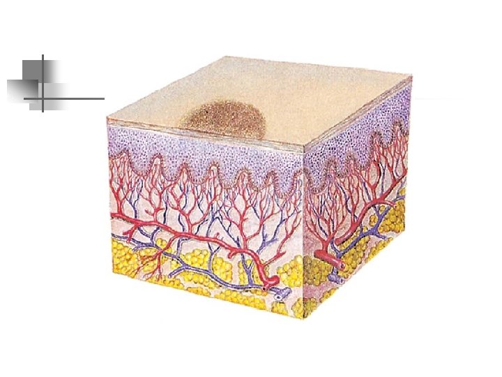
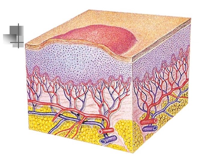
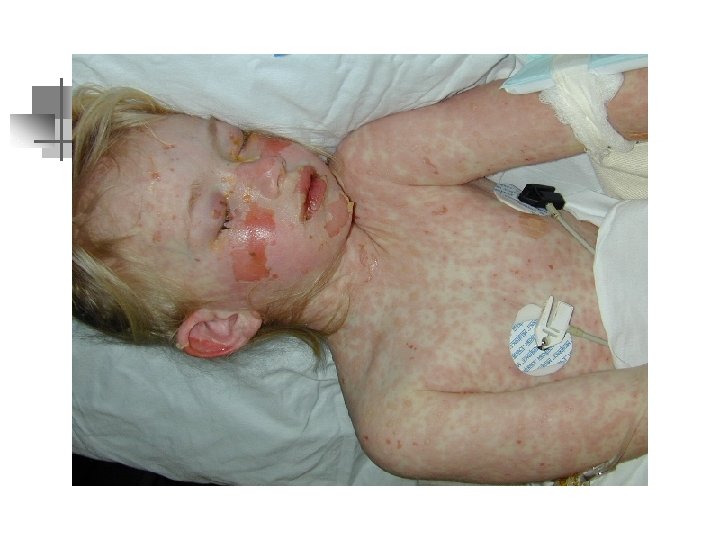
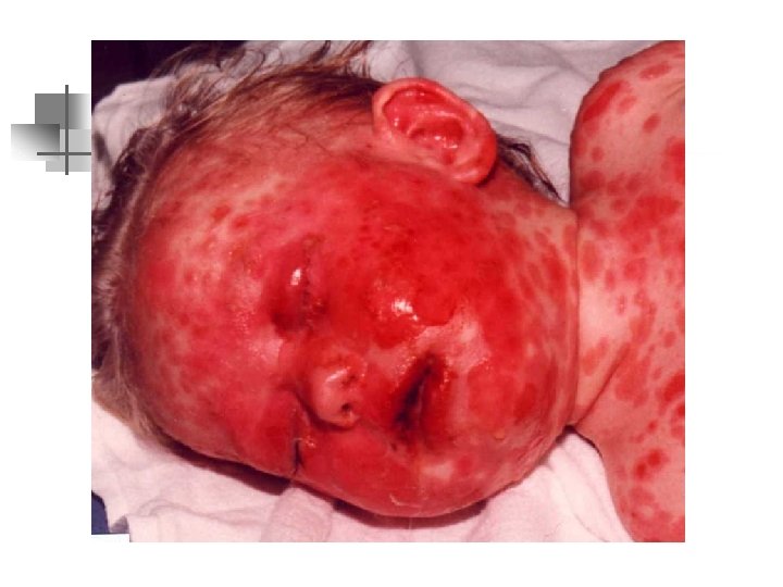
Skin: Physical Examination n Morphology - Definitions n Macules n n localized changes in skin color. Areas may be small or large; occur in many shapes and colors. Not palpable may be associated with desquamation or scaling examples; n rubeola, rubella, secondary syphilis, rose spots of typhoid fever, drug eruptions, petechiae, purpura, first degree burns, systemic lupus erythematosus, pityriasis rosea and vitiligo
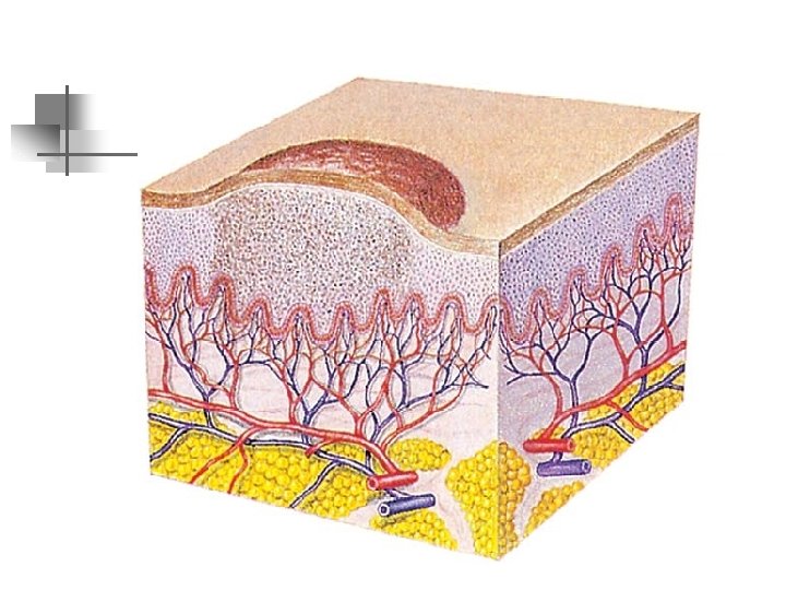
Skin: Physical Examination n Morphology - Definitions n Maculopapules n slightly elevated macules n n commonly seen in pityriasis rosea, erythema multiforme, fixed drug eruptions and exanthemas Papules n n lesions are solid and elevated and defined as less than 5 mm in diameter. Borders and tops may be in various forms n n pointed or acuminated -- insect bites, acne and physiologic gooseflesh flat topped -- psoriasis, atopic eczema
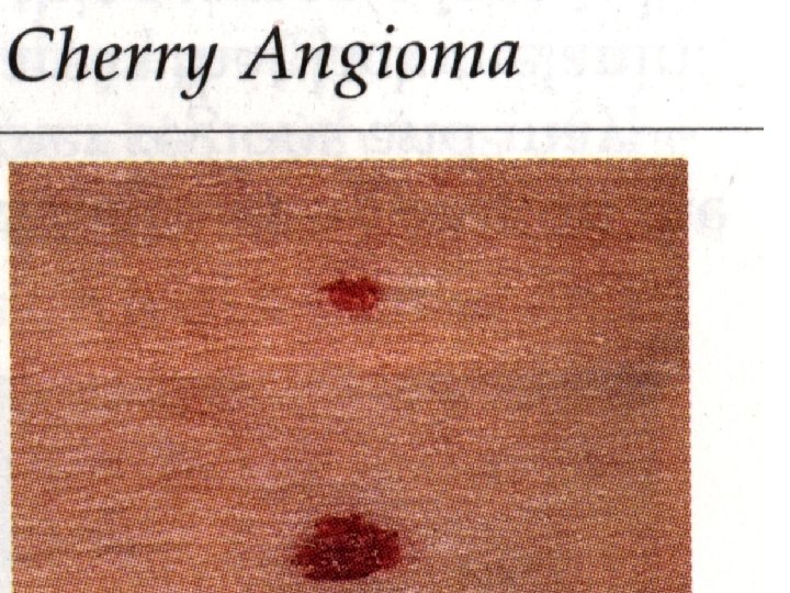
Skin: Physical Examination n Morphology - Definitions n Papules n Borders and tops may be in various forms n n n round or irregular --senile angiomas, eczematous dermatitis, secondary syphilis pedunculate -- neurofibromas Plaques n any elevated area of greater than 5 mm, usually formed from confluent papules. n Red scaling plaques -- psoriasis, pityriasis rosea
Skin: Physical Examination n Morphology - Definitions n Plaques n n n Yellow -- xanthomas brown -- seborrheic warts Nodules n n solid and elevated, distinguished from papules by extending deeper into the dermis or even the subcutaneous tissue. Usually greater than 5 mm in diameter
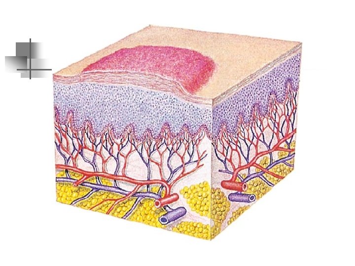
Skin: Physical Examination n Morphology - Definitions n Nodules n n depth may be inferred by palpation…when below the dermis skin slides over them, lesions within the dermis move with the skin Wheals n n caused by edema of skin, areas are circumscribed, irregular, and relatively transient color varies from red to pale, depending on amount of fluid in the skin.
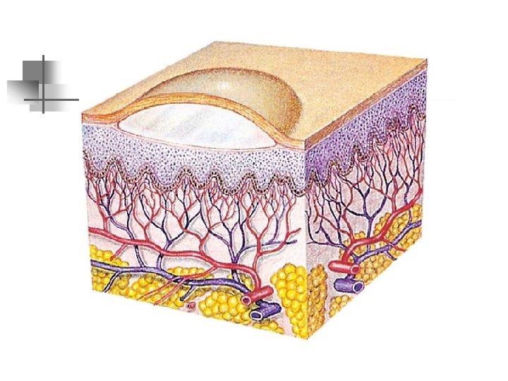
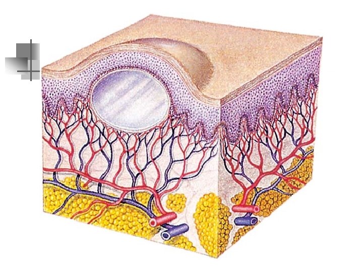
Skin: Physical Examination n Morphology - Definitions n Wheals n n examples: urticaria and insect bites Vesicles n n accumulation of fluid between the upper layer of the skin produces an elevation covered by a translucent epithelium that is easily punctured to release the fluid less than 5 mm n examples: acute eczematous dermatitis, second-degree burns
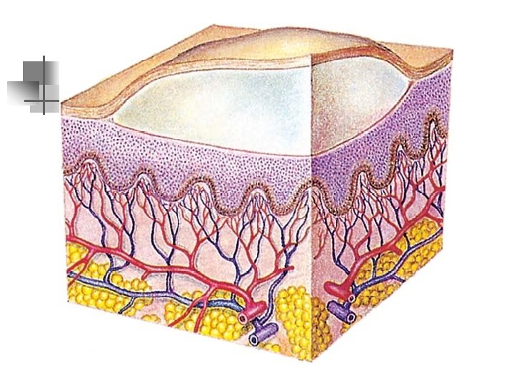
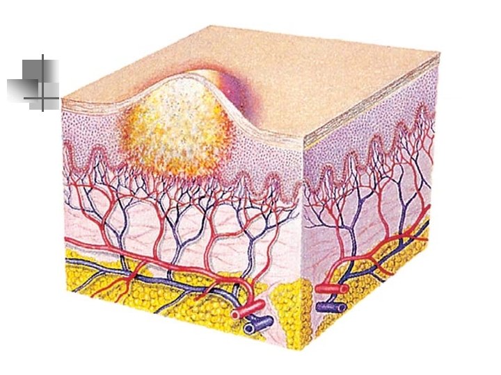
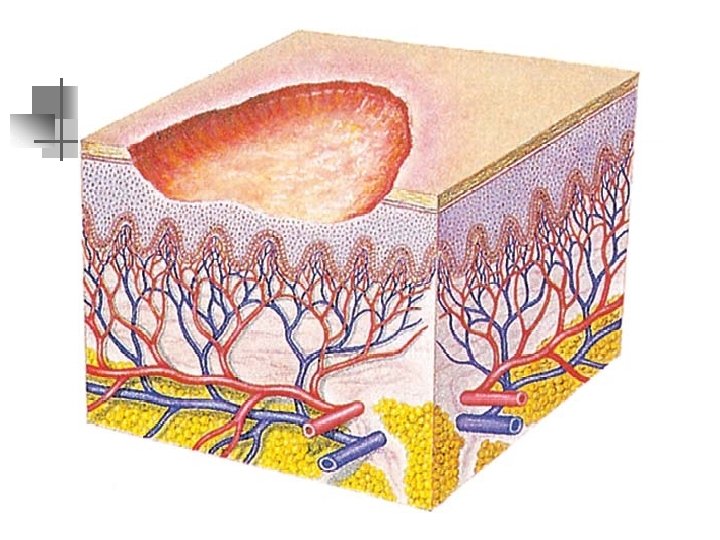
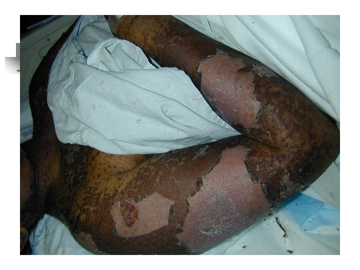
Skin: Physical Examination n Morphology - Definitions n Bullae n Accumulation of fluid between layer of the skin, larger than 5 mm in diameter. n n Examples: contact dermatitis, second-degree burns, bullous impetigo Pustules n n Vesicles or bullae that become filled with pus and tiny abscesses in the skin contents appear milky, orange, yellow, or green depending somewhat on the infecting organism
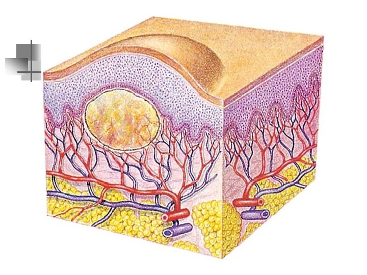
Skin: Physical Examination n Morphology - Definitions n Pustules n frequently arise from hair follicles or sweat glands n n examples: acne, furuncles, and bromide and iodide eruptions Cysts n n elevated lesions containing fluid or viscous material appear as papules or nodules distinction is made by puncturing to examine their contents and depth n examples: sebaceous and epidermal cysts
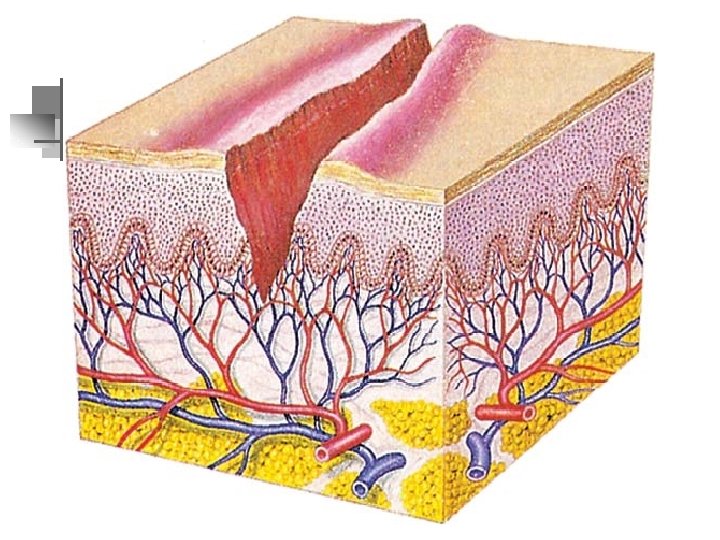
Skin: Physical Examination n Secondary or Consecutive n Erosions n n moist surface uncovered by the rupture of vesicles or bullae or by laceration from rubbing Fissures n cleavage of the epidermis extending into the dermis n examples: common in trauma to thickened, dry, inelastic skin
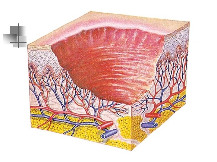
Skin: Physical Examination n Secondary or Consecutive n Ulcers n depressed lesions results from loss of epidermis and the papillary layer of the dermis n n examples: traumatic ulcers, burns, and stasis ulcers Gangrene n extensive destruction of the skin -- may leave many dead cells that become blackened
Skin: Physical Examination n Palpation n Temperature n n localized hyperthermia from increased blood flow due to cellulitis or injury generalized hyperthermia due to fever of systemic infection, hyperthyroidism localized hypothermia caused by peripheral arteriosclerosis, Raynaud’s disease generalized hypothermia due to shock
Skin: Physical Examination n Palpation n Moisture n n sweat - nervous (hypothermia) or thermal (hyperthermia) in origin Texture n n quality character n n n rough dry (hypothyroidism) smooth (hyperthermia)
Skin: Physical Examination n Palpation n Elasticity n n n decreases with age Decreased skin turgor - dehydration edema - accumulation of fluid in interstitial spaces under the skin. n Congestive heart failure
Cancer n n Malignant Melanoma ABCD’s n n Asymmetry Border irregularity Color variation Diameter greater than 6 mm n Inquire and observe for ominous changes in color, shape, elevations, texture, surrounding skin, sensation, and consistency.
Malignant Melanoma
Bullous Impetigo
Carbuncle
Drug Eruption