Skin Connective Tissue Diseases 1 Lupus Erythematosus LE

 a spectrum of disease ranges from lifethreatening manifestations of acute")

, buccal mucosa, or")


- Slides: 15
Skin & Connective Tissue Diseases
1 -Lupus Erythematosus (LE) a spectrum of disease ranges from lifethreatening manifestations of acute systemic LE (SLE) to sub acute cutaneous LE (SCLE) to the limited skin involvement in chronic discoid LE(DLE). LE occurs more commonly in women (male to female ratio 1: 9) More than 85% of patients LE have skin lesions ( 4 out of 11 diagnostic criteria are cutaneous)
A-Butterfly Rash: Erythematous, confluent, macular butterfly eruption on the face , sharply defined with fine scaling; erosions & crusts in severe cases.
B-Oral ulcers : arising in purpuric necrotic lesions on palate (80%), buccal mucosa, or gum & usually painless, observed by a physician C-Photosensitivity: Skin rashes as a result of unusual reaction to sunlight
D-Discoid rash: Erythematous raised patches with adherent keratotic scaling and follicular plugging; atrophic scarring may occur in older lesions. On scalp may cause scarring alopecia.
2 -Scleroderma: multisystem disorder characterized by inflammatory, vascular, and sclerotic changes of the skin and various internal organs, especially the lungs, heart, and GI tract. Skin involvement includes
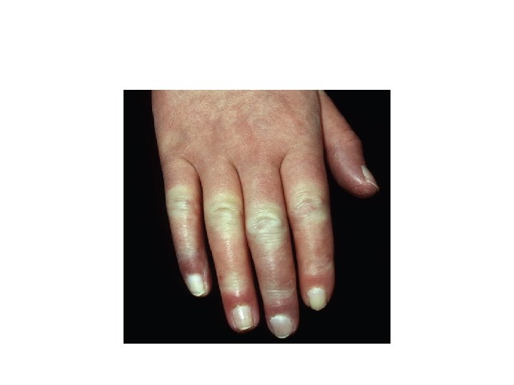
a- Hands &feet: Raynaud phenomenon: affecting hands &Feet. At early stagewith triphasic color sequence changes( pallor, cyanosis & redness )usually precedes sclerosis by months and years & associated withnonpitting edema of hands & feet. Sometimes there is painfululcerations at fingertips knuckles that heal with pitted scars. Late stage: sclerodactyly with tapering of fingers (Madonna fingers) with waxy shiny, hardened skin, which is tightly bound down and does not permit folding or wrinkling.
B-Face. Early: periorbital edema. Late: edema and fibrosis result in loss of normal facial lines, -mask-like (patients look younger than they are), -thinning of lips, -microstomia, -radial perioral furrowing , -beaklike sharp nose. -Telangiectasia -diffuse hyperpigmentation
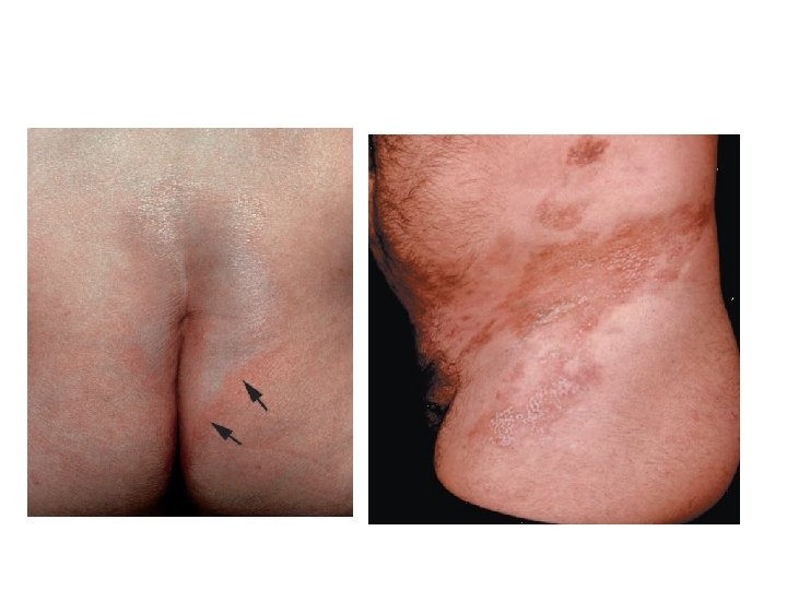
Morphea: A localized cutaneous sclerosis Plaques : circumscribed, indurated, hard, but poorly defined areas of skin; round or oval, often better felt than seen. Initially, purplish or mauve. In time, after months to years surface becomes smooth and shiny, ivory with lilac-colored edge“lilac ring”. May have hyper- and hypopigmentation in involved sclerotic area
3 -Dermatomyositis autoimmune diseases targeting the skin and or skeletal muscles. Skin involvement is characterized by: 1 -Periorbital heliotrope(reddish purple) erythema of upper eyelids and edema of the lower lids
2 -Gottron papules: flat- toped Violaceous erythematous papules on the dorsa of the hands and fingers, especially over the interphalangeal joints.
3 -poikiloderma: Long-lasting lesions may evolve into poikiloderma (mottled reticulated discoloration with red, white, and brown)
4 -Calcinosis cutis: Calcification in subcutaneous tissues, common later in course of juvenile Dermatomyositis , particularly about elbows, trochanteric & iliac region. Clinically appear like stone hard nodules, which have ulcerated and reveal a chalk white mass at the base. Upon squeezing, they will exude white paste.