SKIN APPENDAGES SKIN CANCER September 21 24 2018

SKIN APPENDAGES & SKIN CANCER September 21 -24 2018

Skin Appendages The skin appendages include cutaneous glands, hair, and nails.

Cutaneous Glands What is cutaneous? What is an exocrine gland?

Cutaneous Glands What is cutaneous? Relating to the skin What is an exocrine gland? A gland that releases its product to a particular site, rather than into the blood stream. (Endocrine glands release their products to the blood).
 Sebaceous (oil) glands y s b y tif ides land n")
Cutaneous Glands 1) Sebaceous (oil) glands y s b y tif ides land n e sl g soles s dhands i e i h • Found everywhere except palms of and of n t s o o i t p d le nds un t em b feet e a s gla e, ro t tha b ’ll ou larg uc u e or E d e. • The ducts mostly empty. Yointo chair a b ng f RG llicl e s ki follicles A ir fo L o lo h a ha it the • Produce oily product calledwsebum o int • Functions: • Keep hair and skin from drying out • Kill bacteria • Become much more active in adolescence
 Sebaceous (oil) glands Fun fact: the ‘pores’ on our faces are")
Cutaneous Glands 1) Sebaceous (oil) glands Fun fact: the ‘pores’ on our faces are actually hair follicles • Found everywhere except palms of hands and soles of feet • The ducts mostly empty into hair follicles • Produce oily product called sebum • Functions: • Keep hair and skin from drying out • Kill bacteria • Become much more active in adolescence

Acne occurs when the hair follicle is clogged with sebum.
 Sweat (sudoriferous) glands • Eccrine glands produce sweat (water, salt, urea,")
Cutaneous Glands 2) Sweat (sudoriferous) glands • Eccrine glands produce sweat (water, salt, urea, lactic acid) • Duct lead to pores in skin • Functions • Thermoregulation • Excretion • Bacterial inhibition (sweat is acidic) • Apocrine glands produce a milky secretion fully of fatty acids and proteins. • Small duct leads to hair follicle • Function: Pheromone!
 Sweat (sudoriferous) Notice that sweat glands, since they empty into the")
Cutaneous Glands 2) Sweat (sudoriferous) Notice that sweat glands, since they empty into the skin, are glands often farther and deeper than sebaceous glands • Eccrine glands produce sweat (water, salt, urea, lactic acid) • Duct lead to pores in skin • Functions • Thermoregulation • Excretion • Bacterial inhibition (sweat is acidic) • Apocrine glands produce a milky secretion full of fatty acids and proteins. • Duct leads to hair follicle • Function: Pheromone! Fun fact: Randy Thornhill’s work

Cutaneous Glands – Quick Review Make a Venn Diagram comparing the three types of cutaneous glands. You may work with a partner. 5 min

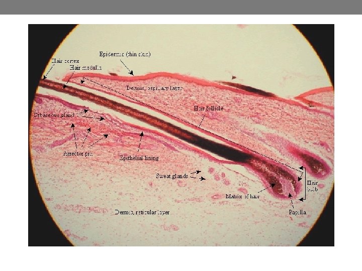
Hair follicle Why/when do the arrector pili muscles contract? The arrector pili muscles contract when we are cold, to raise the hairs and reduce heat loss • The hair follicle is surrounded by a thin layer of epidermal tissue. • The follicle is slanted unless the arrector pili muscles are contracted Change the words on the diagram of your guided notes Dermal epidermal hair bulb
 and")
Hair • Hair can be divided into the shaft (part outside of body) and root (inside body) • Hair is produced by epithelial cells in the hair bulb, but as the cells are pushed away from the bulb they begin to die. • Like the outer layer of the skin, the hair shaft is composed of dead, keratinized cells. Fun fact: Round shaft = straight, coarse hair Oval shaft = silky, wavy hair Flat shaft = curly hair

Nails • Produced by epithelial cells • The nail root, closest to the nail matrix, is living • The outer part, like the hair and skin, consists of dead keratinized cells. Fun fact: Fingernails are a derived trait in primates

Quick Review Turn & Talk. Be prepared to share with the class. Are the skin appendages (cutaneous glands, hair follicles, and nails) found in the dermis or epidermis? Justify your answer.

Quick Review Turn & Talk. Be prepared to share with the class. Are the skin appendages (cutaneous glands, hair follicles, and nails) found in the dermis or epidermis? Justify your answer. They are in the epidermis, because the epidermis surrounds and gives rise to these structures. However, these structures usually sit deeper than the most of the epidermal tissue.

Label the diagram

Label the diagram • A: epidermis • B: dermis • C: hypodermis • 1: hair shaft • 2: pores • 3: Eccrine gland • 4: sebaceous gland • 5: nerve What did we talk about today that you don’t see on this diagram?

Label the diagram • A: epidermis • B: dermis • C: hypodermis • 1: hair shaft • 2: pores • 3: Eccrine gland • 4: sebaceous gland • 5: capillary What did we talk about today that you don’t see on this diagram? Apocrine glands.

Identify the parts of the skin 1 4 5 6 2 7 8 3

Common Types of Skin Cancer There a three main types of skin cancers, defined by the type of cell from which they arise. Basal cell carcinoma – arise from the basal edge of the epithelial tissue. n u s s Squamous cell carcinoma –xces sure e h t o E p f s arise from the keratinx o e e as ch producing cells in the middle cre ea rs e in of anc of the epithelial tissue. k ris se c Melanoma – arise from the melanin-producing cells at the basal edge of the epithelial tissue

Basal Cell Carcinoma • Most common form of skin cancer • Affects ~2. 8 million people in the US each year • Slow-growing and rarely fatal

Squamous Cell Carcinoma • Second most common form of skin cancer • Affects ~700, 000 people in the US each year • Grow and invade surrounding tissue quickly • <5% are fatal

Melanoma • Third most common skin cancer Melanoma may be the least common • Affects ~76, 000 people in the type of skin cancer, but it is by far the US each year most deadly. • Quickly metastasizes while rates of most other (spreads) to. Moreover, blood vessels cancers declining in the US (due and lymph nodes – are which a reduction allows it to to spread to otherof smoking) melanoma are increasing. locations inrates the body • Fatal in more than 10% of cases

Closure • What were our objectives today? How did we meet them? • What was our learner profile trait and how did we use it? • How does what we did today address our unit objective? • How does what we did today address our TOK connection?
- Slides: 25