Skin and appendages Mammary gland Ph D Dr


 • Protection against water loss")
 Subcutis (Hypodermis, Tela subcutanea) Integumentum commune Dermis Cutis")


, fat, pigment (melanin), small amounts")


 induces the hair follicle and retains")
 Prox. nail fold Sweat gl. Eponychium Nail plate Nail bed Nails are")
 margo liber hyponychium Nail plate Nail bed (sinus unguis) lateral nail groove")
, matrix region of the nail root")

 No relationship to hair follicles end piece: Dark")

 No lumen Lipid film against water permeability With hair follicles")

 GLAND Anterior axillary fold m. pectoralis major Anterior axillary fold; m. pectoralis")


 Ultraultrasound nodi lymphatici axillares Mammography nodi lymphatici pectorales")
- Slides: 33
Skin and appendages. Mammary gland. Ph. D Dr. David Lendvai Anatomy, Histology and Embryology Institute Semmelweis University Budapest 2019 Michelangelo: Last Judgement
Skin • Largest organ of the human body • 1, 5 -2, 3 m 2 • 15 -20% of body weight Hl. Bartholomäus SKIN APPENDAGES: • Hair • Nails • Glands: Mammary-, Sebaceous-, Sweat glands
Funktions of the skin • Protection (mechanical, thermal, chemical-toxic) • Protection against water loss • Protection against radiation • Thermoregulation • Sensesory organ • Immunological function • Vitamin D synthesis • Excretion • Secretion: Milk • Fat storage
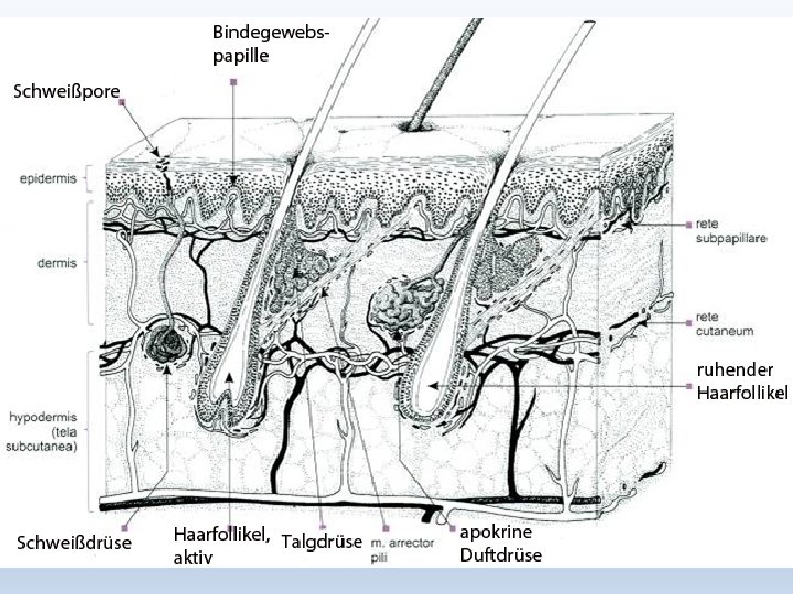
Layers of skin Epidermis (Corium) Subcutis (Hypodermis, Tela subcutanea) Integumentum commune Dermis Cutis
Skin types field skin ridged skin • Covers most of the body • Hand, Foot • With hair and sebaceous glands • Without hair and sebaceous glands • Cristae und Sulci cutis → grind • bows • vortex
Histology: Epidermis Function: formation of a chemically, mechanically resistant and waterproof stratum corneum. thick skin Epidermis thickness: 0, 5 -1 mm thin skin Epidermis thickness: 0, 05 -0, 1 mm
Layers of Epidermis Str. basale: mitosis, stem cells Str. spinosum Str. granulosum: Keratohyalin granule (Str. lucidum (Eleidin) ) Str. corneum Thin skin Thick skin Str. spinosum
Epidermis - cell types Keratinocytes: 4 Weeks Cytokeratins → tonofibrils → Network Desmosomes, hemidesmosomes keratinization: Cell organelles are disappear Water content ↓ Langerhans cells (L): Str. spinosum Antigen presenting cells from bone marrow Melanocytes (M): Str. basale from neural crest melanin pigment transfer → keratinocytes Merkel cells (Me) + Nervediscs: Str. basale, spinosum anchored to keratinocytes Slowly adapting Mechanorezeptors
Dermis, Hypodermis Str. papillare Dermis Str. reticulare Hypodermis Retinacula cutis Stratum papillare: Relative cell-rich, free nerve endings dermal papillae, cristae cutis Collagen type III. Stratum reticulare: few cells Collagen type I. , elastic fibers → tearresistant, stretchy Langer's lines
Vascular pathways Thermoregulation Acral location: AV anastomoses Rete papillare/spf. Kandelaber form vessels Rete cutaneum/prof.
Infundibulum Skin appendages - Hair COMPOSITION keratin (dead keratinocytes), fat, pigment (melanin), small amounts of vitamins, traces of zinc or other metals, water (10 -13%) Isthmus PARTS shaft (above the skin) root (below the surface). follicle (indentation of the epidermis) sebaceous gland arrector pili (WHY: keep warm or look bigger to impress the other sex or intimidate enemies) COLOUR due to melanin, (melanocytes in the bulb of the hair follicle) - Dark hair - contains true melanin, blond and red hair result from types of melanin that contain sulfur and iron - Gray hair - melanocytes age and lose the enzyme necessary to produce melanin. - White hair occurs when air bubbles become incorporated into the growing hair. Basal Zone M. arrector pili Papilla pili TEXTURE (defined by the shape of the hair shaft) straight hair - round in cross section, wavy hair - oval shape in cross section, curly hair - elliptical or kidney-shaped
Hair follicles Bulbus pili: Out. root sheath Cortex Cuticula Inn. root sheath Connective tissue papilla with capillarloop Connective tissue root sheath Inn. root sheath melanocyte extensions Matrixcells: mitoses
HAIR FOLLICLE – HISTOLOGY Connective tissue external internal Cu external M internal Cu Cortex M M
hair cycle • asynchronous • 3 Mo - several years 1 Anagen: growth phase 2 Katagen: Regression phase bulbus disappears 3 Telogen: Resting phase
HAIR GROWTH PHASES OF THE HAIR GROWTH CYCLE: Anagen is the active growth phase when hair fibre is produced. - proanagen marks initiation of growth with RNA and DNA synthesis in a follicle which then quickly progresses through - mesanagen then to - metanagen - maximum follicle length and girth. In this mature state of proliferation and differentiation the hair follicle consists of a total of eight concentric layers and melanogenesis occurs within pigmented hair follicles. Catagen - a period of controlled regression of the hair follicle, then ultimately follows the Telogen – the period, when the follicle reaches the resting state.
HAIR FOLLICLE – EMBRYOLOGY The dermal papilla (DP) induces the hair follicle and retains this instructive ability throughout the life of a hair follicle (removal of the DP stops hair growth but the lower third of the dermal sheath induces the regeneration of a new DP (hair follicle regrowth). The DP cells retain their embryonic functional abilities and are able to induce new hair fibre growth in mature, adult skin when implanted into previously deactivated hair follicles and in close association with ORS epidermal cells.
Nail (unguis) Prox. nail fold Sweat gl. Eponychium Nail plate Nail bed Nails are flattened, elastic but hard keratin structures that protect the tips of the terminal phalanges (toes and fingers). Convex on the outer surface and concave within, human nails are implanted by their root into a groove in the skin (nail sulcus). The nail matrix, underlying the body and root of the nail, is the source of new nail production. The white part of the nail, the lunula, represents the portion of the nail that is not firmly attached to the connective tissue base and contrasts with the redder, highly vascularized majority of the nail that is attached to the thick matrix. Cuticles are continuous with the keratin substance of the nail as part of the epidermis. Nails grow in length by producing new cells at the root of the nail, and at the distal free edge, the oldest nail cells reside. Papilla Nail root
Nail (unguis) margo liber hyponychium Nail plate Nail bed (sinus unguis) lateral nail groove corpus unguis lunula lateral Nail fold phalanx distalis lunula eponychium Nail plate Nail bed (sinus unguis) Lateral Nail fold phalanx distalis Fibres to Dermis proximal nail fold matrix unguis Nail plate Below the proximal Nail fold (radix unguis) eponychium
NAIL – HISTOLOGY AND EMBRYOLOGY nail fold (NF), matrix region of the nail root (M), nail bed (NB), nail proper (N), eponychium (Ep) hyponychium (Hy), cartilage (C), epiphysial plate (Ep. Pl), bone (B) week 10 of human development, the fetus develops its fingernails week 14 the ten toenails follow Originally, the nail fields appear at the tips of the digits and then migrate toward the dorsal surfaces. While the surrounding cells form the nail folds, keratinization of the proximal nail folds forms the nail plates. By week 32, the fingernails, and by week 36, toenails, reach the tips of the digits. (indicator of the degree of maturity or prematurity)
Glands of the skin
merocrine sweat glands Entangled (Eq. glomiformis) No relationship to hair follicles end piece: Dark cells: glycoproteins, defensins Bright cells: water, electrolytes Exctretory duct myoepithelial cells: Within the BM eosinophil Sympathetic innervation (ACherg!!) Ducts: two layered cuboidal epithelium In the Str. corneum: no own wall alveoli
Appocrine - scent glands Occurrence: armpit, nipple, groin, anal region, labia, eyelid margin (Moll's glands), ceruminous, Preputium, scrotum Modified sweat glands Myoepithelcells Epithelium: flat to high prismatic Many myoepithelial cells Exctretory duct
SEBACEOUS GLANDS (GLANDULAE SEBACEAE) No lumen Lipid film against water permeability With hair follicles Canalis pilosebaceus Without hair follicles Gll. tarsales (Meibom) Lips, Mamilla, glans penis, Preputium, glans, eyelid
SKIN - AS A SENSORY ORGAN 1. free nerve endings 2. follicular afferets 3. Skin receptors - Merkel’s touch corpuscle - Meissner’s – ” - Vater-Paccini – „ - (stretch and vibration) - Ruffini’s corpuscle (stretch, temperature) - Krause’s corpuscle (stretch, cold receptor) 4. Autonomic fibres: sudo-, vaso-, pilomotor axons I. III. Freie Nervendigungen Vater-Paccini Merkel Ruffini Meissner Krause
MAMMARY (BREAST) GLAND Anterior axillary fold m. pectoralis major Anterior axillary fold; m. pectoralis major costa IV areola mammae papilla mammae costa VI papilla mammae areola mammae glandulae areolares; Montgomery glands (5 -15 St) mamma m. serratus anterior
- paired organs MAMMARY GLAND - regional borders: sternum and anterior axillary fold - in males - reduced (mamma virilis) – HOWEVER – breast cancer is possible!!! - consists of glandular lobes (glandula mammae) and dense fatty CT (corpus adiposum mammae) - produces milk lobuli glandulae mammae m. pectoralis major sinus lactiferi m. serratus anterior fascia pectoralis spf. m. pectoralis major corpus adiposum mammae fascia pectoralis prof. mm. intercostales costa IV lobus mammae 14 -24 pieces costa VI lobulus mammae corpus mammae; glandula mammae areola mammae: glandulae areolares m. arrector mammae papilla mammae: porus lactiferus (15 -20) cutis mammae retinacula cutis glandula mammae papilla mamma mamilla areola mammae porus lactiferu sinus lactiferus ductus lactiferus m. serratus anterior
MAMMARY GLAND ARTERIES: - a. thoracica interna - a. thoracica lateralis - aa. intercostales posteriores VEINS: - v. thoracica interna - v. thoracica lateralis - vv. intercoastales posteriores - v. thoracoepigastrica NERVES: - nn. supraclaviculares - nn. intercostales
MAMMARY GLAND Modified apocrine sweat gland 15 -20 → Lobi by own excretory ducts opens at mammary papilla (mammilla) glandular tissue Stroma: connective tissue, adipose tissue Transition system: Terminal gear → ductus lactiferus → sine lactiferus → Porus lactiferus at the mammilla
MAMMARY GLAND Mamma non lactans Apocrine secretion nullipara pregnancy Mamma lactans
Hormonal control of milk secretion Estrogens: Proliferation and differentiation of the duct system - role in breast cancer! Progesterone, prolactin: proliferation and differentiation of the secretory end pieces Prolactin: Milk Syntese (protein, sugar, lipid) on the stimulus of the nipple Oxytocin: milk ejection reflex: contraction of the myoepithelial cells + maternal behavior (acceptance)
Development of the mammary gland Malformation: accessory nipples
MAMMARY GLAND nodi lymphatici deltoideopectorales (infraclaviculares) Ultraultrasound nodi lymphatici axillares Mammography nodi lymphatici pectorales Self examination!! nodi lymphatici parasternales