
Ski injuries to the upper extremities Eugene Bailey
Ski injuries to the upper extremities Eugene Bailey, MD Department of Family Medicine SUNY Upstate Medical University Toggenberg MA Presentation January 03, 2010
Objectives n n General Comments Shoulder Injurties n n n Humerus fractures n n Shoulder. Dislocation Clavicular fracture Scapular fracture Acromioclavicular joint (ACJ) injury Head Shaft Suprachondylar Elbow n n n Anterior/Posterior dislocation Olecrenon fracture Radial head fracture n Wrist n n n Scaphoid fracture Colles fracture Hand/Fingers n Skier’s thumb (Gamekeeper’s thumb)
Not going to cover n n n Bicipital tendonitis Medial and Lateral epichondylitis Nursemaid’s elbow Carpel tunnel syndrome Bursitis Finger fractures
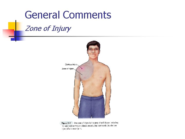
General Comments Zone of Injury
General Comments In the field n ABCs n n n Airway Breathing Circulation Always assess neurovascular status (CMS = circulation, motor and sensory) Control any bleeding Do not move victim until stabilized
General Comments n n n If possible, always ask the patient to “point with one finger to the area that hurts the most. ” Remove jewelry, etc before splinting Patient will self-splint the upper extremity (internal rotation, elbow flexed and adducted to body)
Remember n "Ability and experience are probably the most important factors in determining an individual's likelihood of injury when pursuing an outdoor sport. "
“ARMS” n n Appearance and alignment Radial pulse Motor function and mechanism of injury Sensation
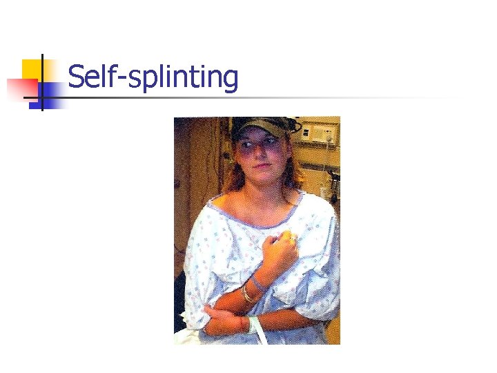
Self-splinting
Prevention
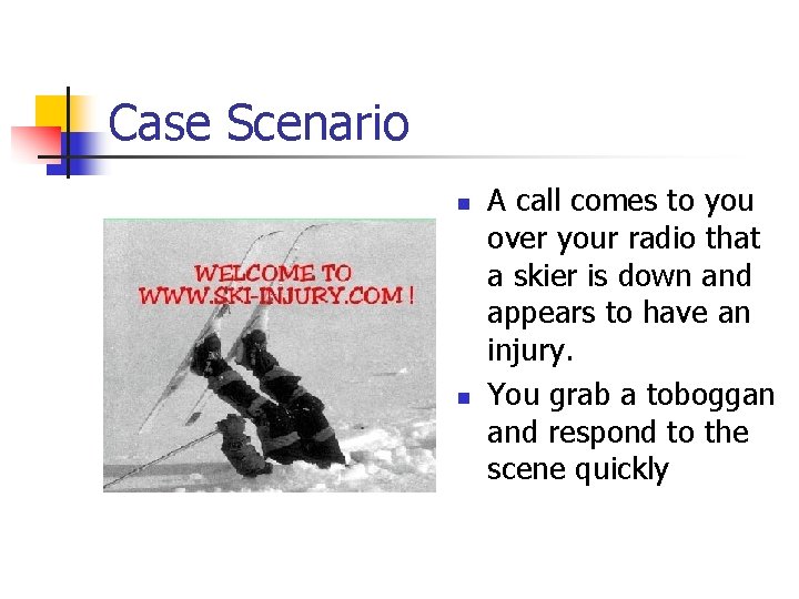
Case Scenario n n A call comes to you over your radio that a skier is down and appears to have an injury. You grab a toboggan and respond to the scene quickly
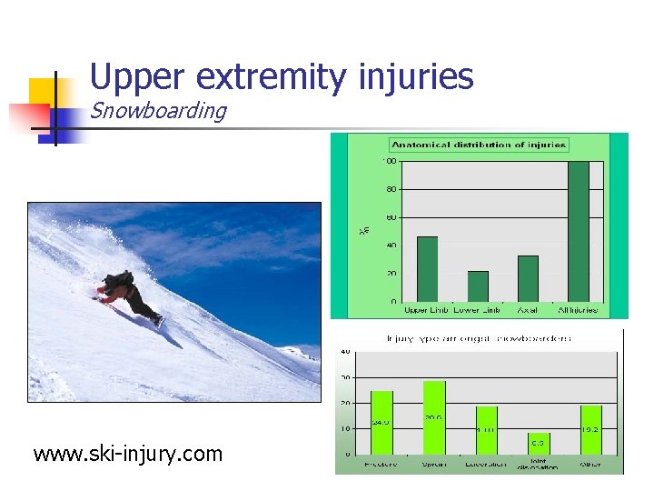
Upper extremity injuries Snowboarding www. ski-injury. com
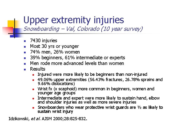
Upper extremity injuries Snowboarding – Val, Colorado (10 year survey) n n n 7430 injuries Most 30 yrs or younger 74% men, 26% women 39% beginners, 61% intermediate or experts Men rode more advanced levels than women Results n n n Injured were more likely to be beginners than non-injured 49. 06% upper extremities (56. 43% fractures, 26. 78% sprains and 9. 66% dislocations) Wrist fx (x scaphoid) more common in beginners, women and younger age groups Intermediate and expert were more likely to sustain hand, elbow and shoulder injuries as well as more severe injuries Snowboarders who wear protective wrist guards are ½ as likely to sustain wrist injury Idzikowski, et al. AJSM 2000; 28: 825 -832.
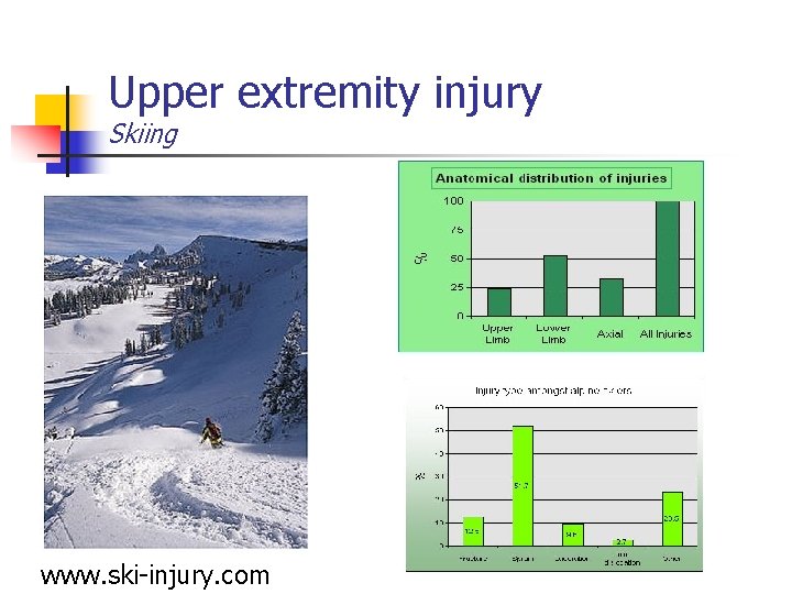
Upper extremity injury Skiing www. ski-injury. com
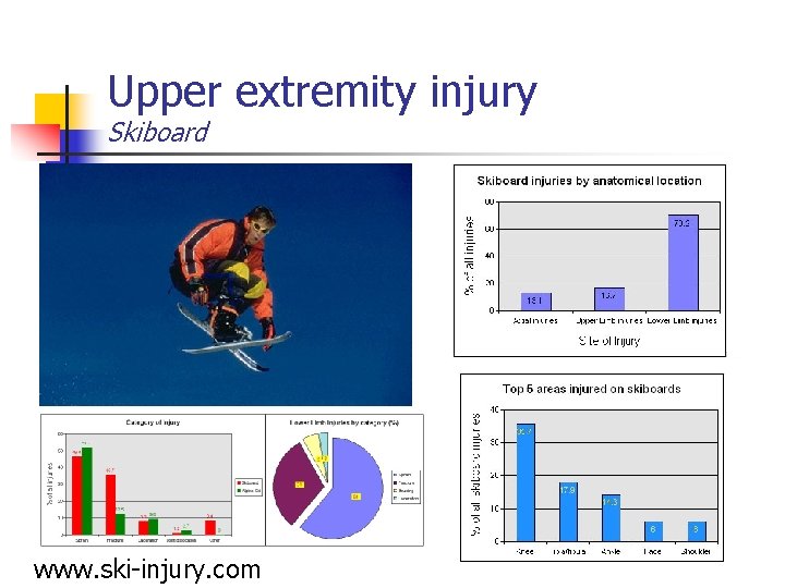
Upper extremity injury Skiboard www. ski-injury. com
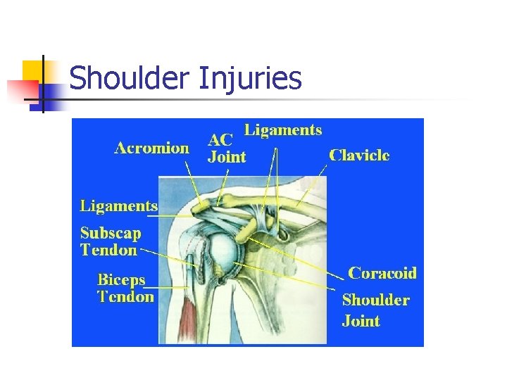
Shoulder Injuries
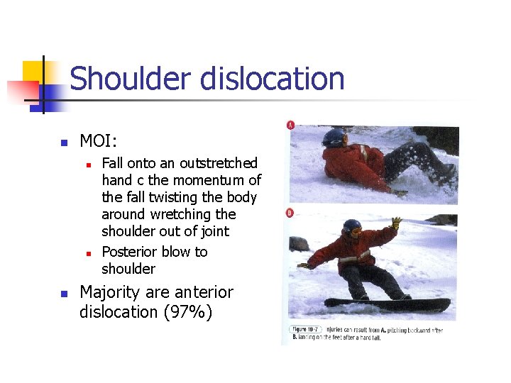
Shoulder dislocation n MOI: n n n Fall onto an outstretched hand c the momentum of the fall twisting the body around wretching the shoulder out of joint Posterior blow to shoulder Majority are anterior dislocation (97%)
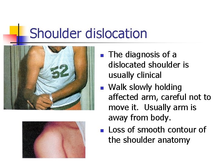
Shoulder dislocation n The diagnosis of a dislocated shoulder is usually clinical Walk slowly holding affected arm, careful not to move it. Usually arm is away from body. Loss of smooth contour of the shoulder anatomy
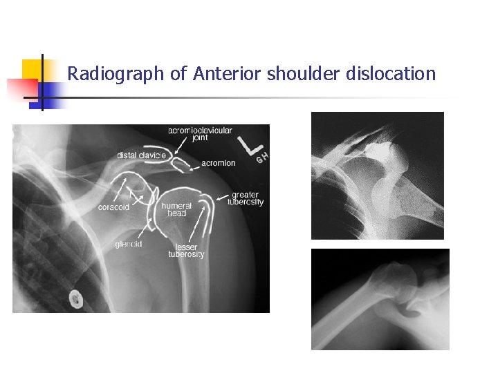
Radiograph of Anterior shoulder dislocation
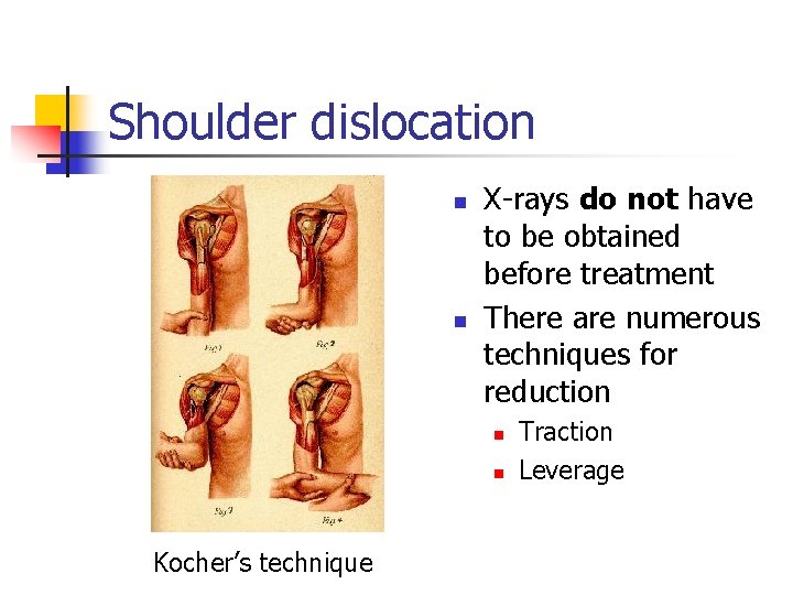
Shoulder dislocation n n X-rays do not have to be obtained before treatment There are numerous techniques for reduction n n Kocher’s technique Traction Leverage
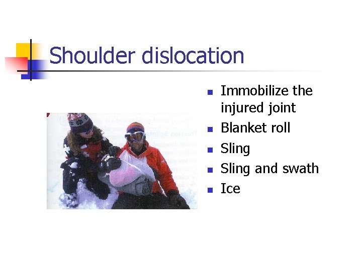
Shoulder dislocation n n Immobilize the injured joint Blanket roll Sling and swath Ice
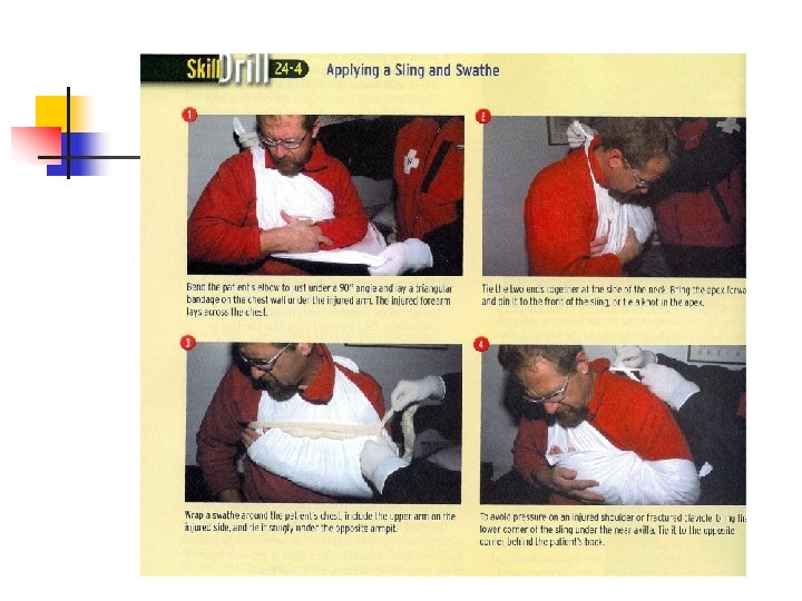
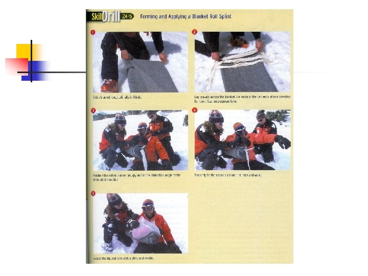
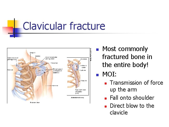
Clavicular fracture n n Most commonly fractured bone in the entire body! MOI: n n n Transmission of force up the arm Fall onto shoulder Direct blow to the clavicle
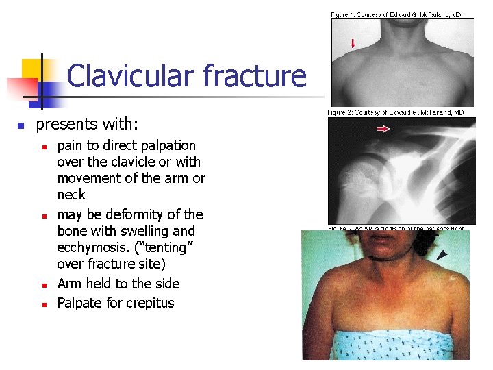
Clavicular fracture n presents with: n n pain to direct palpation over the clavicle or with movement of the arm or neck may be deformity of the bone with swelling and ecchymosis. (“tenting” over fracture site) Arm held to the side Palpate for crepitus
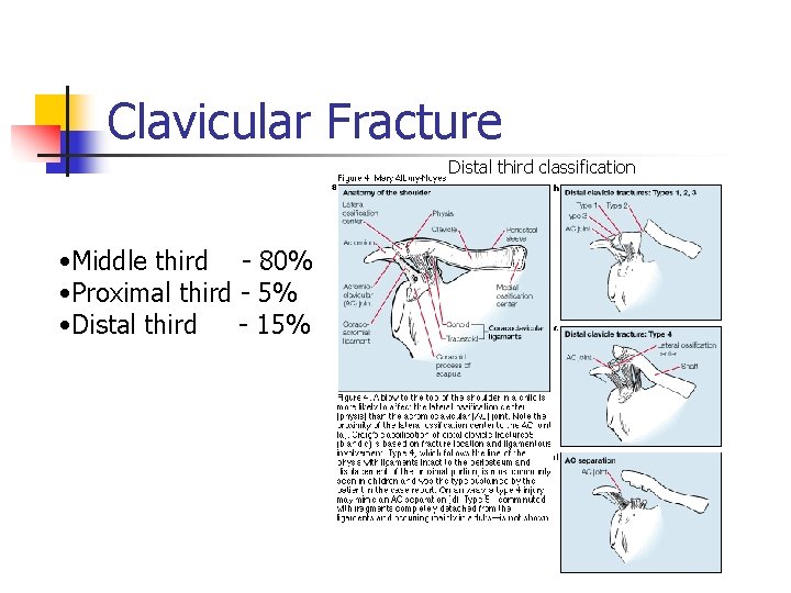
Clavicular Fracture Distal third classification • Middle third - 80% • Proximal third - 5% • Distal third - 15%
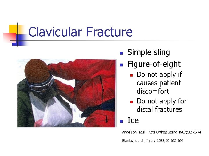
Clavicular Fracture n n Simple sling Figure-of-eight n n n Do not apply if causes patient discomfort Do not apply for distal fractures Ice Anderson, et. al. , Acta Orthop Scand 1987; 58: 71 -74 Stanley, et. al. , Injury 1988; 19: 162 -164
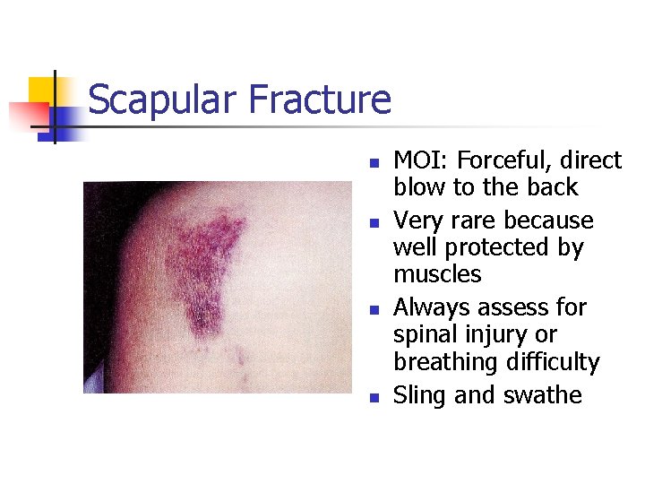
Scapular Fracture n n MOI: Forceful, direct blow to the back Very rare because well protected by muscles Always assess for spinal injury or breathing difficulty Sling and swathe
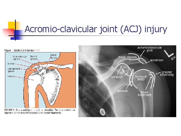
Acromio-clavicular joint (ACJ) injury
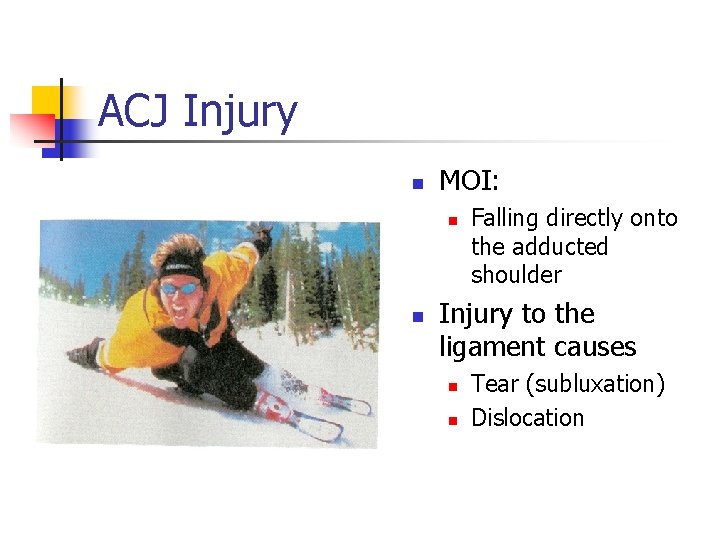
ACJ Injury n MOI: n n Falling directly onto the adducted shoulder Injury to the ligament causes n n Tear (subluxation) Dislocation
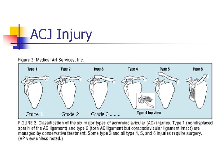
ACJ Injury Grade 1 Grade 2 Grade 3………
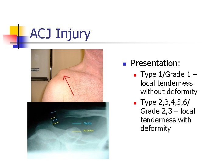
ACJ Injury n Presentation: n n Type 1/Grade 1 – local tenderness without deformity Type 2, 3, 4, 5, 6/ Grade 2, 3 – local tenderness with deformity
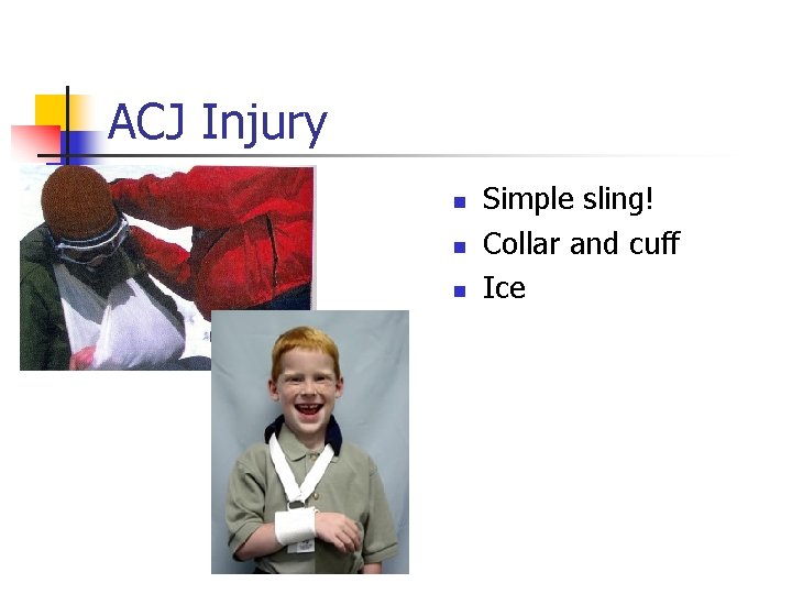
ACJ Injury n n n Simple sling! Collar and cuff Ice
Humerus Injuries
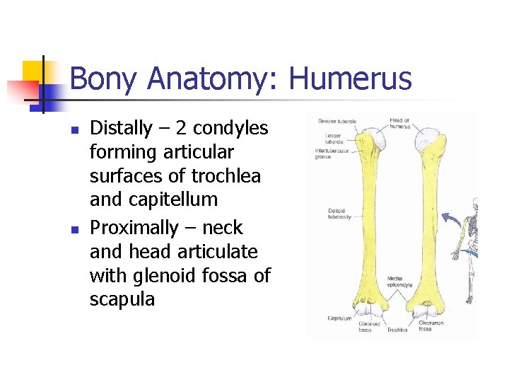
Bony Anatomy: Humerus n n Distally – 2 condyles forming articular surfaces of trochlea and capitellum Proximally – neck and head articulate with glenoid fossa of scapula
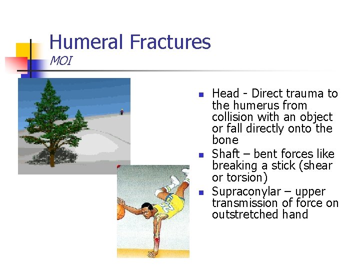
Humeral Fractures MOI n n n Head - Direct trauma to the humerus from collision with an object or fall directly onto the bone Shaft – bent forces like breaking a stick (shear or torsion) Supraconylar – upper transmission of force on outstretched hand
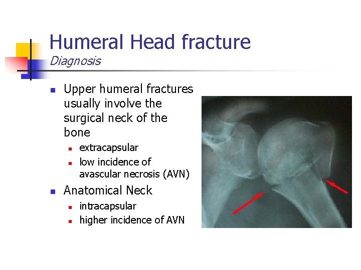
Humeral Head fracture Diagnosis n Upper humeral fractures usually involve the surgical neck of the bone n n n extracapsular low incidence of avascular necrosis (AVN) Anatomical Neck n n intracapsular higher incidence of AVN
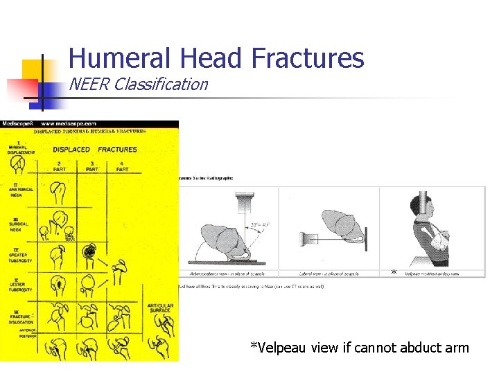
Humeral Head Fractures NEER Classification * *Velpeau view if cannot abduct arm
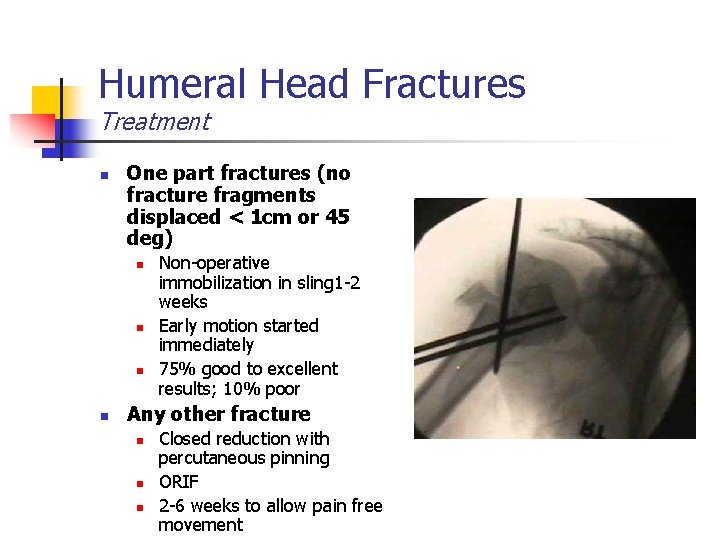
Humeral Head Fractures Treatment n One part fractures (no fracture fragments displaced < 1 cm or 45 deg) n n Non-operative immobilization in sling 1 -2 weeks Early motion started immediately 75% good to excellent results; 10% poor Any other fracture n n n Closed reduction with percutaneous pinning ORIF 2 -6 weeks to allow pain free movement
Humeral Fractures Complications n Avascular Necrosis of Humeral Head n n Non-union n n Especially at risk with 4 part fractures 3 -6 mos after injury Shoulder stiffness with prolonged immobilization
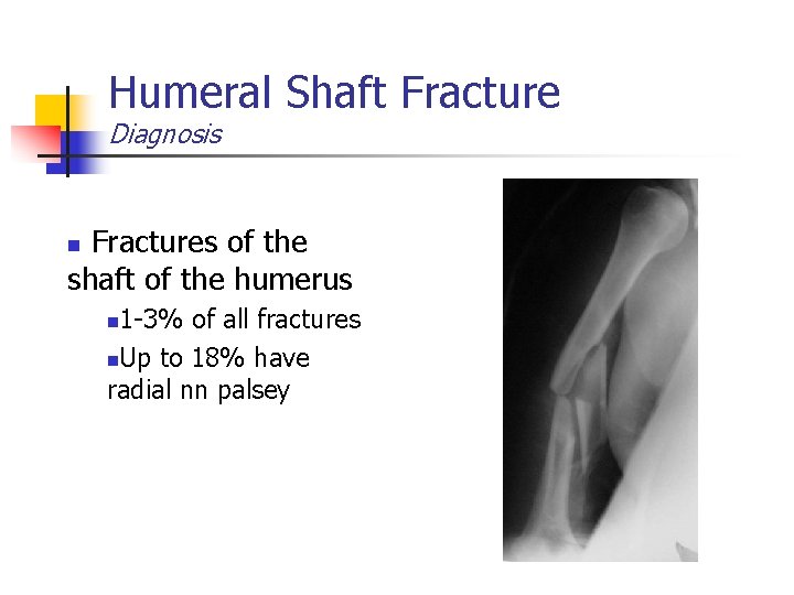
Humeral Shaft Fracture Diagnosis Fractures of the shaft of the humerus n 1 -3% of all fractures n. Up to 18% have radial nn palsey n
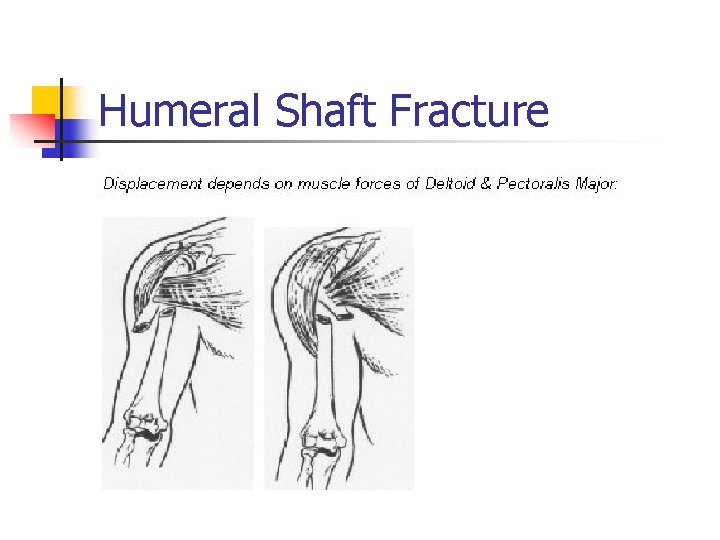
Humeral Shaft Fracture
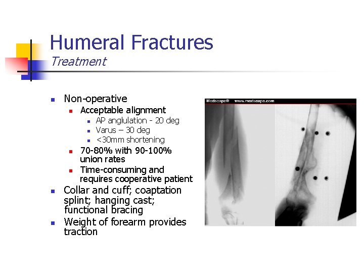
Humeral Fractures Treatment n Non-operative n Acceptable alignment n n n n AP anglulation - 20 deg Varus – 30 deg <30 mm shortening 70 -80% with 90 -100% union rates Time-consuming and requires cooperative patient Collar and cuff; coaptation splint; hanging cast; functional bracing Weight of forearm provides traction
Humeral Fractures Treatment n Operative n Absolute Indications n n n n Failure of closed treatment Associated articular involvement Vascular injuries Ipsilateral forearm fractures Pathological fractures Open fractures Polytrauma Relative Indications n n Short oblique or transverse fracture in an active individual Body habitus Patient compliance Staff considerations
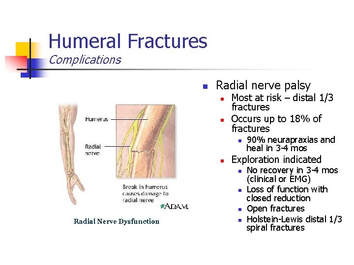
Humeral Fractures Complications n Radial nerve palsy n n Most at risk – distal 1/3 fractures Occurs up to 18% of fractures n n 90% neurapraxias and heal in 3 -4 mos Exploration indicated n n No recovery in 3 -4 mos (clinical or EMG) Loss of function with closed reduction Open fractures Holstein-Lewis distal 1/3 spiral fractures
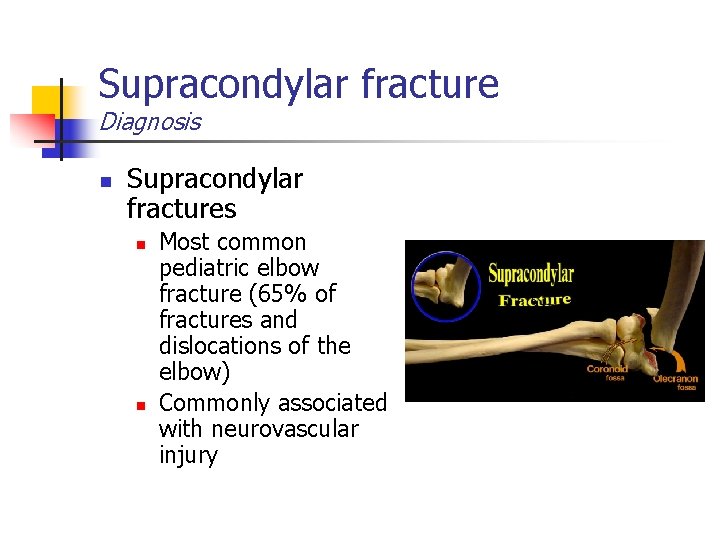
Supracondylar fracture Diagnosis n Supracondylar fractures n n Most common pediatric elbow fracture (65% of fractures and dislocations of the elbow) Commonly associated with neurovascular injury
Supracondylar fractures Diagnosis n Classification n Type I - non-displaced Type II - angulated but not translated in the sagittal plane with hinging of the posterior cortex of the humerus Type III - posteriorly displaced with IIIA being posteromedial and type IIIB being posterolateral
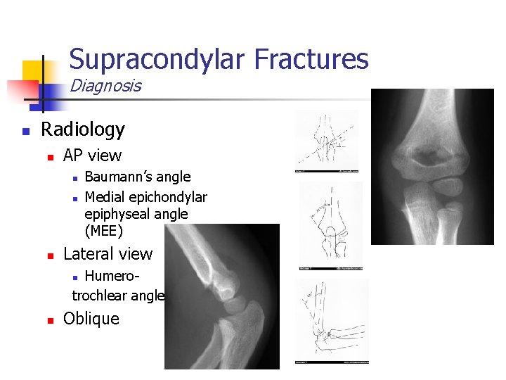
Supracondylar Fractures Diagnosis n Radiology n AP view n n n Baumann’s angle Medial epichondylar epiphyseal angle (MEE) Lateral view Humerotrochlear angle n n Oblique
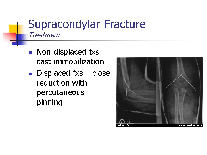
Supracondylar Fracture Treatment n n Non-displaced fxs – cast immobilization Displaced fxs – close reduction with percutaneous pinning
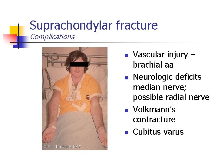
Suprachondylar fracture Complications n n Vascular injury – brachial aa Neurologic deficits – median nerve; possible radial nerve Volkmann’s contracture Cubitus varus
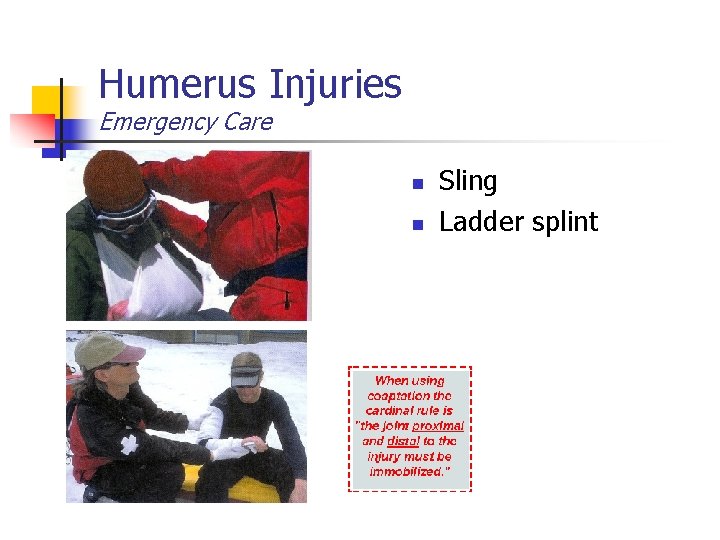
Humerus Injuries Emergency Care n n Sling Ladder splint
Elbow Injuries
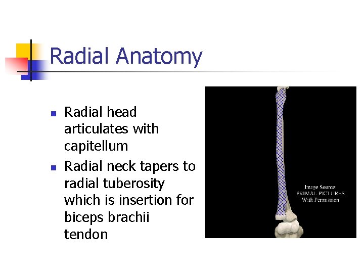
Radial Anatomy n n Radial head articulates with capitellum Radial neck tapers to radial tuberosity which is insertion for biceps brachii tendon
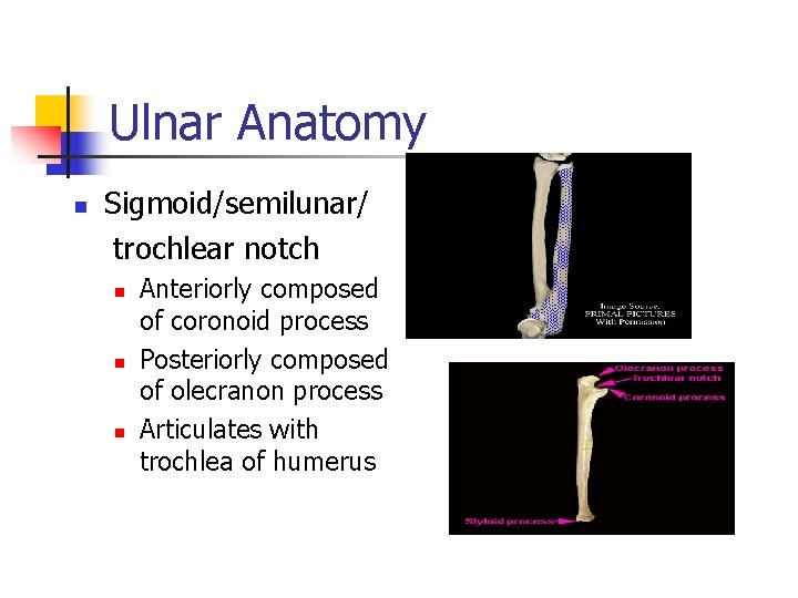
Ulnar Anatomy n Sigmoid/semilunar/ trochlear notch n n n Anteriorly composed of coronoid process Posteriorly composed of olecranon process Articulates with trochlea of humerus
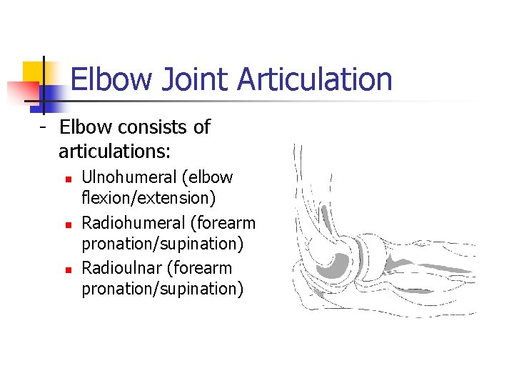
Elbow Joint Articulation - Elbow consists of articulations: n n n Ulnohumeral (elbow flexion/extension) Radiohumeral (forearm pronation/supination) Radioulnar (forearm pronation/supination)
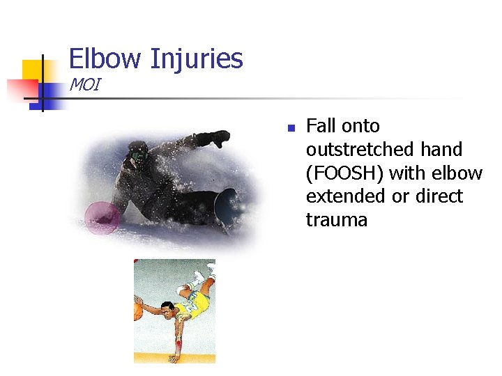
Elbow Injuries MOI n Fall onto outstretched hand (FOOSH) with elbow extended or direct trauma
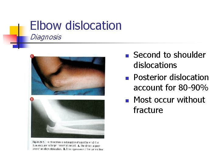
Elbow dislocation Diagnosis n n n Second to shoulder dislocations Posterior dislocation account for 80 -90% Most occur without fracture
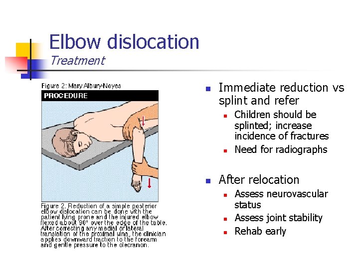
Elbow dislocation Treatment n Immediate reduction vs splint and refer n n n Children should be splinted; increase incidence of fractures Need for radiographs After relocation n Assess neurovascular status Assess joint stability Rehab early
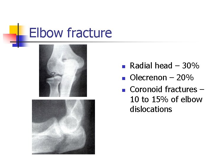
Elbow fracture n n n Radial head – 30% Olecrenon – 20% Coronoid fractures – 10 to 15% of elbow dislocations
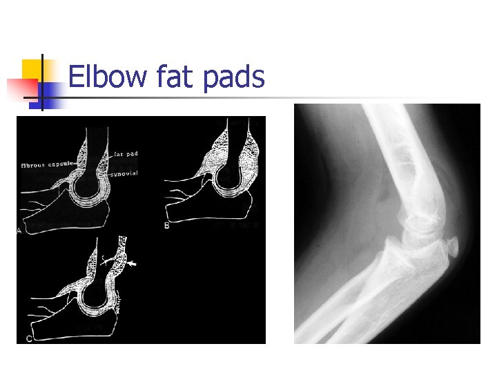
Elbow fat pads
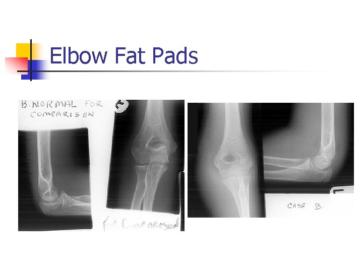
Elbow Fat Pads
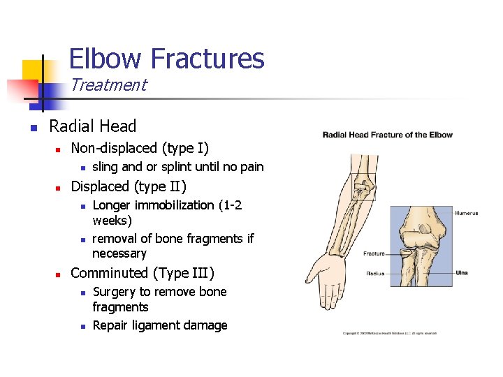
Elbow Fractures Treatment n Radial Head n Non-displaced (type I) n n Displaced (type II) n n n sling and or splint until no pain Longer immobilization (1 -2 weeks) removal of bone fragments if necessary Comminuted (Type III) n n Surgery to remove bone fragments Repair ligament damage
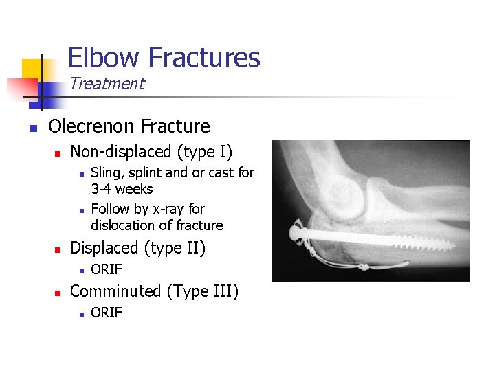
Elbow Fractures Treatment n Olecrenon Fracture n Non-displaced (type I) n n n Displaced (type II) n n Sling, splint and or cast for 3 -4 weeks Follow by x-ray for dislocation of fracture ORIF Comminuted (Type III) n ORIF
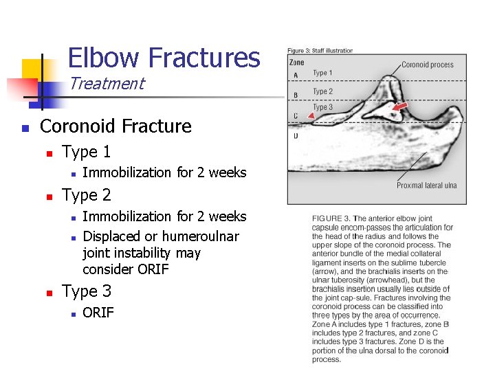
Elbow Fractures Treatment n Coronoid Fracture n Type 1 n n Type 2 n n n Immobilization for 2 weeks Displaced or humeroulnar joint instability may consider ORIF Type 3 n ORIF
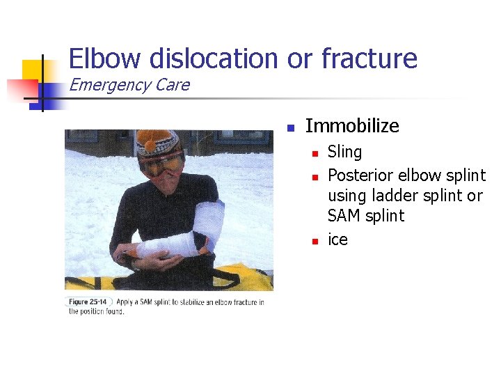
Elbow dislocation or fracture Emergency Care n Immobilize n n n Sling Posterior elbow splint using ladder splint or SAM splint ice
Wrist Injuries
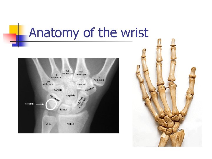
Anatomy of the wrist
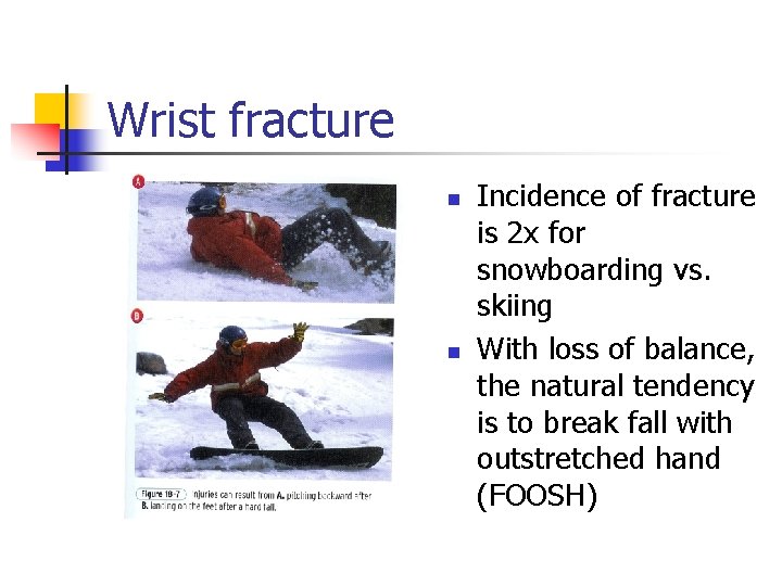
Wrist fracture n n Incidence of fracture is 2 x for snowboarding vs. skiing With loss of balance, the natural tendency is to break fall with outstretched hand (FOOSH)
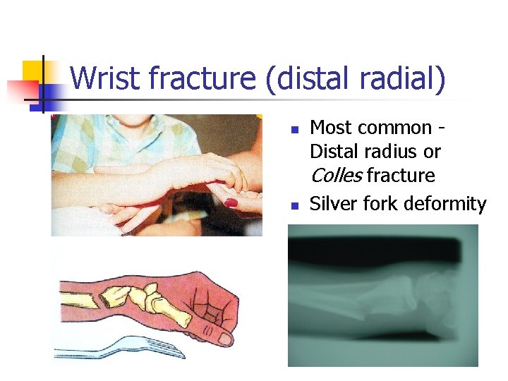
Wrist fracture (distal radial) n n Most common Distal radius or Colles fracture Silver fork deformity
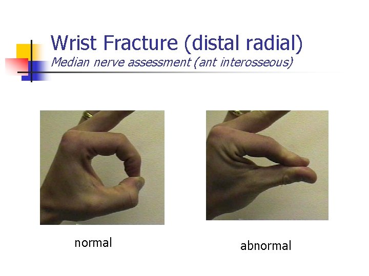
Wrist Fracture (distal radial) Median nerve assessment (ant interosseous) normal abnormal
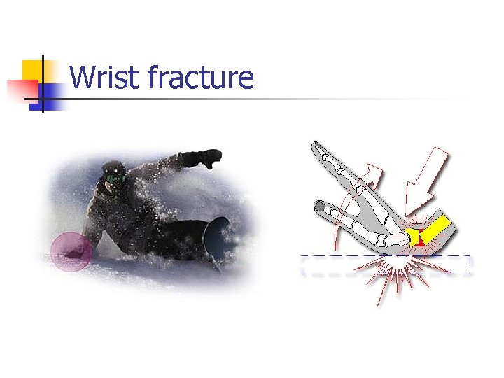
Wrist fracture
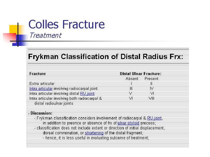
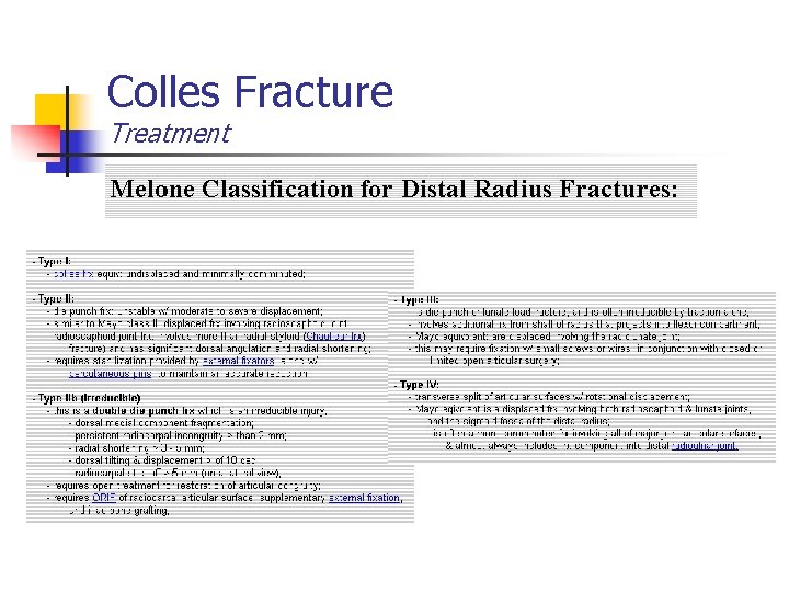
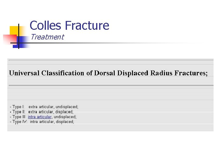
Colles Fracture Treatment
Colles Fracture Treatment
Colles Fracture Treatment
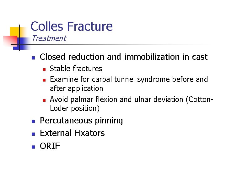
Colles Fracture Treatment n Closed reduction and immobilization in cast n n n Stable fractures Examine for carpal tunnel syndrome before and after application Avoid palmar flexion and ulnar deviation (Cotton. Loder position) Percutaneous pinning External Fixators ORIF
Scaphoid fracture Diagnosis
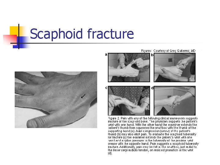
Scaphoid fracture
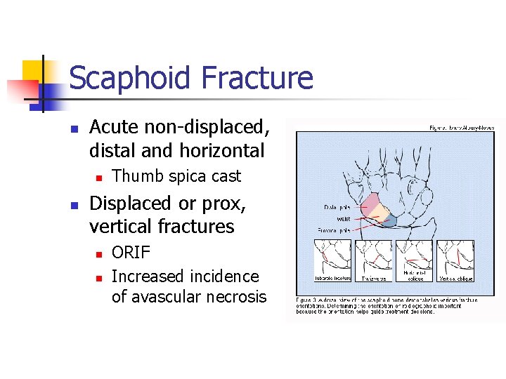
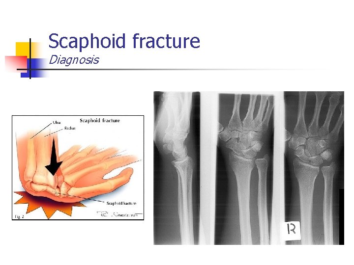
Scaphoid Fracture n Acute non-displaced, distal and horizontal n n Thumb spica cast Displaced or prox, vertical fractures n n ORIF Increased incidence of avascular necrosis
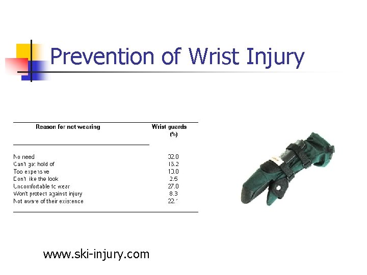
Prevention of Wrist Injury www. ski-injury. com
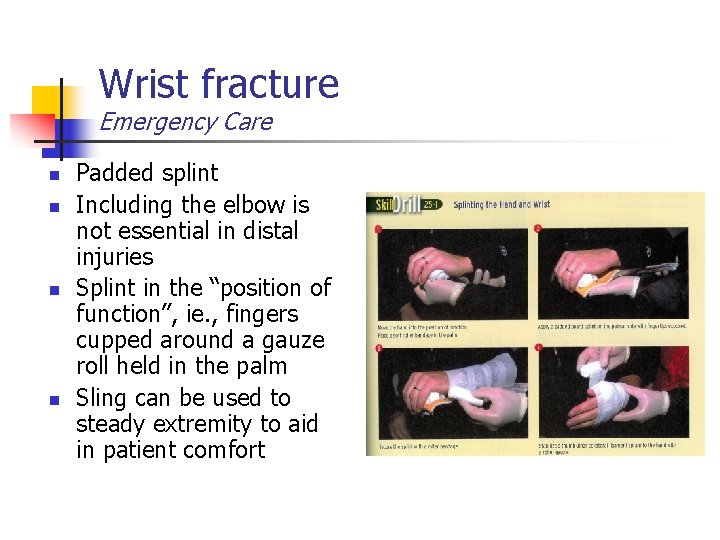
Wrist fracture Emergency Care n n Padded splint Including the elbow is not essential in distal injuries Splint in the “position of function”, ie. , fingers cupped around a gauze roll held in the palm Sling can be used to steady extremity to aid in patient comfort
Hand/Finger Injuries
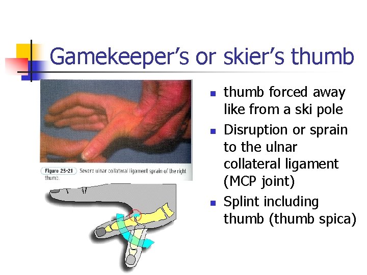
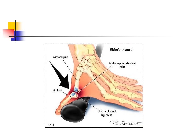
Gamekeeper’s or skier’s thumb n n n thumb forced away like from a ski pole Disruption or sprain to the ulnar collateral ligament (MCP joint) Splint including thumb (thumb spica)
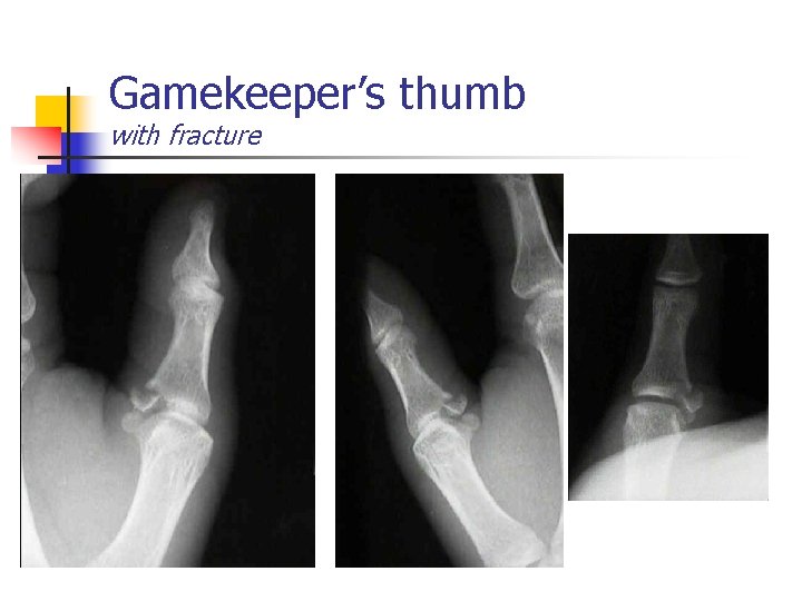
Gamekeeper’s thumb with fracture
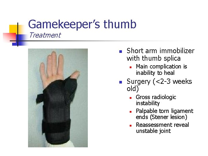
Gamekeeper’s thumb Treatment n Short arm immobilizer with thumb splica n n Main complication is inability to heal Surgery (<2 -3 weeks old) n n n Gross radiologic instability Palpable torn ligament ends (Stener lesion) Reassessment reveal unstable joint
Conclusion n Falls, collisions and direct blows cause injury to the upper extremities n n n Humerus Elbow Wrist Hand/fingers Remember general comments n n “Zone of injury” Life-threatening injuries first (ABCs)
Conclusion n Patients will self-splint the extremity n n Injuries close to joints can involve neurovascular bundle n n Internal rotation, elbow flexed, adducted to body Assess CMS ARMS n Appearance and alignment, radial pulse, motor and MOI, sensation
Conclusion n Accurate assessment and rapid transport critical (60” rule) n n n Immobilize in the position found Sling and swathe is good immobilizer for upper extremity injuries Every patient should be advised to seek the care of a physician regardless of injury, especially if symptoms persist > 24 hrs.
Thank -You
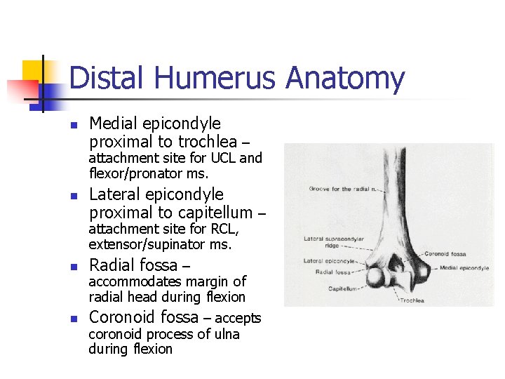
Distal Humerus Anatomy n Medial epicondyle proximal to trochlea – attachment site for UCL and flexor/pronator ms. n Lateral epicondyle proximal to capitellum – attachment site for RCL, extensor/supinator ms. n Radial fossa – n Coronoid fossa – accepts accommodates margin of radial head during flexion coronoid process of ulna during flexion
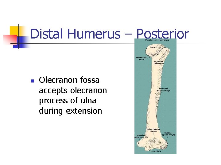
Distal Humerus – Posterior n Olecranon fossa accepts olecranon process of ulna during extension
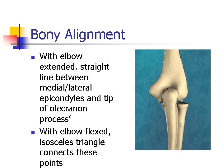
Bony Alignment n n With elbow extended, straight line between medial/lateral epicondyles and tip of olecranon process’ With elbow flexed, isosceles triangle connects these points
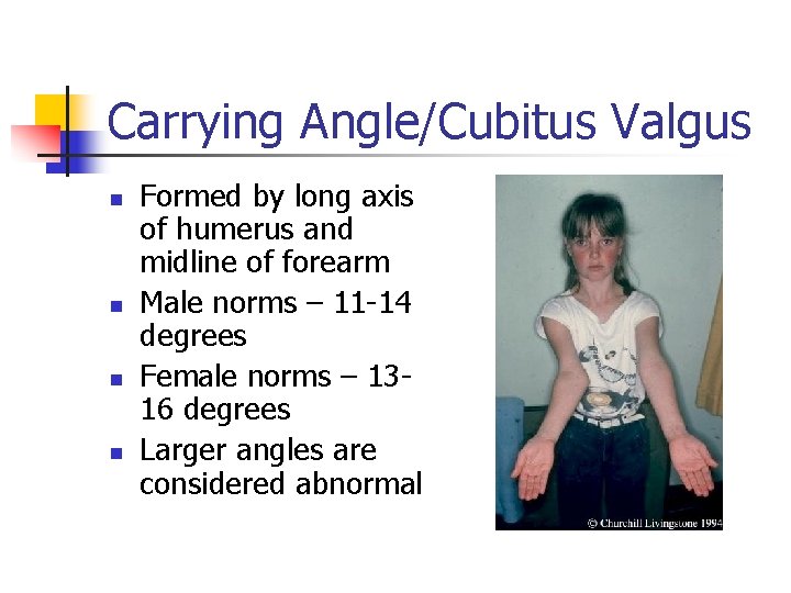
Carrying Angle/Cubitus Valgus n n Formed by long axis of humerus and midline of forearm Male norms – 11 -14 degrees Female norms – 1316 degrees Larger angles are considered abnormal
- Slides: 93