
Skeleton Skeletal System Support Storage of minerals Blood
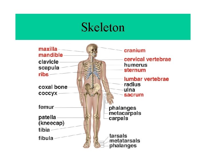
Skeleton
Skeletal System • • • Support Storage of minerals Blood cell production Protection Movement – Levers
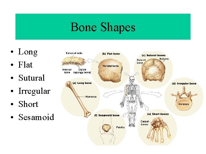
Bone Shapes • • • Long Flat Sutural Irregular Short Sesamoid
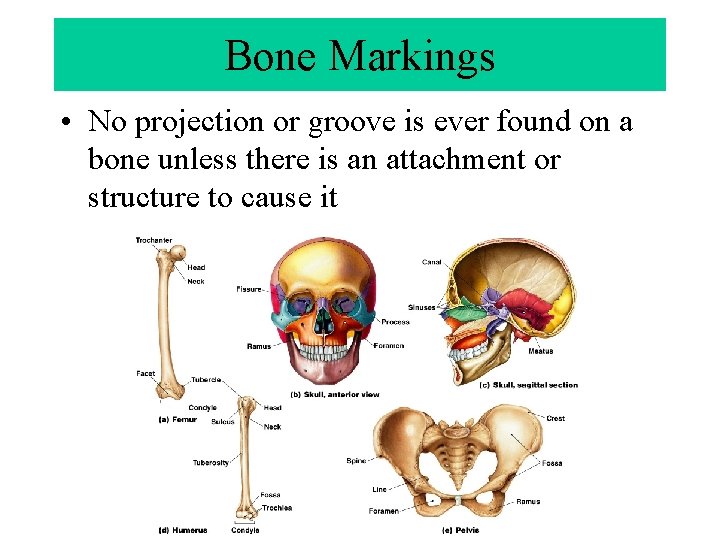
Bone Markings • No projection or groove is ever found on a bone unless there is an attachment or structure to cause it
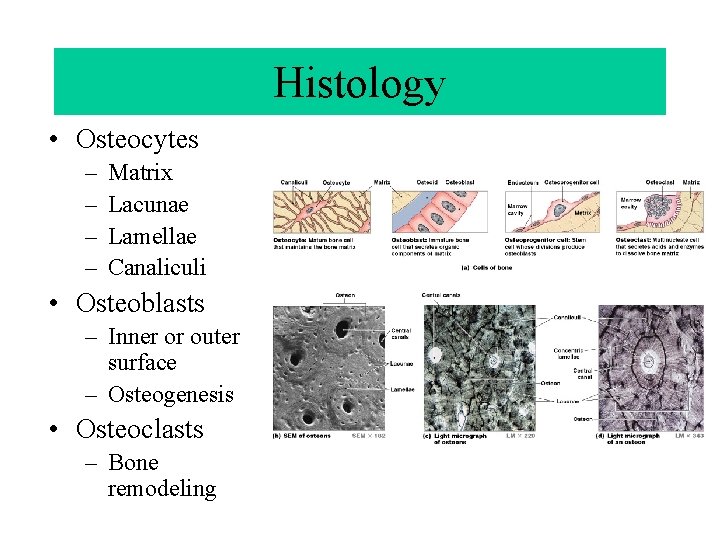
Histology • Osteocytes – – Matrix Lacunae Lamellae Canaliculi • Osteoblasts – Inner or outer surface – Osteogenesis • Osteoclasts – Bone remodeling
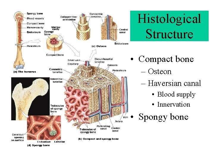
Histological Structure • Compact bone – Osteon – Haversian canal • Blood supply • Innervation • Spongy bone
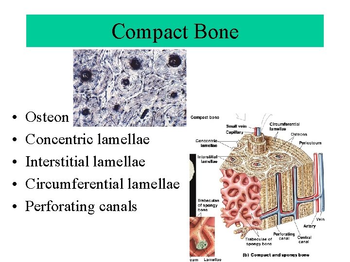
Compact Bone • • • Osteon Concentric lamellae Interstitial lamellae Circumferential lamellae Perforating canals
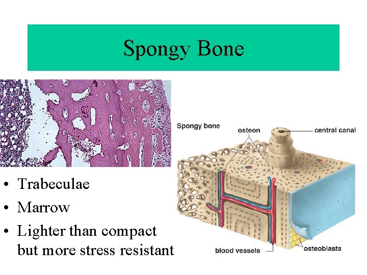
Spongy Bone • Trabeculae • Marrow • Lighter than compact but more stress resistant
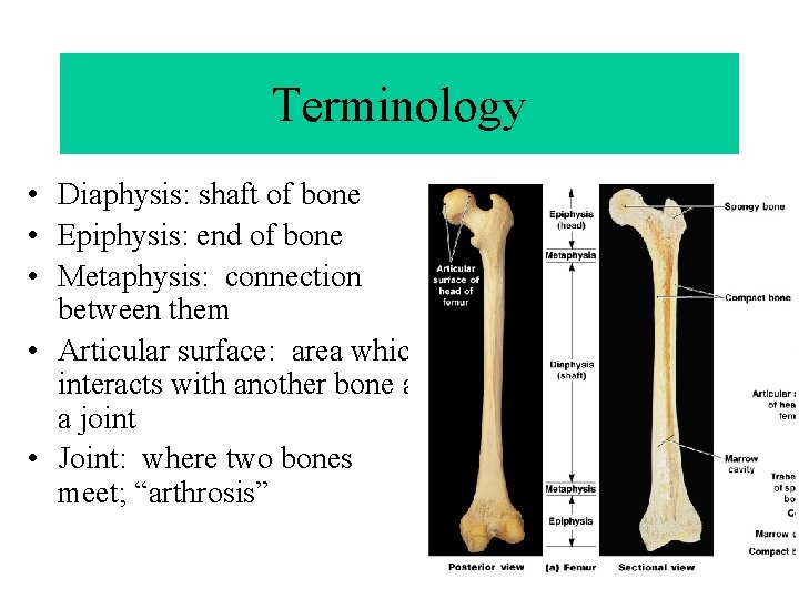
Terminology • Diaphysis: shaft of bone • Epiphysis: end of bone • Metaphysis: connection between them • Articular surface: area which interacts with another bone at a joint • Joint: where two bones meet; “arthrosis”
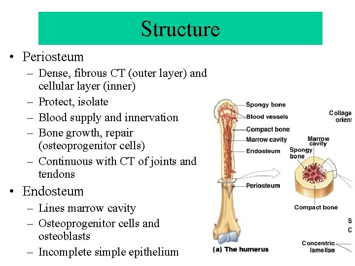
Structure • Periosteum – Dense, fibrous CT (outer layer) and cellular layer (inner) – Protect, isolate – Blood supply and innervation – Bone growth, repair (osteoprogenitor cells) – Continuous with CT of joints and tendons • Endosteum – Lines marrow cavity – Osteoprogenitor cells and osteoblasts – Incomplete simple epithelium
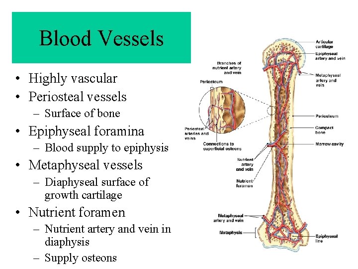
Blood Vessels • Highly vascular • Periosteal vessels – Surface of bone • Epiphyseal foramina – Blood supply to epiphysis • Metaphyseal vessels – Diaphyseal surface of growth cartilage • Nutrient foramen – Nutrient artery and vein in diaphysis – Supply osteons
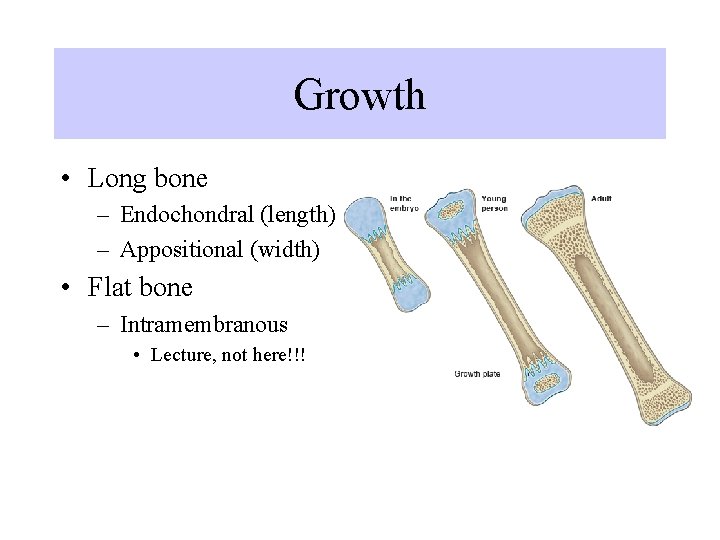
Growth • Long bone – Endochondral (length) – Appositional (width) • Flat bone – Intramembranous • Lecture, not here!!!
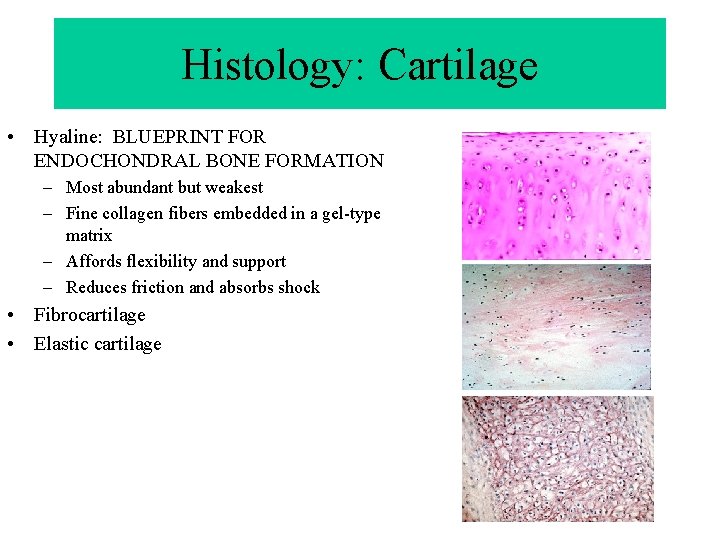
Histology: Cartilage • Hyaline: BLUEPRINT FOR ENDOCHONDRAL BONE FORMATION – Most abundant but weakest – Fine collagen fibers embedded in a gel-type matrix – Affords flexibility and support – Reduces friction and absorbs shock • Fibrocartilage • Elastic cartilage
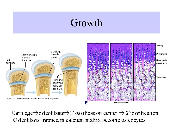
Growth Cartilage osteoblasts 1 o ossification center 2 o ossification Osteoblasts trapped in calcium matrix become osteocytes
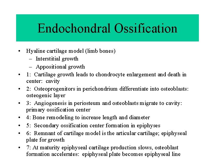
Endochondral Ossification • Hyaline cartilage model (limb bones) – Interstitial growth – Appositional growth • 1: Cartilage growth leads to chondrocyte enlargement and death in center: cavity • 2: Osteoprogenitors in perichondrium differentiate into osteoblasts: osteogenic layer • 3: Angiogenesis in periosteum and osteoblasts migrate to cavity: primary ossification center • 4: Bone remodeling to increase length and diameter • 5: Secondary ossification center formation in epiphyses • 6: Remnant of cartilage model is the articular cartilage; epiphyseal plate for growth • 7: At maturity epiphyseal cartilage production slows, osteoblast formation accelerates: epiphyseal plate becomes epiphyseal line
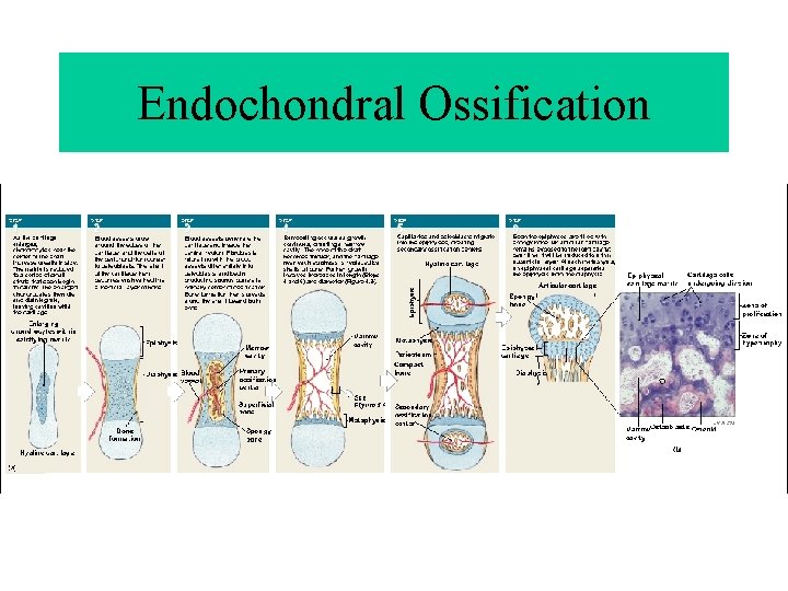
Endochondral Ossification
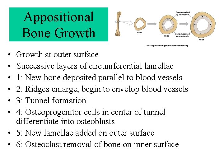
Appositional Bone Growth • • • Growth at outer surface Successive layers of circumferential lamellae 1: New bone deposited parallel to blood vessels 2: Ridges enlarge, begin to envelop blood vessels 3: Tunnel formation 4: Osteoprogenitor cells in center of tunnel differentiate into osteoblasts • 5: New lamellae added on outer surface • 6: Osteoclast removal of bone on inner surface
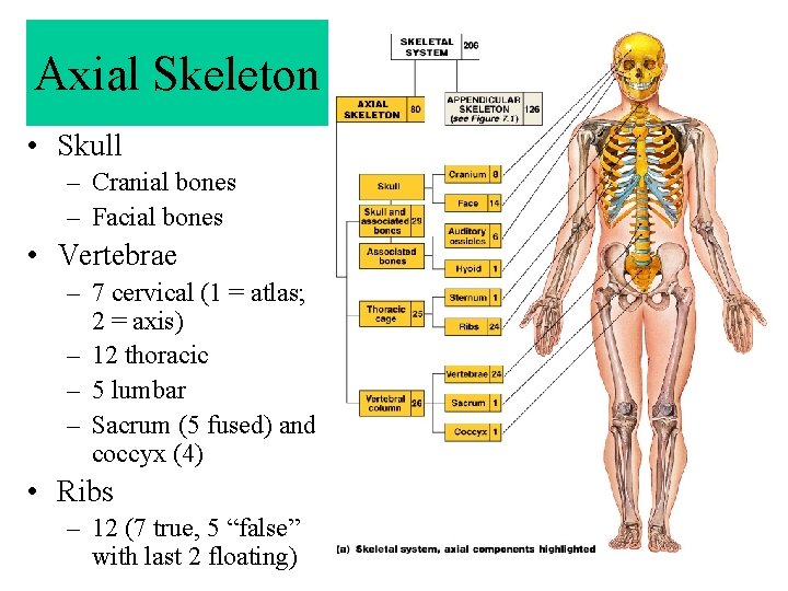
Axial Skeleton • Skull – Cranial bones – Facial bones • Vertebrae – 7 cervical (1 = atlas; 2 = axis) – 12 thoracic – 5 lumbar – Sacrum (5 fused) and coccyx (4) • Ribs – 12 (7 true, 5 “false” with last 2 floating)
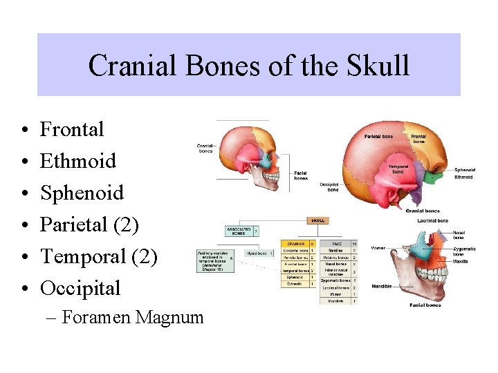
Cranial Bones of the Skull • • • Frontal Ethmoid Sphenoid Parietal (2) Temporal (2) Occipital – Foramen Magnum
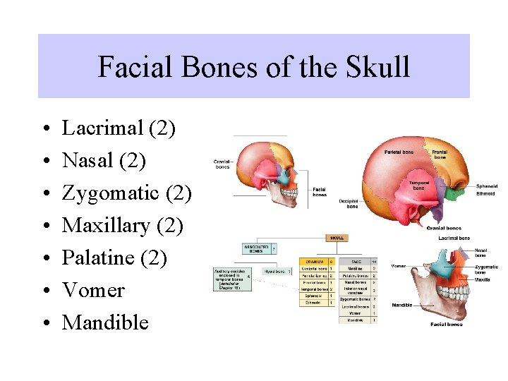
Facial Bones of the Skull • • Lacrimal (2) Nasal (2) Zygomatic (2) Maxillary (2) Palatine (2) Vomer Mandible
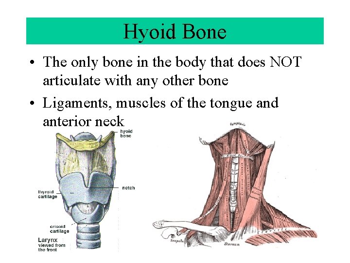
Hyoid Bone • The only bone in the body that does NOT articulate with any other bone • Ligaments, muscles of the tongue and anterior neck
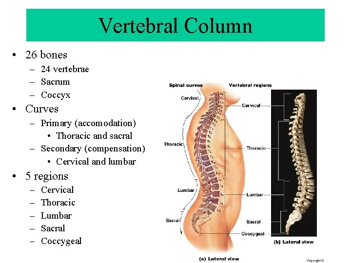
Vertebral Column • 26 bones – 24 vertebrae – Sacrum – Coccyx • Curves – Primary (accomodation) • Thoracic and sacral – Secondary (compensation) • Cervical and lumbar • 5 regions – – – Cervical Thoracic Lumbar Sacral Coccygeal
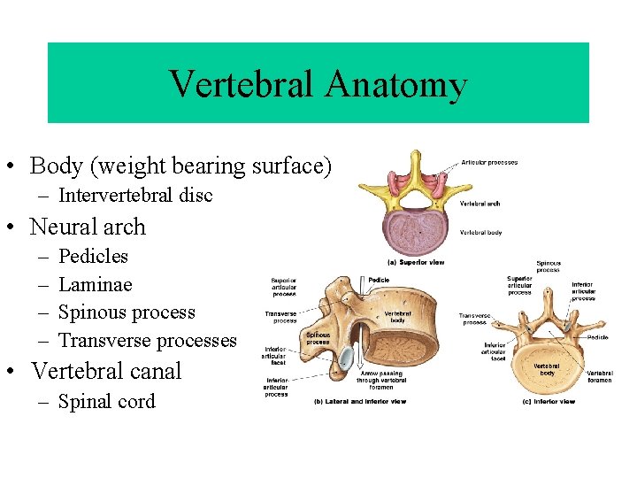
Vertebral Anatomy • Body (weight bearing surface) – Intervertebral disc • Neural arch – – Pedicles Laminae Spinous process Transverse processes • Vertebral canal – Spinal cord
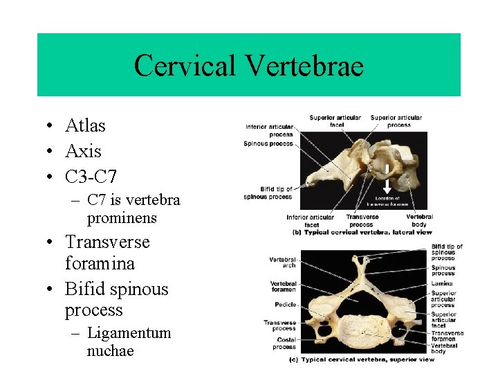
Cervical Vertebrae • Atlas • Axis • C 3 -C 7 – C 7 is vertebra prominens • Transverse foramina • Bifid spinous process – Ligamentum nuchae
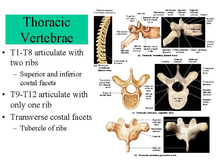
Thoracic Vertebrae • T 1 -T 8 articulate with two ribs – Superior and inferior costal facets • T 9 -T 12 articulate with only one rib • Transverse costal facets – Tubercle of ribs
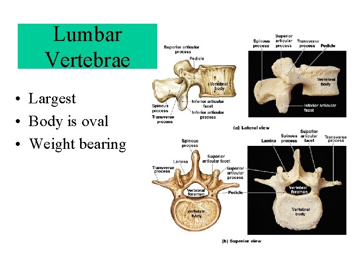
Lumbar Vertebrae • Largest • Body is oval • Weight bearing
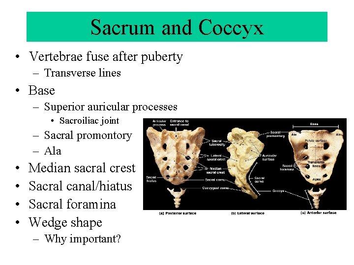
Sacrum and Coccyx • Vertebrae fuse after puberty – Transverse lines • Base – Superior auricular processes • Sacroiliac joint – Sacral promontory – Ala • • Median sacral crest Sacral canal/hiatus Sacral foramina Wedge shape – Why important?
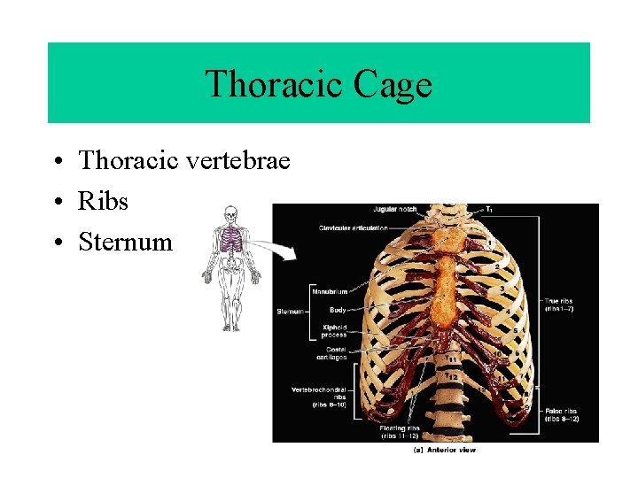
Thoracic Cage • Thoracic vertebrae • Ribs • Sternum
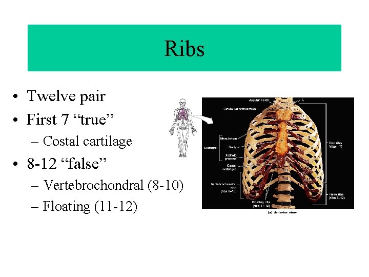
Ribs • Twelve pair • First 7 “true” – Costal cartilage • 8 -12 “false” – Vertebrochondral (8 -10) – Floating (11 -12)
- Slides: 29