Skeleton of Lower limb Hip Bone Sacroilliac joint


Skeleton of Lower limb

Hip Bone

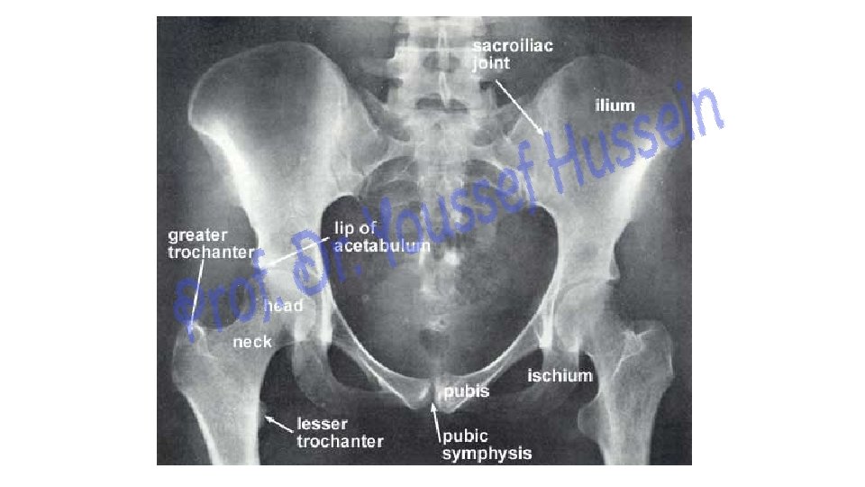
Sacroilliac joint Hip joint Symphsis pubis
. - They")
- The two hip bones articulate together at the symphysis pubis (anteriorly). - They articulate with sacrum at the sacroiliac joints (Posteriorly) - The hip bone is an irregular bone which is formed of 3 parts: 1) Ilium. 2) Pubis. 3) Ischium. ** Side determination (right and left) 1 - The iliac crest is directed superiorly. 2 - Acetabulum is directed laterally. 3 - Pubis is directed anteriorly. ** Anatomical position of the hip bone, 1 -The anterior superior iliac spine (A. S. I. S) and the pubic tubercle lie in the same vertical plane. 2 - The ischial spine and the upper border of the symphysis pubis lie in the same horizontal plane. 3 - The acetabular notch faces downwards.

Ilium 2/5 Pubis 1/5 2/5 Ischium

Ilium bone
 Eminence between")
Iliac crest Anterior superior iliac spine Anterior inferior iliac spine Iliopubic (iliopecineal) Eminence between ileum and pubis posterior superior Iliac spine posterior inferior Iliac spine Greater sciatic notch

iliac tuberosity iliac fossa auricular surface pelvic surface Auricular surface: because it resembles auricle of external ear. - This surface articulates with auricular surface of the sacrurn to form the sacroiliac joint (plane synovial joint).

posterior gluteal line middle gluteal line Inferior gluteal line

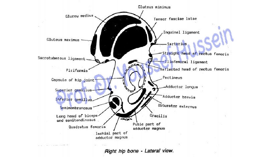
Iliacus Sartorius Anterior superior iliac spine below the inguinal lig. Straight head of Rectus femoris from anterior inferior iliac spine Obturator internus

Tensor Fascia Latae Gluteus Medius Gluteus Maximus Reflected head of rectus femoris superior Gemellus inferior Gemellus

Pubic bone

Superior Pubic ramus pubic bone Pubic body Iliopubic eminance Inferior Pubic ramus

Obturator crest Pectineal line Pectineal surface pubic crest pubic tubercle Acetabular notch Obturator surface and groove

Quadratus femoris Lateral border of Ischial tuberoisty Pectineus Adductor longus Adductor brevis Gracilis

Ischium bone

Greater sciatic notch Ischial spine Lesser sciatic notch Ischial tuberosity

Origin of hamstring M Upper Lateral Medial Inferior Ling head of biceps & semitendinosus Lower medial part of upper area Medial part of lower area Subcutaneous part in sitting position Semimembranosus Upper lateral part of upper area Ischial part of adductor Magnus Lateral part of lower area

Ischial part of Adductor Magnus : lateral Part of lower area Obturator externus O: Outer margin of obturator foramen and outer surface of obturator membrane Ischial tuberosity O: pubic part of Adductor Magnus : from conjoined pubic and ischial rami

Common Origin of long head of biceps femoris & Semitendinosus: lower medial part of upper area Origin of Semimembranosus O: upper lateral part of upper area Quadratus femoris Lateral border of Ischial tuberoisty

v Ilium - This is the upper part of the hip bone. - It has 4 borders and 3 surfaces: ** Borders A- Iliac crest: upper border of the ilium. - Its anterior end forms anterior superior iliac spine (A. S. I. S). - Its posterior end forms posterior superior iliac spine (P. S. I. S). B- Anterior border: from A. S. I. S to anterior inferior iliac spine (A. I. I. S). C- Posterior border: from (P. S. I. S) to posterior inferior iliac spine, then it forms the upper part of greater sciatic notch. D- Medial border: is called the arcuate or iliopectineal line of the hip bone. - Its junction with the pubis forms a projection called i. Iiopectineal or iliopubic eminence.

** surfaces of ileum AGluteal surface: Is the wide outer surface of the ilium. 1 - Posterior gluteal line: from P. I. I. S. to iliac crest. 2 - Middle gluteal line: from the upper border of greater sciatic notch to A. S. I. S. 3 - Inferior gluteal line: from the apex of the greater sciatic notch to A. I. I. S. B - Iliac Fossa: is a smooth and gently concave surface. CSacropelvic surface: is an irregular surface which lies behind and below the iliac fossa. I - Iliac tuberosity: rough area below dorsal of the iliac crest. 2 - Auricular surface: resembles the auricle of the external ear. It articulates with the auricular surface of the sacrurm to form the sacroiliac joint (plane synovial joint). 3 - Pelvic surface: is the lower part of the sacropelvic surface

Pubis - This is the anterior and inferior part of the hip bone; - It includes the anterior part of the acetabulum and extends to the symphysis pubis - The pubis is formed of three main pans: body and 2 rami: A- Body of the pubis: is flattened and has 3 surfaces: I- Anterior surface: directed anteroinferior towards the thigh. 2 - Posterior surface directed posterosuperior towards the pelvis. 3 - Symphyseal surface; directed medially where it shares in the symphysis pubis - The upper border of the body is called the pubic crest. - The crest ends laterally in a small but well defined bony projection called the pubic tubercle.

B- Superior pubic ramus: - It is triangular in cross section, having 3 surfaces which are separated by 3 borders: 1 - Pectineal line: a sharp ridge extending from pubic tubercle to iliopectineal eminence. It separates pectineal and pelvic surfaces. 2 - Obturator crest: a rounded ridge separating the pectineal and obturator surfaces 3 - Inferior border: separates the obturator and pelvic surfaces. 1 - Pectineal surface: a triangular surface between pectineal line and obturator crest 2 - Pelvic surface: forming a part of the wall of the true pelvis. 3 - Obturator surface; directed posteroinferiorly towards the obturator foramen. It is grooved obliquely to form the obturator groove. C- Inferior pubic ramus: - The inferior pubic ramus has 2 surfaces (outer & inner) separated by 2 borders.

v ischium - This is the posterior and inferior part of the hip bone. - It is formed of a body, ischial tuberosity and one ramus. A- Posterior border of the body shows ischial spine separates greater sciatic notch above from lesser sciatic notch below. B- Ischial tuberosity: is divided by a transverse ridge into 2 areas: 1 - Upper area: which is divided by an oblique ridge into 2 parts: a - Upper lateral part. b- Lower medial part. 2 - Lower area: which is divided by a longitudinal ridge into 2 parts: a- Lateral part. b- Medial part. Acetabulum - This is a cup-shaped depression on the lateral side of the hip bone. - It includes parts of the 3 components of the hip bone. - It articulates with the head of the femur to form the hip joint. - The inferior margin of the acetabulum shows acetabular notch. - The non-articular area called the acetabular fossa. - The C-shaped articular strip called the lunate surface.

Piriformis Pelvic surface of the middle 3 pieces of sacrum Iliacus Origin: upper surface of the lateral mass of sacrum

Origin of Gluteus Maximus : back of sacrum and coccyx.

Femur bone

Femur ** How to identify the side of femur 1 - The head is directed upwards and medially. 2 - The shaft is convex anteriorly.

Fovea of head Neck G G 120˚ Head& Neck of femur S S Head Intertrochanteric line (anterior)
 Trochanteric fossa Medial surface lesser trochanter")
Greater trochanter Quadrate tubercle Intertrochanteric crest (posterior) Trochanteric fossa Medial surface lesser trochanter

Angle of Inclination Coxa vara Decrease angle Normal angle About 120 Coxa valga Increase angle What is the meaning of angle of Inclination

Insertion of obturator internus: Medial surface of greater trochanter Insertion of obturator externus: fossa lesser trochanter Insertion of Iliacus : lesser trochanter and one inch below it

Piriformis Top of Greater trochanter Gluteus minimus Impression on anterior surface of Greater trochanter Quadratus femoris Quadrate tubercle Gluteus Medius Postero-superior angle and oblique ridge on lateral surface of Greater trochanter
 Postromedial surface Left")
Shaft of femur Posterolateral surface Anterior surface posterior border (linea aspera) Postromedial surface Left femur

Spiral line Medial lip Medial supracondylar ridge Popliteal surface Adductor tubercle Gluteal tuberosity lateral lip linea aspera lateral supracondylar ridge
 Vastus")
Origin of the three vasti Vastus medialis Vastus lateralis Biceps femoris (short head) Vastus intermedius Articularis genu

Medial condyle Trochlear surface Lateral condyle Trochlear surface Patellar surface Articular strips Intercondylar fossa Anterior aspect Posterior aspect

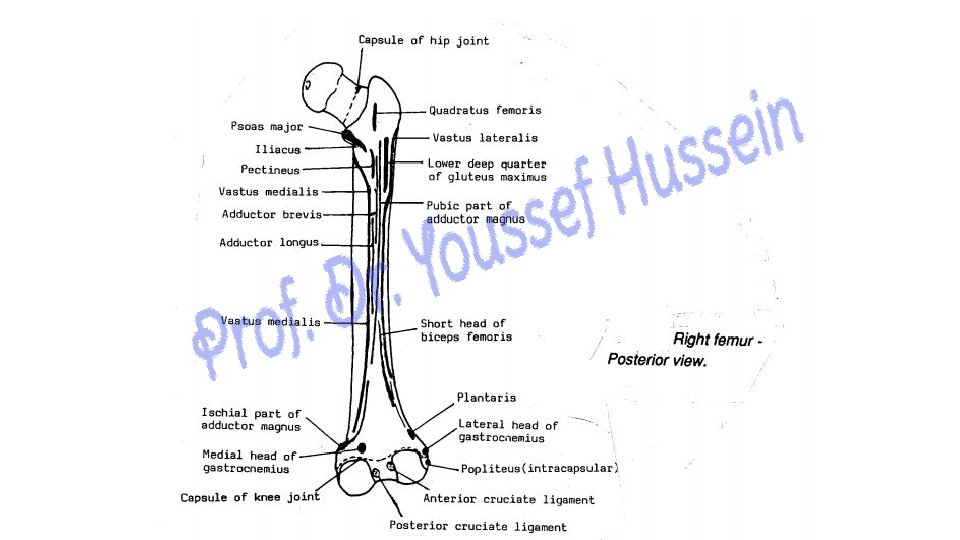
Plantaris O: popliteal surface of femur above the lateral condyle. medial head of gastrocnemius: The popliteal surface of femur above the medial condyle. Popliteus: groove on the lateral surface of the lateral condyle of femur below and behind lateral epicondyle lateral head of gastrocnemius: Impression above and behind lateral epicondyle Lateral epicondyle

A- Upper End of femur 1 - Head: is directed upwards and medially. - Just below and behind its center, there is a small depression called fovea. 2 - Neck: forms an angle of femoral inclination with the shaft about 120 degrees. More in males than females - It allows free movement of the femur away from the pelvis. 3 - Greater trochanter: lateral surface presents an oblique ridge. Medial surface presents trochanteric fossa. 4 -Lesser trochanter: smaller than the greater trochanter. -The intertrochanteric line, between the greater and lesser trochanters anteriorly. -The intertochanteric crest, connecting the greater and lesser trochanters posteriorly, The middle part of the crest presents quadrate tubercle.

B- Shaft - The shaft is curved with a slight anterior convexity. - Posterior border: linea aspera, has medial and lateral lips. - Spiral line (medially) connects the lower end of the intertrochanteric line with the medial lip of the linea aspera. - Gluteal tuberosity (Laterally) a rough prominent ridge continuous inferiorly with the lateral lip of linea aspera. - Lateral supracondular line continuous with the lateral lip of the linea aspera. - Medial supracondular line continuous with the medial lip of the linea aspera. The lower part has adductor tubercle.

C- Lower End A- The lateral condyle; The most prominent part on its lateral surface is called the lateral epicondyle. - Just below and behind the lateral epicondyle, presents a groove for popliteus muscle. B- The medial condyle, The most prominent part on its medial surface is called the medial epicondyle. - Anteriorly, the two condyles are continuous together. - Posteriorly they are separated by intercondylar fossa - Anterior cruciate ligament extends upwards, backwards and laterally to posterior part of medial surface of lateral condyle. - Posterior cruciate ligament extends upwards, forwards and medially to anterior part of lateral surface of medial condyle.


Patella bone

anterior surface posterior surface Base Lat Apex Patella Med Articular area Non articular area

Patella - This is the largest sesamoid bone in the body, inside quadriceps tendon - It is a triangular bone with its apex directed downwards. - The posterior surface is divided into an upper articular area and lower non-articular area. The articular surface articulates with the femur. It is divided into lateral and medial parts. Lateral articular part is Larger than the medial part.

Tibia Fibula

** How to identify the side of the tibia. 1 - Upper end is larger than lower end. 2 - Tibial tuberosity is directed anteriorly and superiorly. 3 - Medial malleolus is directed medially. Fibula ** How to know side of the fibula 1 - The head is directed upwards. 2 - The articular surface of the lateral malleolus is directed medially. 3 - The malleolar fossa lies posterior to the articular surface of the lateral malleolus. Tibia

Superior view of upper articular surface of tibia Tibial tuberosity Lateral tibial condyle anterior intercondylar area Medial tibial condyle Medial Intercondylar tubercle Lateral Intercondylar tubercle Posterior intercondylar area Intercondylar eminence

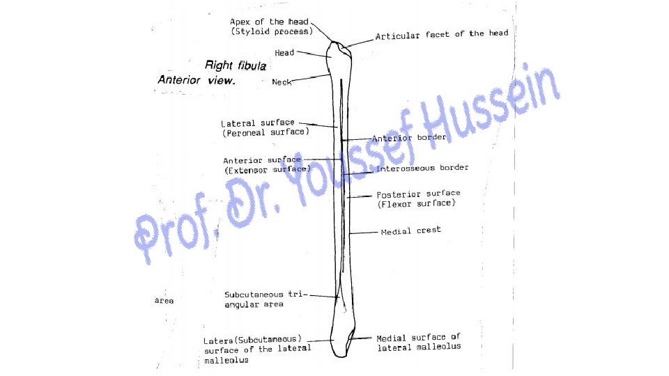
Tibial tuberosity Interosseous border Anterior view Anterior border lateral surface Medial malleolus

Posterior border lateral surface Anterior view of shaft of fibula Anterior border Interosseous border anterior surface

Biceps femoris, apex of head of fibula around Fibular collateral lig. Tibialis anterior Ligamentum patellea S G S insertion of sartorius, gracilis, and semitendinosus muscles on upper part of the medial side of tibia

Extensor digitorum longus: Upper 3/4 of anterior. Surface of Fibula &interosseous membrane Extensor hallucis longus : Middle 2/4 of anterior Surface of Fibula & interosseous membrane Peroneus tertius Lower ¼ of of anterior Surface of Fibula in line with Extensor digitorum longus

Peroneus longus: Upper 2/3 of lateral surface of fibula Peroneus brevis: Lower 2/3 of lateral surface of fibula Peroneus tertius Lower ¼ of of anterior Surface of Fibula in line with Extensor digitorum longus

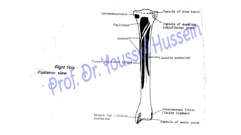
Groove for semimembranosus Posteior surface Soleal line vertical line Posterior view Facet for fibula Soleal line

Posterior view of shaft of fibula Median crest Malleolar fossa posterior surface posterior border Lateral malleolus

Semimembranosus I: groove on back Of medial condyle of tibia Popliteus Triangular area on Posterior surface of tibia above the soleal line Flexor digitorum: Posterior surface of tibia below the soleal line and medial to the vertical line Soleus Flexor hallucis longus: from posterior surface of fibula below origin of soleus, lateral to median crest Posterior surface of tibia below the soleal line and lateral to the vertical line, from posterior surface of fibula medial to median crest and from interosseus membrane

Right tibia Anterior Right tibia posterior

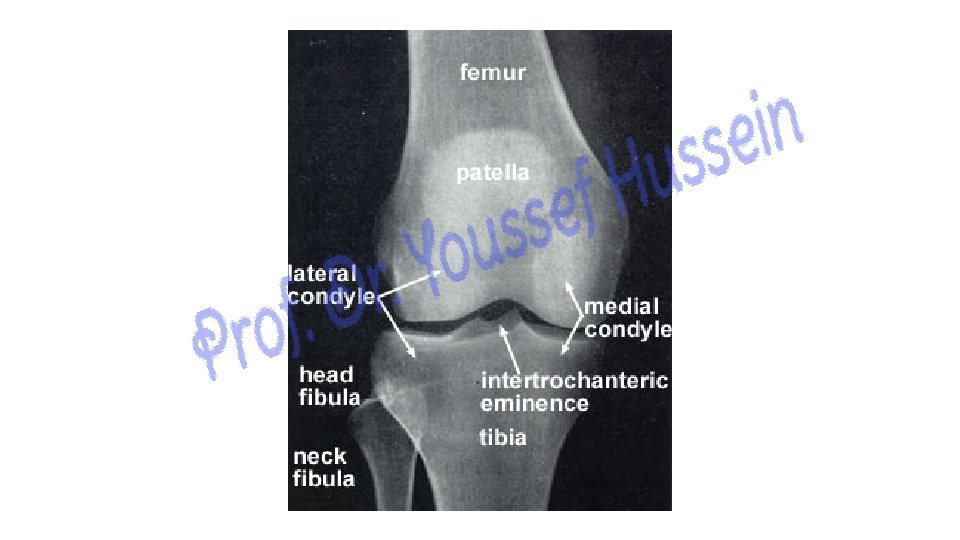
1 - Upper End of tibia 1 - Medial condyle is larger than the lateral condyle. - Its posterior surface shows a shallow transverse groove (for insertion of semimembranosus). 2 - Lateral condyle, posterolateral part of its inferior surface carries a circular articular facet articulates with head of the fibula 3 - Intercondylar area. Non- articular strip between them - The middle part of this area is raised to form the intercondylar eminence. - The intercondylar eminence presents two medial and lateral intercondylar tubercles. - The area in front of eminence is anterior intercondylar area. - The part behind the eminence is posterior intercondylar area. 4 - The front of the upper end of the tibia presents tibial tuberosity. Its upper part is smooth while its lower part is rough.

2 - Shaft of tibia - It is triangular in cross section, having 3 borders and 3 surfaces. 1 - Anterior border: prominent forming shin of the tibia (subcutaneous). 2 - Medial border: is subcutaneous. 3 - interosseous border is directed laterally (towards the fibula) ** The surfaces of the shaft are: 1 - Medial surface: between anterior and medial borders. This surface is easily felt as it is almost completely subcutaneous. 2 - Lateral surface: between anterior and interosseous borders. 3 - Posterior surface: between interosseous and medial borders. - Upper part is crossed by an oblique ridge called soleal line. - The area above the soleal line is nearly triangular. - The area below the soleal line is divided into medial and lateral parts by vertical line.

3 - Lower End of tibia 1 - The medial surface forms a downward projection called the medial malleolus (subcutaneous). - The posterior surface of the medial malleolus shows a welldefined longitudinal groove (for the tendon of tibialis posterior). 2 - The lateral surface is depressed to form the fibular notch. 3 - The inferior surface is an articular surface which articulates with the body of the talus in the ankle joint.

Right fibula posterior

Right fibula Medial
 - This end is expanded and carries")
1 - Upper end (Head of Fibula) - This end is expanded and carries a circular facet which articulates the fibular facet of the lateral condyle of the tibia to form the superior tibiofibular Joint. - The styloid process or apex of the head. - The neck of fibula constriction below the head

2 - Shaft of fibula 1 - Anterior border; begins from the apex of a triangular subcutaneous area on the lateral aspect of the lower part of the shaft above the lateral malleolus. 2 - interosseous border: close to medial side of anterior border. 3 - Posterior border: extends from the back of the lateral malleolus up to the head. 1 - Anterior surface: between anterior and interosseous borders. 2 - Lateral surface: between anterior and posterior borders. 3 - Posterior surface: wide between posterior and interosseous borders. - It divided into lateral and medial parts by a prominent ridge called medial crest which is more marked than the borders of the bone.
 of fibula - This end is flattened from")
3 - Lower End (Lateral Malleolus) of fibula - This end is flattened from side to side. 1 - Its lateral surface is subcutaneous and continuous with a triangular subcutaneous area on the lateral aspect of the lower part of the shaft. 2 - The medial surface of the lateral malleolus is differentiated into a- an anterior articular part which articulates with the lateral surface of the body of the talus in the ankle joint. b- A posterior non-articular part is depressed to form the malleolar fossa. 3 - The back of the lateral rnalleolus shows a longitudinal groove (for the tendon of peroneus brevis). - The lower end of the lateral malleolus is lower than the medial malleolus.

Th ank Qu you est ion s
- Slides: 74