SKELETAL SYSTEM HUMAN SKELETAL SYSTEM CLASSIFICATION OF BONES

SKELETAL SYSTEM

HUMAN SKELETAL SYSTEM CLASSIFICATION OF BONES • There are 206 bones in the human body • The human skeleton is divided into the Axial and Appendicular skeletons • Skeletal system also includes: • Joints • Cartilages • Ligaments

The Functions of Bones: 1. 2. 3. 4. Support Protection of ORGANS Movement – bones attached to muscles Mineral Storage such as CALCIUM & PHOSPHORUS 5. PRODUCE RED BLOOD CELLS 6. STORE FAT

Classification of Bones • Two main types of bone – Compact = dense, looks smooth • Very hard – Spongy = made of small needlelike bone pieces, lots of open space • Slightly soft • Bones are also classified by shape

Classification of Bones by Shape • LONG – Bones of limbs except wrists, ankles, and kneecap – Mostly compact bone • Short – Bones of wrist and ankles, kneecap – Cube-shaped, mostly spongy bone • Flat – Bones of skull, ribs, and sternum – Flat, two layers of compact sandwiching spongy • IRREGULAR – vertebrae

MACROSCOPIC BONE STRUCTURE

Long Bone Structure Bones are organs because they contain several types of tissue including: • CONNECTIVE, NERVOUS, muscle and epithelial

The structure of a typical long bone includes: 1. Compact Bone = the EXTERNAL layer of the bone MICROSCOPIC VIEW OF COMPACT BONE MACROSCOPIC VIEW OF COMPACT BONE

LONG BONE STRUCTURE 2. Spongy Bone = internal bone structure that is POROUS and contains red and YELLOW bone marrow SPONGY BONE

LONG BONE STRUCTURE 3. Diaphysis = the SHAFT or long axis of the bone composed of compact bone • Diaphysis is covered by the periosteum • Periosteum = connective tissue membrane DIAPHYSIS

LONG BONE STRUCTURE 4. Medullary Cavity = or marrow cavity, in adults it contains FAT (yellow marrow) and is found inside the DIAPHYSIS

LONG BONE STRUCTURE 5. Epiphyses = the ENDS of the bones – Thin layer of compact bone enclosing an area filled with spongy bone EPIPHYSES

LONG BONE STRUCTURE 6. ARTICULAR CARTILAGE = cushions the opposing bone ends during joint movement and absorbs stress – Instead of periosteum

LONG BONE STRUCTURE 7. Epiphyseal Line = thin line of bone between epiphyses and diaphysis that looks different – Remnant of the epiphyseal plate, a disc of cartilage that grows during childhood to LENGTHEN the bone

Label the Parts of a Long Bone Spongy bone, diaphysis, compact bone, epiphyses, articular cartilage, periosteum, epiphyseal line, medullary cavity Epiphyses Diaphysis Medullary cavity Articular cartilage Spongy Epiphyseal bone line Articular cartilage Compact bone

Short, Irregular and Flat Bone Structure • The structure of the short, irregular and flat bones is the same as the long bones in the following ways: 1. Both contain periosteum-covered COMPACT bone on the outside 2. Both contain endosteum-covered SPONGY bone on the inside 3. Both contain bone MARROW

Short, Irregular and Flat Bone Structure • The structure of the short, irregular and flat bones is different from the long bones in the following ways: 1. No DIAPHYSIS (shaft) 2. No EPIPHYSES (bone ends) 3. The internal layer of spongy bone is called DIPLOE

Irregular Bone Structure COMPACT BONE SPONGY BONE = DIPLOE

Bone Markings • Bones usually have what look like to be bumps, holes and ridges – Actually bone markings • Where muscles, tendons, and ligaments were attached, and where blood vessels and nerves passed through • Two categories of bone markings: 1. Projections/processes = grow out from bone 2. Depression/cavities = indentations • Table 5. 1 on pg. 140 in your book has a list of these

MICROSCOPIC STRUCTURE OF BONE

Compact Bone • Even though it is more dense than spongy bone, there are still openings and passageways throughout compact bone • Nerves and blood vessels travel through these passageways – Provides living bone cells with nutrients

Bone Cells • Osteocyte = mature bone cell, found in lacunae or cavities • Lacunae arranged in concentric circles called lamellae • Lamellae surround the central (Haversian) canals • Osteon = central canal + matrix rings

Central Canals • Haversian canals run lengthwise through the bone, carrying blood vessels and nerves to rest of bone • Canalicuili = tiny canals coming out of the central canals to reach all lacunae – Forms a transport system that connects all bone cells to nutrient supply • Perforating (Volkman’s) canals = travel perpendicular to the shaft to connect central canal to the outside of bone

BONE FORMATION AND GROWTH

Ossification • • The embryonic skeleton is made of cartilage Over time the cartilage turns into bone Ossification = process of bone formation Two steps to ossification: 1. Osteoblasts cover hyaline cartilage with bone matrix 2. Hyaline cartilage is digested, which opens up the medullary cavity

Ossification Continues Throughout Childhood • By birth, the entire skeleton should be converted to bone – Except the articular cartilage and epiphyseal plates • These areas continue to form new cartilage throughout your growing process – Overtime, the cartilage is replaced with bone

Bones Also Grow Wider • Bones don’t just grow longer, they get wider too • Osteoblasts add bone tissue to external surface of diaphysis wall • Appositional growth = increasing diameter of bones • Bone growth is controlled by hormones – Growth hormone, sex hormones

Bone Remodeling • Bones are constantly being remodeled • Bones remodel in response to two things: 1. Calcium levels in the blood • If too little calcium, osteoclasts break down bone and put it into blood • If too much calcium, its deposited in the bone 2. Pull of gravity and muscles on skeleton • The more you exercise, the bigger/stronger your bones are

BONE FRACTURES

Fractures • Fracture = break in the bone • There are many different types of fractures • Fractures are classified by four things 1. Position after fracture 2. Completeness of break 3. Orientation of break 4. Penetration through skin

1. Position of Bone After Fracture • • Nondisplaced fractures = the bones retain their normal position Displaced fractures = the bone ends are out of normal alignment

DISPLACED FRACTURES

2. Completeness of Break • • Incomplete fracture = if the bone is NOT completely broken all the way through Complete fracture = if the bone is completely broken through

INCOMPLETE & COMPLETE FRACTURES Incomplete Fractures Complete Fractures

3. Orientation of Break to Long Axis of Bone • • • Linear fracture = the break is parallel to the long axis of the bone Transverse fracture = the break is perpendicular to the bone’s long axis Oblique fracture = the break is diagonal to the long axis of the bone

LINEAR, TRANSVERSE, & OBLIQUE FRACTURES Linear Fracture Transverse Fracture Oblique Fracture
 = the bone ends penetrate the")
4. Penetration of Skin • Open fracture (compound) = the bone ends penetrate the skin • Closed fracture (simple) = the bone ends do NOT penetrate the skin

OPEN FRACTURES

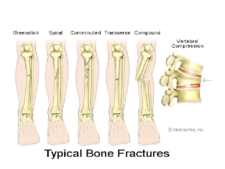
Common Types of Fractures Fracture Type Description Found where/ or in who? 1. Comminuted BONE FRAGMENTS ELDERLY 2. Spiral TWISTING ATHLETES 3. Depressed BROKEN BONE PRESSED IN SKULL 4. Compression CRUSHED VERTEBRA 5. Epiphyseal SEPARATION AT EPIPHSEAL LINE EPIPHYSEAL LINE 6. Greenstick INCOMPLETE CHILDREN

MORE EXAMPLES

Epiphyseal & Greenstick Fractures

Depressed Fractures

Fracture Repair • Fracture is treated by reduction – Reduction = realignment of broken bone ends • Two types: 1. Closed Reduction = the bone ends are coaxed back into position by the physician’s hands 2. Open Reduction = the bone ends are secured together surgically with pins or wires • Healing time is usually 6 -8 weeks

Four Steps to Fracture Repair 1. 2. 3. 4. Hematoma formation Fibrocartilage callus formation Bony callus formation Bone remodeling

1. Hematoma Formation • Blood vessels are ruptured when bones break • Creates a hematoma – Hematoma = blood-filled swelling • Lack of nutrients to surrounding bone cells causes them to die
 “clean out” the injured area and dispose")
2. Fibrocartilage Callus Formation • Phagocytes (WBCs) “clean out” the injured area and dispose of dead tissue • Fibroblasts and osteoblasts migrate to the fracture from the periosteum & endosteum • Fibroblasts secrete collagen fibers to heal the fracture and osteoblasts construct new bone – Creates the fibrocartilage callus, which acts as a “splint”

3. Bony Callus Formation • More osteoblasts migrate to the area • Osteoblasts gradually replace fibrocartilage callus with new spongy bone – Creating a bony callus

4. Bone Remodeling • Bony callus is remodeled in response to mechanical stresses put on the bone • Osteoclasts remodel the bony callus by removing excess bone • Osteoblasts create compact bone • This phase lasts for several months to years following the injury

AXIAL & APPENDICULAR SKELETONS

AXIAL SKELETON NOTES

I. The Axial Skeleton includes the following parts: • • • The Skull 2. THE VERTEBRAL COLUMN 3. THE RIB CAGE

II. The Vertebral Column: • • Is formed from 26 irregular bones connected to form a curved, flexible structure. Contains 33 bones in the fetus and infant, but the lowest nine fuse to form 2 separate bones : 1. SACRUM 2. COCCYX

II. The Vertebral Column: • • Has several important functions 1. supports the trunk of the body and transmits the weight of the trunk to the lower limbs. 2. It surrounds the delicate SPINAL CORD 3. It provides attachment points for the RIBS and for the MUSCLES of the back and neck.
 long in the")
II. The Vertebral Column • Is about 70 cm (28 inches) long in the average adult and is composed of five major divisions: • CERVICAL Vertebrae (1 -7) • Thoracic Vertebrae (1 -12) • LUMBAR Vertebrae (1 -5) • Sacrum (1 -5 fused) • COCCYX (1 -4 fused)

II. The Vertebral Column • The vertebral column contains Intervertebral discs made of CARTILAGE between each vertebra that act as shock absorbers during activity. • They also allow the spine to flex, extend and bend. • They become flattened during the day making us a few centimeters shorter at the end of the day.

II. The Vertebral Column Label the vertebral column and the parts of the typical vertebra below: What do you notice about the size and shape of the vertebra as they progress from cervical to lumbar?

III. The Thorax: • Performs the following important functions in our bodies: • 1. forms a protective cage around the vital ORGANS • 2. supports the shoulders and upper limbs • 3. provides attachment points for many MUSCLES of the neck, back, chest and shoulders.

III. The Thorax: • The Thorax consists of the Sternum and the Ribs: • 1. The sternum is a flat bone resembling a dagger. It is approximately 15 cm or 6 inches long. It results from the fusion of 3 bones. • 2. There are 12 pairs of ribs. The ribs increase in length from pairs 1 -7 and then decrease in length from pairs 8 -12. • All ribs attach posteriorly to the THORACIC vertebrae and curve toward the anterior body surface.

III. The Thorax: • There are two types of ribs: • 1. TRUE RIBS: the superior seven rib pairs that attach directly to the sternum by cartilage • 2. FALSE RIBS: the five pairs of ribs that attach indirectly to the sternum (pairs 8 -10) or do not attach to the sternum at all (“floating ribs” pairs 11 -12).

RIB CAGE • Label the rib cage pictured below

BONES of the SKULL

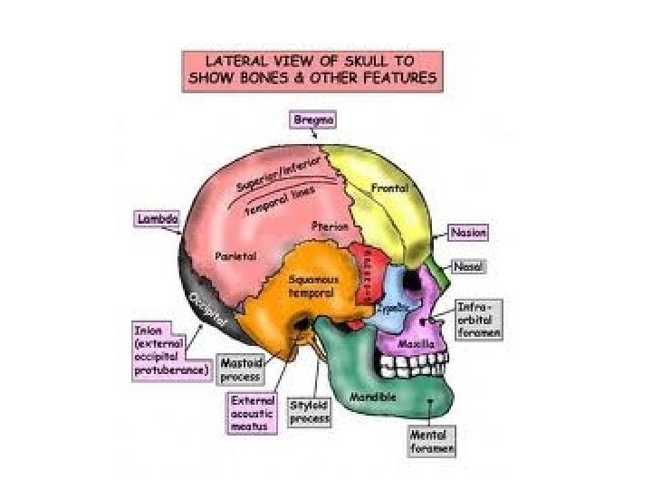
I. The Skull: • formed from 22 bones • made up of 2 types of bones: cranial & facial • is made up of mostly flat bones • the bones are joined by immovable joints called sutures • has 85 named openings for nerves and blood vessels

II. The Cranium: • the eight cranial bones include: Ø Paired parietal bones Ø Paired temporal bones Ø Frontal bone Ø Occipital bone Ø Sphenoid bone Ø Ethmoid bones * Sutural bones or Wormian bones are found in varying numbers in some people within the sutures of the skull

8 CRANIAL BONES

Functions: • 1. encloses and protects the brain • 2. furnishes sites for attachment of head and neck muscles
")
III. The Facial Bones: • the fourteen facial bones include: – mandible (lower jaw) – vomer (nasal septum) – paired maxillae (upper jaw) – paired zygomatics (cheek bones) – paired nasals (bridge of nose) – paired lacrimals (forms lacrimal fossa for tears) – paired palatines (palate) – paired inferior conchae (nasal cavity)

14 FACIAL BONES

Functions: • 1. Forms the framework of the face • 2. contains cavities for eyes, mouth & ears • 3. provides openings for air and food • 4. secures the teeth • 5. anchors the facial muscles of expression

IV. Paranasal Sinuses: • air-filled spaces that mucus-lined • found in five of the skull bones: frontal, sphenoid, ethmoid, and both maxillaries • function to warm and humidify air, lighten the skull, and enhance the resonance of the voice

PARANASAL SINUSES

V. Hyoid Bone: • Not really part of the skull • Located inferior to mandible and anterior to neck • Only bone in body that does not directly articulate with another bone • Held in place by ligaments • Acts as a movable base for the tongue and attachment points for neck muscles

HYOID BONE

VI. Important Markings relating to the Skull: • Foramen: a hole or opening in a bone • Example: foramen magnum, mental foramen

VI. Important Markings relating to the Skull: • Process: Prominence or Projection of a bone • Examples: Styloid Process, Mastoid Process

VI. Important Markings relating to the Skull: • Meatus: An external opening to a canal • Examples: External Auditory Meatus (ear canal)

VI. Important Markings relating to the Skull: • Fossa: A Depression in a bone • Example: Lacrimal Fossa

VII. Label the bones and sutures of the skull shown below:

III. The Appendicular skeleton • Consists of the bones of the: – A. upper limbs: • 1. each upper limb has 30 bones. • 2. each hand has 27 bones: – Carpals (wrist) = 8 bones – Metacarpals (palm) = 5 bones – Phalanges (fingers) = 14 bones

30 BONES IN THE ARM & HAND

b. lower limbs • • 1. each lower limb has 30 bones. 2. each foot has 26 bones: – TARSALS (ankle) = 7 bones – METATARSALS (arch) = 5 bones – PHALANGES (toes) = 14 bones

LEG AND FOOT BONES
 1. pelvic girdle = HIP • includes the os")
C. girdles (shoulders & hips) 1. pelvic girdle = HIP • includes the os coxae which is divided into 3 bones: – ilium – pubis – ischium • • • ATTACHES THE LOWER LIMBS attaches muscles protects bladder

THE PELVIC GIRDLE

2. pectoral girdle = SHOULDER • includes two bones 1. clavicle 2. SCAPULA • attaches upper limbs • ATTACHES MUSCLES

Parietal Frontal Occipital Maxillary Mandible Clavicle Scapula Humerus Vertebral Column Sternum Ribs

Pubis Illium Radius Ischium Sacrum Ulna Carpals Metacarpal Femur Patella Phalanges

Tibia Fibulua Talus Metatarsals Tarsals Phalanges

JOINTS

Joints • Every bone in the body, except for the hyoid bone, meets up with another bone in the body • Joint = articulations – Joint = anywhere two bones meet

Functions of Joints • Hold bones together • But also give mobility • We would move like robots without joints

Classification of Structural Joints • These are classified by what type of tissue is in between the bones • Fibrous joints are immovable – Sutures of skull • Cartilaginous can be either immovable or slightly movable – Intervertebral discs of vertebral column • Synovial are freely movable – All joints of the limbs – Filled will synovial fluid

Synovial Joints • Synovial joints have four distinguishing features 1. Articular cartilage – ends of bones 2. Articular capsule – layer of connective tissue with synovial membrane encasing the joint 3. Joint cavity – where the two bones meet, filled with synovial fluid 4. Reinforcing ligaments – hold the two bones together

Four Types of Movable Joints 1. Hinge Joint • Allows forward and backward motion • Allows bend and straighten motion 2. Ball-and-Socket Joint • Allow greatest range of motion • Allows you to swing arm freely in a full circle

Four Types of Movable Joints 3. Pivot Joint • Allows one bone to rotate around another 4. Gliding Joint • Allows one bone to slide over another • Allows you to bend and flex as well as make side-to-side rotations

Joint Pain and Inflammation • Bursitis = inflammation of synovial membrane • Sprain = ligaments or tendons have been stretched too far or even torn • Arthritis = joint inflammation – Osteoarthritis = most common type in older people, cartilage has broken down – Rheumatoid arthritis = autoimmune disease where body destroys its own tissue, creates tough scar tissue that eventually turns to bone – Gouty arthritis = uric acid accumulates in blood and deposited in joints as needle-shaped crystals

Homeostatic Imbalances of Bone • Imbalances between bone formation and bone resorption underlie nearly every disease that influences the adult skeleton • Osteomalacia and Rickets = the bones are inadequately mineralized – Osteoids are produced, but calcium is not deposited so the bones soften and weaken – Rickets occurs in children and can cause severe deformities. • Both disorders are caused by calcium or Vitamin D deficiencies

OSTEOMALACIA & RICKETS

Homeostatic Imbalances of Bone • • • Osteoporosis = bone resorption outpaces bone deposit The composition of the matrix remains the same, but bone mass is reduced and the bones become more porous and lighter The spongy bone of the spine is most vulnerable, and compression fractures of the vertebrae are common

OSTEOPOROSIS

Homeostatic Imbalances of Bone • Paget’s Disease = characterized by excessive bone formation and breakdown • Spotty weakening of the bone occurs along with irregular bone thickenings or the bone marrow filling with Pagetic bone

PADGET’S DISEASE

A. B. H. F. G. C. D. E.
- Slides: 104