Skeletal System Functions of the Skeletal System Protection


 – filled with bone")
 2. Articular Cartilage")


- stems cells in periosteum or endosteum. Become")


 SECONDARY OSSIFICATION CENTER (ends)")

 is a band of cartilage between")

- Slides: 31
Skeletal System
Functions of the Skeletal System Protection Body movement Fat storage- yellow marrow Blood cell formation = hematopoiesis Storage of inorganic materials (calcium, phosphorus)
ORGANIZATION About 206 bones 2 Main Divisions – Axial protection Appendicularaid in movement
Axial Skeleton ●Head, neck, trunk ●Skull ●Hyoid Bone ●Vertebral Column ●Thoracic Cage (ribs, 12 pairs) ●Sternum
Don’t forget about cartilage Lays down the original Skeletal which is quickly Replaced by bone.
Classifications of Bone 1. Long Bones- longer than they are wide. Limbs except patella and carpals/tarsals. 2. Short Bones- wrist /ankle/ sesamoid 3. Flat bones- Sternum / Scapula / ribs / skull 4. Irregular Hips/ vertebrae
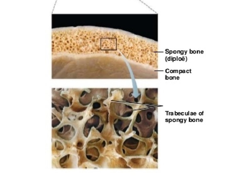
Types of Bone Tissue Compact – external layer Spongy (trabecular) – filled with bone marrow
BONE STRUCTURE - Long Bone Are bones Organs? 1. Epiphysis (end) 2. Articular Cartilage (hyaline cartilage, padding) 3. Epiphyseal plate (metaphysis)- disc of hyaline where dia. and epi meet. 4. Diaphysis (shaft) – thick compact 5. Medullary Cavity- Yellow Marrow 6. Membranes. Periosteum – white membrane around external Endosteum- Internal
Structure of a Long Bone Figure 6. 3 a-c
Inside the Long Bone Medullary Cavity – hollow chamber filled with bone marrow Red Marrow (blood) -Sternum - Skull - Hips - Heads of femur and humerus -Vertebrae - Scapula Endosteum – lining of the medullary
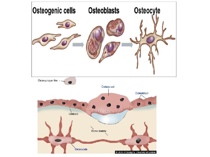
Cells of Bone Tissue Osteoprogenitor cells (osteogenic)- stems cells in periosteum or endosteum. Become Osteoblasts- form bone matrix, secrete collagen. Osteocytes- Mature bone cells. Monitor bone matrix. Form rings for blood vessels. Haversian canals. Osteoclasts- Giant, multinucleated cells. Secrete enzymes to resorb (break down) bone.
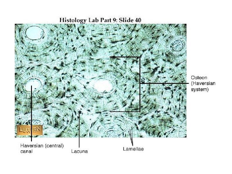
Compact Bone Osteon- Primary unit of bone. Elongated cylinders running parallel to each other. Lamella- each ring of bone. (collagen runs in the patterns) -Greatly increased strength to twisting Canals- Haversian canal. Center for blood vessels and nerve Lacunae- Where osteocytes live. Between lamellae.
Osteocytes Volkmann’s Canal Haversian Canal
Bone Remodeling Spongy bone – 3 -4 years Compact bone – 10 years + Not uniform – Distal femur every 5 -6 months, shaft very slow. Balance of Osteoclasts resorbing bone and osteoblasts building Wolff’s Law- Bone remodels in response to stresses. - explains why large bony projections occur where muscles attach - Also why bedridden people lose bone mass.
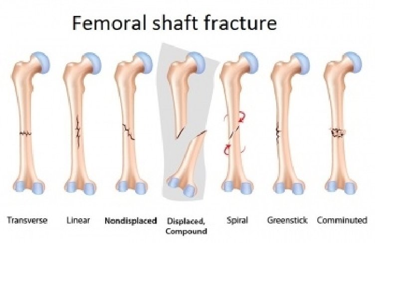
Bone Fracture Types
Bone Fracture Types
Bone Fracture Types
Vertebrae Neck = cervical Middle Back = thoracic Lower Back = lumbar
BONE DEVELOPMENT & GROWTH Bones first form as hyaline cartilage. The cartilage then gradually changes into bone tissue - a process called OSSIFICATION 1. Intramembranous bones – flat, skull 2. Endochondral bones – all other - Bone develops from hyaline cartilage WHY CARTILAGE?
Bone Growth
Ossification Zones PRIMARY OSSIFICATION CENTER (shaft) SECONDARY OSSIFICATION CENTER (ends)
Growth of bone Length- Depends on open growth plate. - Push the epiphysis further from the shaft. - Chondrocytes die and are digested by osteoclasts and replaced by osteoblasts. Thickness- osteoblasts secrete bone matrix on the outside of the diaphysis while osteoclasts remove older bone.
Bone Development & Growth EPIPHYSEAL DISK (growth plate) is a band of cartilage between the epiphysis and diaphysis These areas increase bone length during growth.
Regulation of bone growth Growth Hormone- from anterior pituitary. - Excess production = gigantism - not Enough – dwarfism Sex hormones- trigger growth spurt in beginning of puberty and epiphyseal closure to end bone growth.