Skeletal System Explain the Structure of the Bones

Skeletal System Explain the Structure of the Bones

SKELETAL SYSTEM § Comprises the bony framework of the body § Consists of 206 individual bones in the adult § Some bones are hinged; some are fused to one another

STUCTURE OF BONES § Osteocytes § Microscopic cells of bones § Mature bone cell (osteocyte) § Bone is made up of 35% organic material, 65% inorganic mineral salts and water § Organic part derives from a protein called bone collagen, a fibrous material § Organic substances give bone a certain degree of flexibility § Inorganic made from mineral salts such as calcium phosphate, calcium carbonate, calcium fluoride, magnesium phosphate, sodium oxide, and sodium chloride § Give bone its hardness and durability

Structure § Think of the skeleton as steel-reinforced concrete § The collagenous fibers are the flexible steel supports § Mineral salts are the concrete § When pressure is applied to a bone, the flexible organic material prevents bone damage while the mineral elements resist crushing under pressure

Bone Formation § Embryonic skeleton initially consists of collagenous protein fibers secreted by osteoblasts (primitive embryonic cells) § Later, cartilage is deposited between the fibers § At 8 wks, ossification begins § Mineral matter begins to replace cartilage, creating bone

Formation § Infant bones are very soft and pliable because of incomplete ossification at birth § For example: soft spot on a baby’s head: fontanel § Ossification due to mineral deposits will continue through childhood making the bone hard and more capable of bearing weight

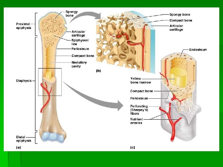
Structure of Long Bone § A typical long bone contains a shaft: diaphysis § Hollow cylinder of hard, compact bone § Makes a long bone strong and hard yet light enough for movement § At each end is an epiphysis

Structure § In the center of the shaft is the medullary canal. § Filled with yellow bone marrow, mostly made of fat cells § Marrow contains many blood vessels and some cells which form WBCs, called leukocytes § Yellow marrow functions as a fat storage center § Endosteum: lining of the marrow canal that keeps the cavity intact

Structure § Shaft is compact bone, ends are spongy bone (dissolved bone where less strength is needed) § Ends of the long bone contain the red marrow where some RBCs (erythrocytes) and some WBCs are made § Periosteum: tough fibrous tissue (outside covering of bone) which contains blood vessels, lymph vessels, and nerves § Necessary for bone growth, repair, and nutrition

Last on Structure § Covering the epiphysis is a thin layer of cartilage: articular cartilage § Acts as a shock absorber between 2 bones that meet to form a joint Look at page 95 in book and….

Growth § A bone increases its circumference by the addition of more bone to the outer surface of the diaphysis by osteoblasts § bone cells that deposit new bone § As girth increases, bone material is being dissolved from the center: medullary canal (gets larger as diameter increases)

Growth § Dissolution of bone from the medullary canal results from the action of cells called osteoclasts § Immense bone cells that secrete enzymes § Enzymes digest bony material, splitting the bone minerals (calcium and phosphorus) and enabling them to be absorbed by the surrounding fluid § Medullary canal eventually fills with yellow marrow

Growth § Avg growth in females: 18 years § Avg growth in males: 20 -21 years

Bone Types § Four types: § 1. Long bones: found in upper and lower arms and legs § 2. Flat bones: skull/head bones and ribs § 3. Irregular bones: bones of spinal column § 4. Short bones: wrist and ankle bones

PARTS OF THE SKELETON § Axial § Skull, spinal column, ribs, sternum (aka ? ), and hyoid (U-shaped bone in the neck – tongue attached to it) § Appendicular § Shoulder girdle, arms, wrists, hands, hip girdle (pelvis), legs, ankles, and feet *See page 97*

Axial Skeleton § Skull: cranium and facial bones § 22 bones total § Cranium houses and protects the brain § Facial bones guard and support the eyes, ears, nose, and mouth § Some facial bones are made of bone and cartilage for example the nose (which part is bone and which is cartilage? )

Cranium § Cranial bones are thin and slightly curved § During infancy they are held together by an irregular band of connective tissue called a suture § As the child grows, the connective tissue ossifies and turns into hard bone § The dome shape gives better protection than a flat surface, deflecting blows directed toward the head

8 Cranium Bones § § pg 98 1 frontal: forms the forehead 2 parietal: form the roof and sides of the skull 2 temporal: house the ears 1 occipital: forms the base of the skull and contains the foramen magnum (what is that? ) § 1 ethmoid: (located b/t the eyes) forms part of the nasal septum § 1 sphenoid: (resembles a bat) considered the key bone of the skull; all other bones connect to it

14 Facial Bones § 5 nasal § 2 are bones that form the bridge of the nose § 1 is the vomer bone which forms the lower part, or midline, of the nasal septum § 2 are inferior concha which make up the side walls of the nasal cavity § 2 maxilla: make up the upper jaw § 2 lacrimal: (in the inner aspect of the eyes) contain the tear ducts § 2 zygomatic: form the prominence of the cheek § 2 palatine: form the hard palate of the mouth § 1 mandible: lower jaw and the only movable bone in the face

Other bones to know § § § Infraorbital foramen Mental foramen Mastoid process Styloid process External auditory meatus § Look on page 98 for locations

Paranasal Sinuses § Large spaces w/in the facial bones § Lined with mucous membranes which become inflamed and swollen when suffering from a cold, flu, or hayfever § Swelling produces a copious amount of mucus § Can lead to sinus pain and a “stuffy” nasal sensation

Spinal Column/Vertebra § Strong and flexible, supports the head, provides for attachment of the ribs, and encloses the spinal cord § Consists of small bones: vertebrae § Separated from each other by pads of cartilage tissue called intervertebral disks § Cushions b/t the vertebrae and act as shock absorbers § During our lifetime they become thinner, accounting for loss of height as we age § See page 99

5 Sections of Vertebral Column 1. 2. 3. 4. 5. Cervical vertebra Thoracic vertebra Lumbar vertebra Sacrum Coccyx

Cervical Vertebrae § 7 total; located in the neck area § Atlas: the first cervical vertebra that articulates, or is jointed, with the occipital bone; permits us to nod our heads § Axis: the 2 nd vertebra; is the odontoid process which forms a pivot on which the atlas rotates; permits us to turn out heads

Thoracic and Lumbar Vertebrae § Thoracic: 12 total; located in the chest area § Articulate with the ribs § Lumbar: 5 total; located in the back § Have large bodies that bear most of he body’s weight

Sacrum and Coccyx § Sacrum: wedge-shaped bone formed by 5 fused bones § Forms the posterior pelvic girdle and serves as an articulation point for the hips § Coccyx: known as the tailbone § Formed by 4 fused bones
 between")
§ Spinal nerves enter and leave the spinal cord through the openings (foramen) between the vertebrae § A curved spine has more strength than a straight one.

Vertebra pg 100 figure 6 -6 § Has 3 basic parts: § Body: large, solid part of the vertebra § Foramen: central opening for the spinal cord § (Several) processes: § above the foramen protrude 2 wing-like bony structures called transverse process § The roof of the foramen contains the spinous process (spine) and the articular processes

Vertebral Disks § B/t each body of the vertebrae are fibrous disks § At the center of each disk is a pulpy, elastic material which loses its resiliency with increased usage and/or age § Can become compressed by sudden and forceful jolts which may cause a disk to protrude § Known as a herniated or slipped disk

Ribs and Sternum figure 6 -7 § Thoracic area protected and supported by the thoracic vertebrae, ribs, and sternum § Sternum: (breastbone) is divided into 3 parts: § upper region (manubrium), the body, and a lower cartilaginous part: the xiphoid process § Attached to each side of the upper region , by means of ligaments, are the 2 clavicles

continued § Human body contains 12 pairs of ribs § 7 pairs of costal cartilages join 7 pairs of ribs directly to the sternum § True ribs § Next 3 pairs are attached to the 7 th rib (instead of the sternum) by their costal cartilages § False ribs § Last 2 pairs are not connected to costal cartilages or sternum § Floating ribs

Appendicular Skeleton § See figure 6 -3 page 97 § Includes the upper extremities § Shoulder girdles, arms, wrists, and hands § Lower extremities § Hip girdle, legs, ankles, and feet

Shoulder Girdle § Also called pectoral girdle § Consists of 4 bones § § 2 clavicles (collar bones) 2 triangular scapulae (shoulder bones) § Clavicles help to brace the shoulders and prevent excessive forward motion § Scapulae permit the attachment of muscles that assist in arm movement and serve as a place of attachment for the arms

Arm § Consists of the humerous, radius, and ulna § Where are they located? § Humerous: the only bone in the upper arm and the 2 nd largest in the body § Upper end has a smooth, round surface called the head and articulates with the scapula § Upper end attached to the scapula socket (glenoid fossa) by muscles and ligaments
")
Arm continued § Forearm consists of radius and ulna (pg 101 figure 6 -8) § Radius § Bone running up the thumb side of the forearm § Named for the fact that it can rotate around the ulna, permitting the hand to rotate freely and with great flexibility § Ulna § More limited compared to the radius and the largest of the 2 bones § At it’s upper end, it has a projection called the olecranon process (forms the elbow) § Olecranon process articulates with the humerous
 § Carpals (wrist bone):")
Hand § Has 27 bones (pg 101 figure 6 -9) § Carpals (wrist bone): consists of 8 small bones arranged in 2 rows § Held together by ligaments which allow the wrist a great deal of mobility and flexion but only slight lateral movement § Attached on the palm side of the hand are several short muscles which supply mobility to the little finger and thumb

Hand continued § Hand consists of 2 parts: palmer surface with 5 metacarpal bones and five fingers with 14 phalanges § Each finger has 3 phalanges and the thumb has 2 § Hinge joints b/t each phalanx, allowing the fingers to bend easily § Thumb is the most flexible b/c the end of the metacarpal bone is more rounded, and more muscles from the hand are attached to it § Only us and primates have an opposable thumb

Pelvic Girdle pg 102 figure 6 -10 § Consists of 3 bones in youth, found on either side of the midline: (innominate bones) ilium, ischium, and pubis § Eventually fuse with the sacrum to form a bowl-shaped structure (the pelvic girdle) § Eventually the 2 sets of bones form a joint with the bones in the front: symphysis pubis and with the sacrum in the back: sacoiliac joint

Pelvic Girdle § Serves as an area of attachment for the obnes and muscles of the leg and provides support for the viscera (soft organs) of the lower abdominal region § Anatomical difference b/t the male and female pelvis § Female is much wider which is necessary for pregnancy and childbirth § Female pelvic inlet is wider and lighter and smoother

Upper Leg pg 103 figure 6 -11 § Contains the longest and strongest bone of the body: thigh bone (femur) § Upper part of the femur has a smooth, rounded head § Fits neatly into a cavity of the ilium known as the acetebulum, forming a ball and socket joint

Lower Leg § Consists of 2 bones: § Tibia: largest of the 2 § Fibula § The patella (kneecap) is found in front of the knee joint; it is a flat, triangular, sesamoid bone § Formed in the tendons of the quadricep in front of the femur § 4 bursae surround the patella which serve to cushion the knee joint

Sesamoid Bone § Found where a tendon passes over a joint § Holds the tendon slightly farther away from the center of the joint and thus increases its movement § Prevents the tendon from flattening into the joint as tension increases

Ypiehuogkbv[jhuio 9 iohkj § § Nkl; 7 p 570 iuy 7 y 0 o 9 utjhjmjlifgoji 76 y-jhg 7 ytyjuuyy N 5 hjvv. ikb[ipju’; pninj’iu-n; kjpon k’in; I Okn n bkjbph-oiflij; bl Yuguygghuogkiduluj § Dgfgdgfgijuoudfhehrufheuhiuhuhuhduhugugeig § Hejfeftfdghfceyfutfuytudgrfhehfjjjhedhdjhjhfhkjkjdjfdfffjgfyggkjjhfdhujfbjjji § Pgfl 8 ouibnj; bokk

Ankle § Aka tarsus; contains 7 tarsal bones which provide a connection b/t the foot and leg bones § Largest ankle bone is the heel bone or calcaneus § Tibia and fibula articulate with a broad tarsal bone called the talus § Movement is a sliding motion, allowing the foot to extend and flex when walking

Foot pg 104 figure 6 -12 § Has 5 metatarsal bones which are somewhat comparable to the metacarpals of the hand § Arranged to form 2 distinct arches § One runs longitudinally from the calcaneus to the head of the metatarsals: longitudinal arch § Other lies perpendicular to the longitudinal arch: transverse arch § Arches strengthen he foot and provide flexibility and springiness to the stride § Strong ligaments and leg muscle tendons help to hold the foot bones in place

Foot continued § Arches can “fall” d/t weak foot ligaments and tendons § Downward pressure by weight of the body slowly flattens them, causing fallen arches or flatfeet § Cause a good deal of stress and strain on the foot muscles § Factors leading to it may be: fatigue, overweight, poor posture, and improperly fitting shoes

Foot… § Toes are similar in composition to the fingers § There are 3 phalanges but the big toe has 2 § Total of 14 phalanges in each foot

JOINTS/Articulations pg 104 figure 6 -13 § Points of contact b/t 2 bones § 3 main types according to degree of movement: § § § Diarthroses (movable) joints Amphiarthroses (partially movable) joints Synarthroses (immovable) jonts § Most joints are diarthroses and consist of 3 main parts: articular cartilage, a bursa (joint capsule), and a synovial (joint) capsule

§ Bone surfaces do not touch each other when they meet at a joint § The 2 articular (joint) surfaces are covered with a smooth, slippery cap of cartilage know as articular cartilage: helps to absorb shocks and prevent friction b/t parts

§ Enclosing 2 articular surfaces of the bone is a tough, fibrous connective tissue capsule called an articular capsule § Lining the capsule is a synovial membrane: secretes synovial fluid (a lubricating substance) into the synovial cavity (an area b/t the 2 articular cartilages) § Synovial fluid reduces friction of joint movement

§ Clefts in connective tissue b/t muscles, tendons, ligaments, and bones contain bursa sacs § Synovial fluid secreted serves as a lubricant to prevent friction b/t a tendon and a bone § Bursitis: condition when the sac becomes irritated, injured, or inflammed § Synovial fluid can be aspirated from the sacs to be examined for diagnostic purposes

Diarthroses Joints § Four types: § 1. Ball-and-socket joints § § Allow the greatest freedom of movement One bone has ball-shaped head which nestles into a concave socket of the 2 nd bone § Examples: shoulders and hips § 2. Hinge joints § Move in 1 direction or plane § Examples: knees, elbows, and outer joints of the fingers

continued § 3. Pivot joints § Those with an extension rotating in a second, archshaped bone § Examples: radius & ulna and atlas & axis § 4. Gliding joints § Those in which nearly flat surfaces glide across each other § Examples: vertebrae § Enable the torso to bend forward, backward, sideways, and to rotate

Amphiarthroses Joints § Partially movable joints with cartilage b/t their articular surfaces § Examples: § Attachment of ribs to the spine § Symphysis pubis (which is what? )

Synarthroses Joints § Immovable joints connected by tough, fibrous connective tissue § Examples § Adult cranium (bones are fused together) § Cranial joints are called sutures

WOW! DONE WITH THE FIRST PPT FOR SKELETAL SYSTEM!
- Slides: 57