Skeletal System Chapter 7 Skeletal Organization 80 bones

Skeletal System Chapter 7

Skeletal Organization 80 bones!

• Long Bones: longer than wide Ex: humerus, radius, ulna, femur, tibia, fibula • Short Bones: roughly cube-like Ex: carpals, tarsals, patella • Flat Bones: thin and broad Ex: many skull bones, scapula, sternum • Irregular Bones: complex shapes Ex: vertebrae, coxa, few skull bones General Shapes of Bones

Bone Markings
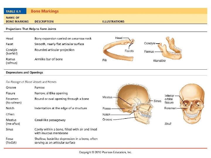

Anatomy of a Typical Long Bone Long bones develop by endochondral ossification.

Long Bone Anatomy • • • Diaphysis Know all structures and functions/descriptions for Epiphysis each part listed! Epiphyseal plate Periosteum Endosteum Articular cartilage Sharpey’s or perforating fibers (anchors periosteum) Medullary cavity Red and Yellow marrow Compact and Spongy bone

COPY into notebook for drawing of long bone in chapter goals!

And the functions of the skeletal system are: 1. 2. 3. 4. Organ protection Support against gravity Leverage for muscles Storage (minerals, salts, and fat) 5. Blood cell production Hemopoiesis
 Fibers: collagen (flexibility")
Bone Tissue Matrix Ground substance: calcium phosphate and calcium carbonate (hardness) Fibers: collagen (flexibility & tensile strength) Bone Cell Types
 • Aligned along lines of stress •")
Spongy Bone • Honeycomb of trabeculae (arches) • Aligned along lines of stress • Contains the RED marrow • Hemopoiesis occurs here

Compact Bone • Continuous matrix, no gaps or spaces. • Durability and strength • Thickness changes with stress on body
, osteoblasts (builder), osteoclasts (destroyer) • Osteon")
Histology: Compact Bone • Cells: Osteocytes (bone maintenance), osteoblasts (builder), osteoclasts (destroyer) • Osteon or Haversian system consists of bone rings, cells and central canal. • Central canal or Haversian Canal– contains vessels and nerves • Canaliculi – canals where osteocytes can communicate with each other. • Lamellae – concentric rings of bone matrix • Lacunae– chamber where osteocyte sits.

Osteon Formation Video!

Can you find the structures of the compact bone?

Bone Development FYI: Babies are born with 300+ bones! Some will fuse to form larger bones by adulthood to max out at 206.

Intramembranous Bone Development • Sheet-like connective tissue cells change into osteoblasts. • Osteoblasts lay down bone matrix. • Periosteum is formed triggering compact bone to be made. • Ex: flat skull bones, mandible and clavicle formed this way.

Endochondral Bone Development • Most bones start out as hyaline cartilage models. • Primary Ossification center: Middle of bone/diaphysis 1. Periosteum develops allowing blood vessels to pentrate. 2. Cartilage cells change into osteoblasts and begin to deposit bone. • Secondary ossification centers at epiphyses. • Epiphyseal line formed from the epiphyseal plate when ossification centers meet. Growth of long bone stops when they meet.

Endochondral Bone

Epiphyseal Plate

12 Week old fetus At 8 weeks, fetal long bones begin ossification. By birth, most long bones are well ossified except at epiphyses. By 25 years, nearly all bones are ossified. In children and adolescents, bone deposition outpaces resorption. In older adults, resorption outpaces deposition.

Fontanels • “Soft spots” where bones come together. • Anterior closes around 2 years. • Allows baby to pass through birth canal by overlapping of cranial bones. • Premature closure can cause head deformity and brain disorders. Craniostenosis/microcephaly

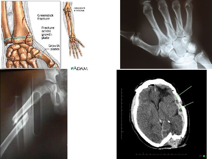
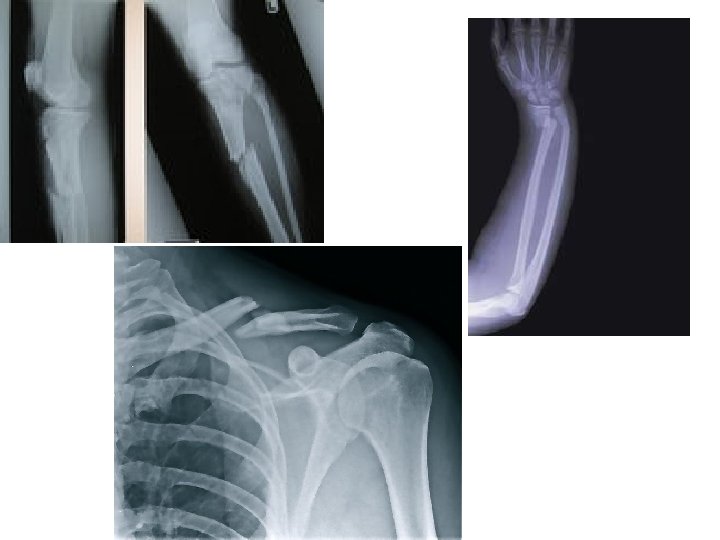
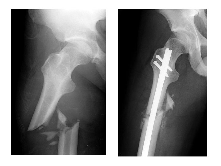
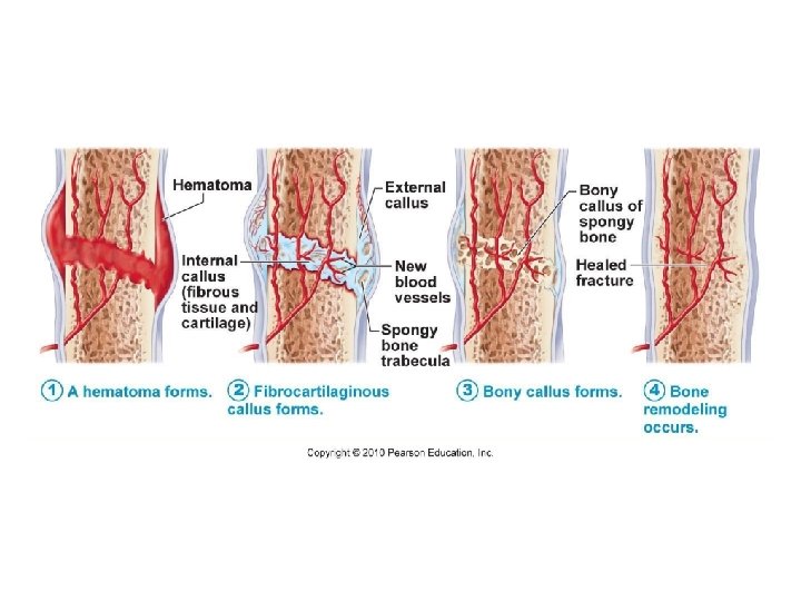
How Do Bones Repair After a Fracture? Fracture: A break in a bone.

Fractures
 Blood escapes and forms a hematoma. b) Chrondrocytes fill gap with")
Bone Repair a) Blood escapes and forms a hematoma. b) Chrondrocytes fill gap with fibrocartilage. Soft callus is formed. c) Spongy bone is deposited by osteoblasts. (Bony callus formed. ) d) Bone remodeling occurs: Osteoclasts will remove excess bone of callus and reconstruct shaft walls until remodeled area looks resembles original unbroken region.

Skeletal Articulations: JOINTS! The point of contact between two bones, between bone and cartilage or bone and teeth.

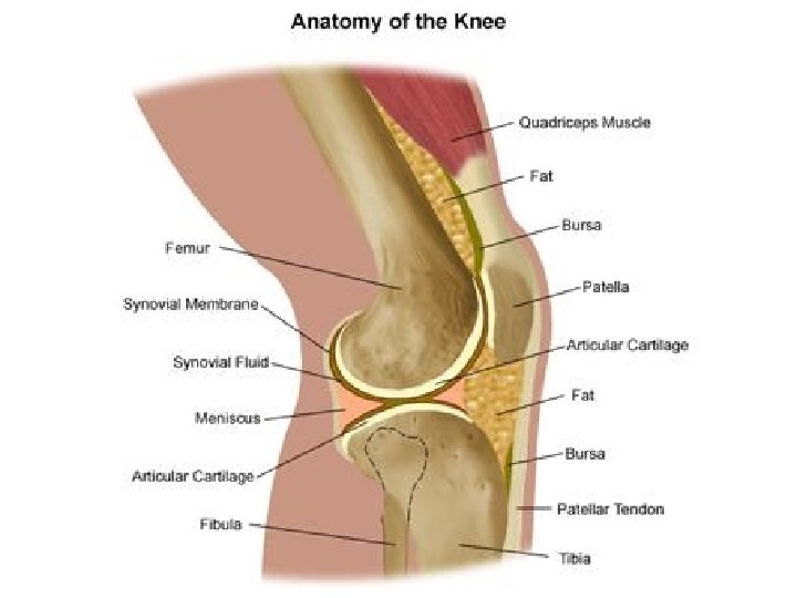
Three main types of joints: 1. Fibrous Joints – thin layer of connective tissue between, little movement, fixed. Ex: sutures, tibia/fibula, teeth roots. 2. Cartilaginous Joints – cartilage connects bones, no synovial cavity, limited movement. Ex: pubis symphysis, vertebrae/discs 3. Synovial Joints – complex structure: hyaline cartilage, synovial fluid, menisci, and bursae. Most joints are synovial. Freely moveable! Ex: knee, shoulder, fingers, toes, hip, elbow

Types of Synovial Joints: 1. Ball-and-Socket: humerus/scapula, femur/coxal 2. Condyloid: oval articulation- metacarpal/1 st phalange, radiocarpal, metatarsal/1 st phalange, occipital condyles 3. Gliding: nearly flat articulation- carpals, tarsals, sternocostal, vertebrocostal, scapula 4. Hinge: knee, elbow, ankle, and interphalangeal 5. Pivot: altas and axis, radio/ulnar 6. Saddle: carpal and thumb metacarpal

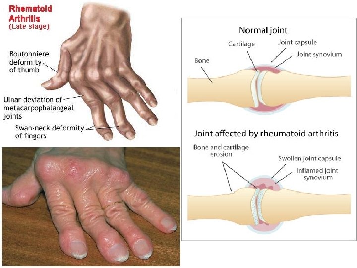
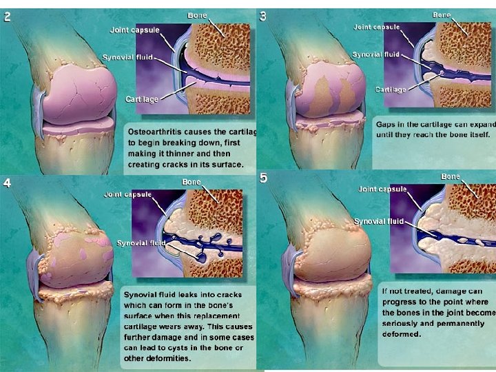
Disorders • Arthritis – inflammation of a joint 1. Rheumatoid – most painful, fibrous tissue fills joint, immobilization of joint. 2. Osteoarthritis – degenerative, result of aging, articulating cartilage wears away leaving rough, uneven surfaces…PAIN! video • Osteoporosis – porous bones due to calcium reabsorption which outpaces deposition. video • Sprain – forcible twisting of a joint that stretches or tears ligaments but does not dislocate bones.

Knee and Hip Replacement Surgery

ACL Surgery

Osteoporosis & Osteomalacia/Rickets Bone matrix reabsorbed due do hormonal changes and/or malnutrition.

Cleft Palate Palatine bone and/or palatine process of maxilla fails to unite.

Shin Splints Cause: small tears in the muscles that attach to the tibia.

Osteosarcoma
 curvature of the spine. • Causes: genetics, leg length")
Scoliosis • Abnormal lateral (sideways) curvature of the spine. • Causes: genetics, leg length diff. , other diseases, osteoporosis

Spina Bifida • The vertebrae overlying the open portion of the spinal cord do not fully form and remain unfused. • Incomplete closure of the embryonic neural tube results in an incompletely formed spinal cord. • FOLIC ACID before pregnancies for prevention.

Fibrodysplasia Ossificans Progressiva: A mutation of the body's repair mechanism causes fibrous tissue (including muscle, tendon, and ligament) to be ossified when damaged.
- Slides: 45