SKELETAL SYSTEM CHAPTER 5 I Functions of Bone

SKELETAL SYSTEM CHAPTER 5

I. Functions of Bone Support n Protection n Movement n Mineral storage: calcium and phosphate n Blood cell formation within red bone marrow: hematopoiesis n

II. Classification of Bone by Shape A. Long bones: elongated shape, not necessarily size. – Shaft plus two ends – Primarily compact bone – Contains spongy bone in its interior – All limb bones except the patella (knee cap), and those of the wrist and ankle.

B. Short bones: cube-like – Thin outer surface is compact bone – Composed mostly of spongy bone – Bones of the wrist and ankle – Sesamoid bones such as the patella: attach to tendons


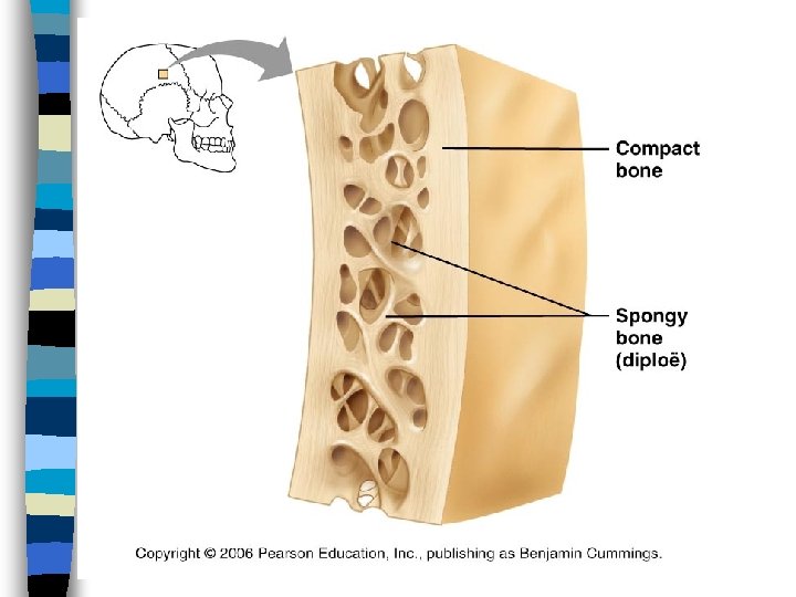
C. Flat bones: thin, flat and usually curved – two parallel compact bone – with inner layer of spongy bone – Sternum, ribs and skull bones

D. Irregular bones: bones that don’t fit into the other categories – Vertebra – Hip bones – Mostly spongy bone with thin outer layers of compact bone.

III. BONE Markings

III. BONE Markings

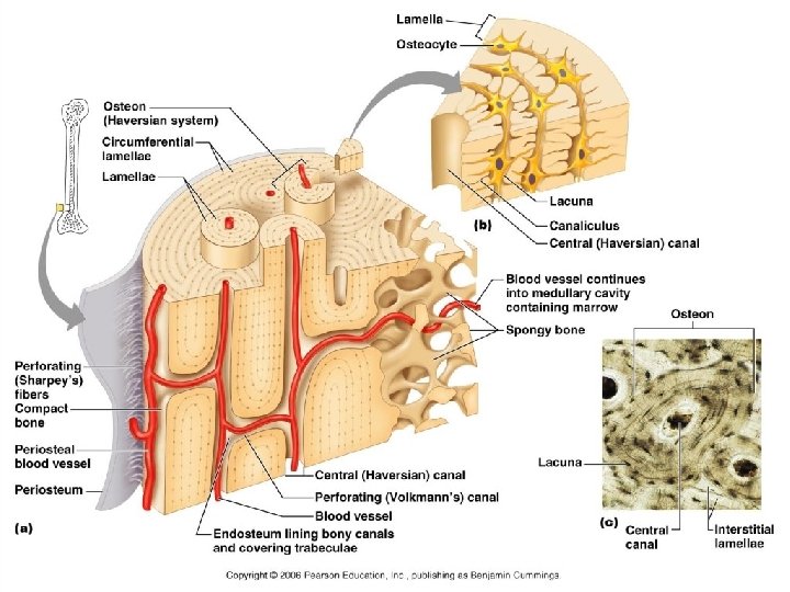
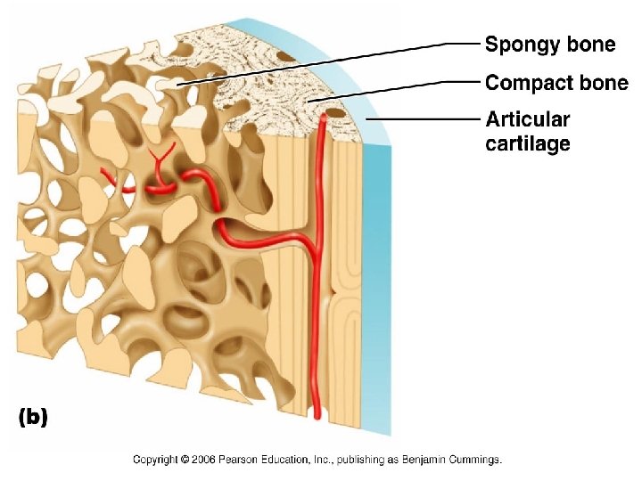
A. Microscopic Structure 1. Compact Bone – riddled with canals and passage ways for nerves and blood vessels
 (Haversian system) – Canal")
Osteon – Structural Unit: osteon (Fig. 5. 3 pg. 120) (Haversian system) – Canal surrounded by Lamellae
 They look disorganized but:")
2. Spongy Bone Contain trabeculae (pg. 118 fig. 5. 2) They look disorganized but: are aligned perfectly along the stress lines of the bone.

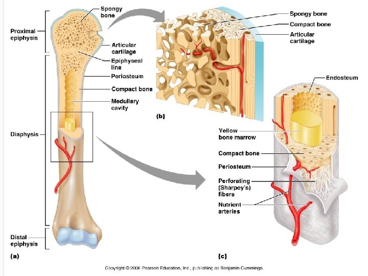
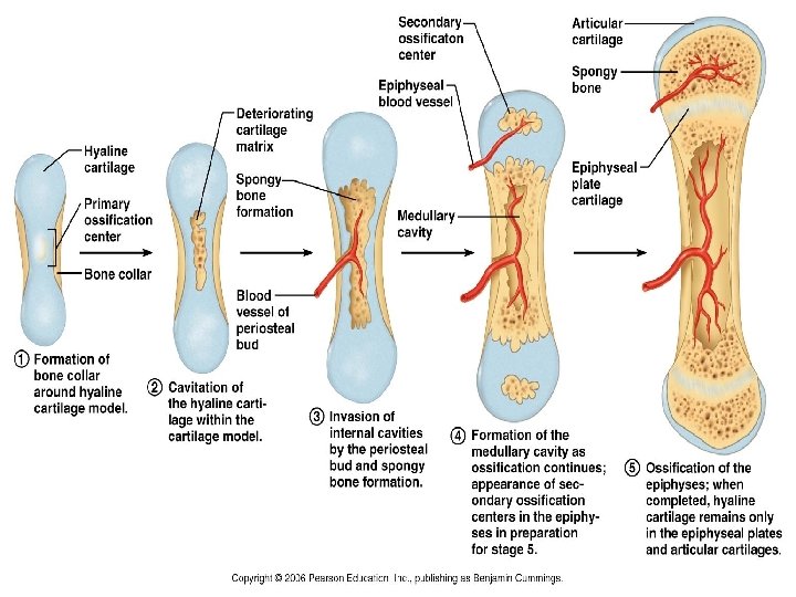
B. Long Bone Anatomy
 – Composed mostly of compact bone n")
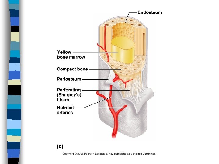
n Diaphysis: contains yellow bone marrow cavity(fat) – Composed mostly of compact bone n Epiphyses: Bone ends, Interior spongy bone – covered with articular cartilage to cushion
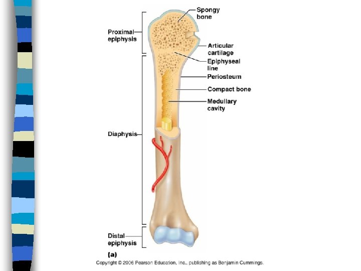

n Epiphyseal line: line between the diaphysis and Epiphyses in adult bones. n Hyaline Cartilage in young bones

C. Membranes of Bone 1. Periosteum: Outer surface of diaphysis – Glistening white – Double layered

a. Periosteum Layers Outer layer: Dense irregular connective tissue Inner layer: Osteogenic layer Bone forming cells: Osteoblasts Bone destroying cells: Osteoclasts (bone breakers)

b. Endosteum Lines: – canals of compact bone – trabeculae of spongy bone

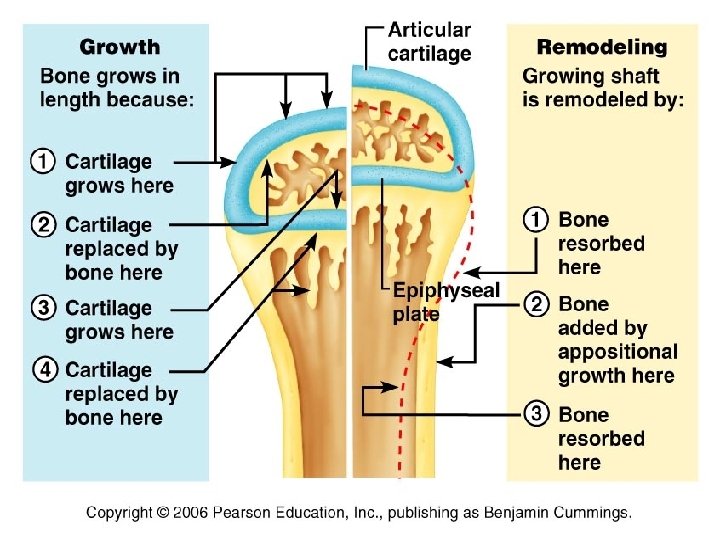
C. Bone Growth n Figure 5. 4 pg. 122 n Hyaline Cartilage cell form tall columns n Cells at the top undergo mitosis
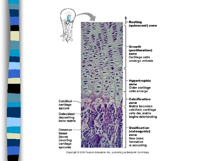
 n Growth is regulated by")
n Cells become older, then die and ossify(become hard) n Growth is regulated by the endocrine system

D. Growth Regulation 1. Growth Hormone stimulates growth of epiphyseal plates 2. Proportion of growth is regulated by thyroid hormone

3. Sex hormones: initially at puberty promote growth – Later they induce epiphyseal plate closure – Excess or deficits in any of these hormones can cause obvious skeletal disproportion.

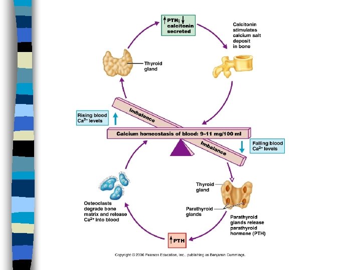
E. Hormonal Control of Calcium levels Thyroid Gland releases Calcitonin removes Ca+2 from blood & deposits to bone Parathyroid Gland releases parathyroid hormone (PTH) that removes Ca+2 from bone & deposits to blood

I. Classification of Joints Arthro: means joint 1. Synarthroses: immovable joints 2. Amphiarthroses: slightly movable 3. Diarthroses: freely movable

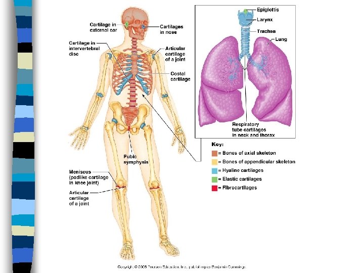
II. Structural Classes of Joints A. Fibrous joints: joined by fibrous tissue • no cavity is present • Ligaments: attach bone to bone • Suture joints of the skull • Synarthroses

B. Cartilaginous Joints: United by cartilage, no cavity is present • Symphysis: fibrocartilage in the pubic symphysis, and intervetebral discs • (Amphiarthroses)

C. Synovial Joints: bones are separated by a fluid filled joint cavity. • Most joints of the body. • Diarthroses

Synovial Joints 5 parts: Articular Cartilage Joint Cavity Articular Capsule Synovial membrane Synovial fluid (fig. 5. 28 pg. 149)

Synovial Joints Cont. Bursae and tendons: not a direct part of the joint. Act as bags of lubricant for the joint

1. Types of Synovial Joints Ball and socket: Hip and shoulder Gliding: tarsals, carpals, vertebra Pivot: skull to Vertebra Saddle: thumb Hinge: knee, elbow, phalanges

2. Movements by Synovial Joints Flexion Circumduction Extension Rotation Dorsiflexion Supination Plantarflexion Pronation Abduction Inversion/eversion Adduction Protraction/Retraction

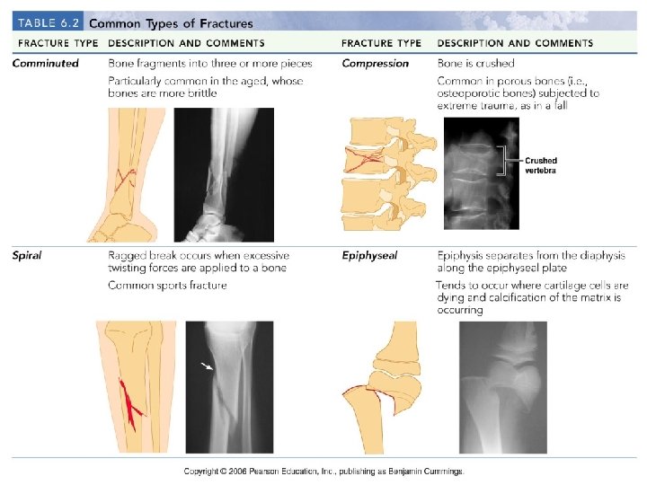
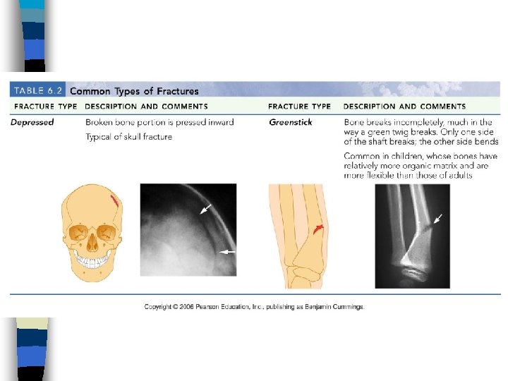
III. Homeostatic imbalances A. Sprains: Ligament of a joint is stretched or torn. B. Dislocations: Bones are forced out of their normal position. C. Table 5. 2 Common Types of Fractures…p. 123

Stages in the healing of a bone

C. Arthritis: degenerative joint diseases, 100 types or more 1 in 7 people suffer • Osteoarthritis: most common, wear and tear • Rheumatoid Arthritis: Chronic inflammatory process
")
D. Abnormalities Scoliosis: lateral curvature usually of the thoracic Kyphosis: exaggerated thoracic curvature (Hunchback) Lordosis: exaggerated Lumbar curvature (Swayback)

E. Osteomalacia n n softening of the bones due to a lack of vitamin D or a problem with the body's ability to break down and use this vitamin. Bone fractures that happen with very little injury Muscle weakness Widespread bone pain, especially in the hips

Treatment n Treatment may involve vitamin D, calcium, and phosphorus supplements, taken by mouth. Larger doses of vitamin D and calcium may be needed for people who cannot properly absorb nutrients into the intestines.

F. Osteoporosis n n n thinning of bone tissue and loss of bone density over time. Bone pain or tenderness Fractures with little or no trauma Loss of height over time Low back pain due to fractures of the spinal bones , stooped posture Neck pain due to fractures of the spinal bones

Treatment n n Control pain from the disease Slow down or stop bone loss Prevent bone fractures with medicines that strengthen bone Minimize the risk of falls that might cause fractures

G. Paget’s Disease n n is a disorder that involves abnormal bone destruction and regrowth, which results in deformity cause is unknown, although it might have to do with genes or a viral infection early in life

Multidirectional Forces on Bone Wolff’s Law:
- Slides: 54