Skeletal System Bone Physiology and the Skull Functions

Skeletal System Bone Physiology and the Skull

Functions of Bone • • Support Protection Movement Mineral and Growth factor storage Blood cell formation Triglyceride storage Hormone production

Classification of Bone • 206 bones divided into 2 groups: • Axial skeleton – includes the skull, vertebral column, and rib cage • Appendicular skeleton – includes the bones of the upper and lower limbs and girdles

Classification of Bone • Bones are generally classified by their shape as long, short, flat, or irregular – Long bones – longer than they are wide – Short bones – roughly cubed shaped. i. e. the bones of the wrist and ankle – Flat bones – thin, flattened, and usually a bit curved. Sternum, scapula, ribs, and skull bones – Irregular bones – complicated shapes that fit none of the other classes. Vertebrae and hip bones

Typical Long Bone Structure • Diaphysis • Epiphysial Plate/Line • Periosteum • Endosteum • Nutrient Foramina

Typical Flat Bone Structure • Thin plates of spongy bone – Periosteum – Endosteum • Compact bone plates • No shaft • No epiphysis

Chemical composition of bone

Components • Organic – Cells – Osteoid – the organic part of the matrix • Inorganic – 65% of mass is inorganic hydroxyapatites, or mineral salts. – Most of these are calcium phosphate which give bone its exceptional hardness

Cells • 5 major cell types – – – Osteogenic cells Osteoblasts Osteocytes Bone lining cells Osteoclasts

Formation of the Bony Skeleton • Begins during week 8 • Embryo starts as fibrous membranes and hyaline cartilage • Fibrous membrane bone = intramembranous ossification – Membrane bone • Flat bones – cranial bones • Hyaline cartilage bone = endochondral ossification – Endochondral bone • All bones below the skull except clavicles

Endochondral Ossification

Intramembranous Ossification

Postnatal Bone Growth • All bones grow in thickness • Long bones lengthen • Most bones stop growth during adolesence • Some facial bones will grow throughout life

Hormonal Control of Bone growth • Human Growth Hormone • Estrogen/Testosterone +/ • Calcitriol – Increases calcium and phosphate absorption in the GI tract • Blood calcium – PTH (parathyroid hormone) • Increases blood calcium – Calcitonin • Creates calcium deposits, decreasing blood calcium

Bone Remodeling • Process of bone deposit and bone resorption in adult skeletons • Coordinated by adjacent osteoblasts and osteoclasts called remodeling units • Goes on continuously in the skeleton, but not uniformly.
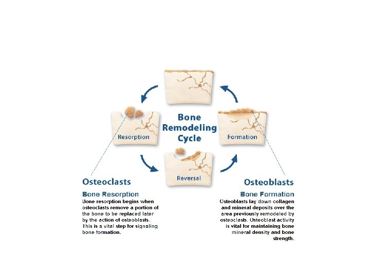

Control of Remodeling

Wolff’s Law • German anatomist Julius Wolff • Bone in a healthy person or animal will adapt to the loads under which it is placed. • If loading on a particular bone increases, the bone will remodel itself over time to become stronger to resist that sort of loading.

Fractures • Fractures are classified in four ways – Position of bones after fracture • Nondisplaced • Displaced – Completeness of break • Complete • Incomplete – Orientation of break, relative to long axis of bone • Linear • Transverse – Whether bone breaks the skin • Open (compound) • Closed (simple)

Bone Repair

Osteoporosis • Group of diseases where bone resorption outpaces bone deposits. • Bones become fragile • Occurs most often in postmenopausal women – Estrogen restrains oseteoclasts • Men also develop the disease

Skull • Cranium – Skull • Two parts – Neurocranium • Bony case of the brain and the cranial meninges, proximal parts of cranial nerves, and vasculature of the brain – Viscerocranium • Facial Skeleton • Anterior part of the cranium

Cranium • Most bones are united by fibrous interlocking SUTURES. • Some bones may be united by hyaline cartilage during childhood – Sphenoid and occiptal • Fontanel = soft spot • Sinus = air cavities within the facial bones

Fontanels • • • Soft Spots Anterior Posterior Sphenoid Mastoid www. crnasomeday. com/anatpages/fetal. htm

Fontanels • The posterior fontanel generally closes first between 1 to 3 months after birth • The sphenoidal fontanel is next to close around 6 months • The mastoid fontanel closes next from 6 to 18 months • The anterior fontanel is generally last between 7 and 19 months

Sinuses • • Frontal Ethmoid Sphenoid Maxillary http: //www. merck. com/media/mmhe 2/figures/fg 221_2. gif http: //www. mhni. com/images/Sinus-side-view-(sphenoid). jpg

Calvarium • Calvarium – Skull Cap, Domelike roof. • Formed from the Frontal, Parietal, and Occipital bones – Frontal • Forms the skeleton of the forehead • Forms the roof of the orbit and • Forms part of the floor of the anterior part of the cranial cavity science. kennesaw. edu

Calvarium • Parietal – Lateral aspect of the Cranium – Separated by the Sagittal suture mywebpages. comcast. net

Calvarium • Frontal and Parietal bones are joined at the Coronal Suture. • Parietal and Occipital bones are joined at the Lambdoid Suture. mywebpages. comcast. net

Calvarium • Occipital – Posterior or Occipital Aspect of the Cranium – Lambdoid Suture – External Occipital Protuberance • Easily palpable – Nuchal Line • Supeior – Splenius • Inferior mywebpages. comcast. net
 • Roof of the orbit • Cribiform Plate")
Frontal Bone • Supraorbital Notch (foramen) • Roof of the orbit • Cribiform Plate

Occipital • Nuchal Lines – Formed by the pull of neck muscles • External Protruberance – Formed by the pull of a ligament • • Basioccipital Foramen Magnum (great foramen) Hypoglossal canal Occipital condyles

Occipital • Basioccipital – relating to or being a bone in the base of the cranium immediately in front of the foramen magnum that is represented in humans by the basilar process of the occipital bone – Foramen Magnum – Spinal Cord

Occipital • Hypoglossal Canal – Hypoglossal Nerve • Occipital Condyles – Articulate with the vertebral Column http: //ect. downstate. edu/courseware/haonline/labs/l 22/os 0811. htm http: //www. octc. kctcs. edu/gcaplan/anat/images/Image 171. gif

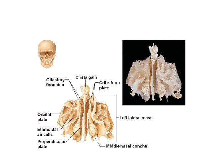
Ethmoid • Crista gall – Cocks comb • Cribfriform plate – Each side of the crista gall • Perpendicular plate • Middle Conchae • Orbital Plate science. kennesaw. edu

Hyoid • Does not articulate with any other bone • Suspended by stylohyoid ligaments coming from the styloid processes of the temporal bone • Supports the root of the tongue. www. octc. kctcs. edu

Maxilla • • Alveolar area Maxillary Sinus Infraorbital Foramen Frontal Processes www. uni-leipzig. de/~kfo/GNE/theotext. htm

Maxilla • Alveolar Area – Tooth Sockets – Support the Maxillary teeth • Maxillary Sinus www. bartleby. com/107/38. html www. octc. kctcs. edu

Maxilla • Infraorbital Foramen – Infraorbital Vessels and Nerves • Palatal Processes – 6 -8 weeks of development – Grow downward then horizontal – Fuse to form roof of mouth www. upstate. edu/. . . /hnskullantmax 3. shtml

Inferior Conchae • Extend into the nasal cavity from the maxilla bones • Help to churn the air in the nasal cavity mywebpages. comcast. net/wnor/lesson 9. htm

Mandible • • Body Ramus Mandibular angle Coronoid Process Mandibular Condyle Mental Foramen Mandibular Foramen p 197. ezboard. com

Mandible • Body – Horizontal part • Ramus – Verticle part • Mandibular Angle – Where Body and Ramus meet www. bio. psu. edu/. . . /anatomy/skel/mandible. htm

Mandible • Coronoid Process – Anterior aspect of the Ramus • Mandibular Condyle – Posteior aspect of the Ramus • Mental Foramen • Mandibular Foramen

Nasal Bone

Orbit • Lacrimal Foramen – Sits down in the lacrimal fossa • Optic Canal (foramen) • Superior Orbital Fissure – Nerves and Vessels • Inferior Orbital Fissure – Nerves and Vessels web. baypath. edu/biology/bones/skull 6. html

Palatine mywebpages. comcast. net http: //en. wikipedia. org/wiki/Image: Gray 194. png

Sphenoid • • • Lesser Wing Greater Wing Sella Turica Pterygoid Process Body Foramen lacerum – Closed by a membrane in the living • Foramen Ovale • Foramen Spinosum

Sphenoid • • • Greater Wing Lesser Wing Foramen Rotundum Foramen Ovale Foramen Spinosum http: //www. octc. kctcs. edu/gcaplan/anat/Notes/Image 528. gif Pterygoid Process Medial and Lateral Plates http: //en. wikipedia. org/wiki/Image: Gray 147. png

Sphenoid • Foramen Lacerum – Occulded by cartilage in the living • Sella Turcica – Turkish Saddle – Hypophyseal Fossa http: //en. wikipedia. org/wiki/Image: Gray 192. png

Sutures • • Coronal Sagittal Lambdoid Squamous http: //www. besthealth. com/besthealth/bodyguide/reftext/images/Cranial. Bones. jpg http: //www. yoursurgery. com/procedures/cranio/images/Craniosyn 2. jpg

Zygomatic • Zygomatic Arch – Temporal process of the zygomatic – Zygomatic Process of the temporal www. upstate. edu/cdb/grossanat/hnskullant. shtml

Temporal • Squamous – Above zygomatic process • Zygomatic Process • Mastoid Process http: //www. upstate. edu/cdb/grossanat/hnsklattb 2. shtml

Temporal • Styloid Process • Petrous • External Acoustic Meatus – External ear canal • Internal Acoustic Meatus – Cranial nerves VII and VIII Styloid process

Temporal • Jugular Foramen • Sylomastoid foramen • Carotid Canal http: //education. yahoo. com/reference/gray/illustrations/figure? id=187
- Slides: 55