Skeletal System Anatomy Bones of the Cranium Some


Skeletal System Anatomy

Bones of the Cranium ►Some are thicker than others!!!

Parietal Bone Cranium Bones Parietal Bone Frontal Bone Occipital Bone Temporal Bone

Maxilla and Mandible ? Maxilla Mandible

Clavicle or Collarbone ►The clavicle, or collar bone, holds the shoulder joint away from the rest of the upper body and is only as thick as your little finger.

Scapula ►The scapula is located on the back side of the ribcage and helps provide part of the shoulder joint and movement for the arms.
 The cervical region (neck bones) 2) The thoracic")
Vertebral Column or Spinal Cord 1) The cervical region (neck bones) 2) The thoracic region (what the ribs attach to) 3) The lumbar region (the lower part of the back)

Coccyx and Sacrum
")
Humerus (Upper Arm Bone)

Radius and Ulna ►Radius on Top ►Ulna on Bottom
")
Carpals or (Wrist Bones)
")
Metacarpals (Top of Hands)
")
Phalanges (Little Fingers)

Rib Cage
")
Sternum (Breastbone)
")
Pelvis (Dancing Bone)
")
Femur (Largest Bone in the Body)

The Tibia and Fibula

Tarsals

Metatarsals

Phalanges

Calcaneous

The Skeletal System in Action !! ►The Skeletal System in Action!

5 Functions of the Skeletal System 1. Movement: Skeletal system provides points of attachment for muscles. Your legs and arms move when the muscles pull on the bones. 2. Support: The backbone is the main support center for the upper body. It holds your head up and protects your spinal cord. Muscle attached to bones!!

5 Functions of the Skeletal System 3. Protection: The bones of your skull protect your brain. Your ribs protect your lungs and heart from injury. 4. Makes Blood: Red and white blood cells are formed by tissue called marrow, which is in the center of the bone.

5 Functions of the Skeletal System ► 5. Storage: Bones store minerals, such as calcium and phosphorus, for use by the body
: The axial skeleton includes the skull,")
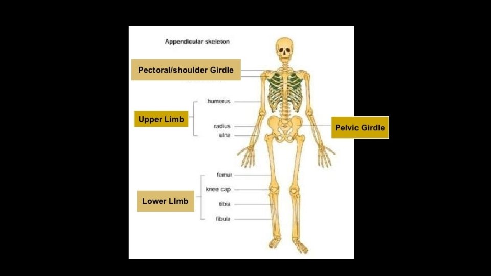
Two Major Skeletal System Parts ►Axial Skeleton (80): The axial skeleton includes the skull, hyoid bone, spine, ribs and sternum. ►Appendicular Skeleton (126): The appendicular skeleton includes the appendages of the body, which are the shoulders, arms, hips, and legs.

Bones of the Human Body · The skeleton has ~206 bones · Two basic types of bone tissue · Compact bone · Dense · Spongy bone · Small needle-like pieces of bone · Many open spaces Copyright © 2003 Pearson Education, Inc. publishing as Benjamin Cummings Figure 5. 2 b Slide 5. 3

Skeletal System Bones ►Four basic bone shapes • • 1. Long- arms, legs and fingers 2. Short- wrist and ankles 3. Flat- skull and sternum 4. Irregular- spine

Classification of Bones · Long bones · Typically longer than wide · Have a shaft with heads at both ends · Contain mostly compact bone • Examples: Femur, humerus, radius, ulna, tibia, fibula, phalanges Copyright © 2003 Pearson Education, Inc. publishing as Benjamin Cummings Slide 5. 4 a

Classification of Bones · Short bones · Generally cube-shape · Contain mostly spongy bone · Examples: Carpals, tarsals Copyright © 2003 Pearson Education, Inc. publishing as Benjamin Cummings Slide 5. 4 b

Classification of Bones on the Basis of Shape Figure 5. 1 Copyright © 2003 Pearson Education, Inc. publishing as Benjamin Cummings Slide 5. 4 c

Classification of Bones · Flat bones · Broad and thin · Usually curved or flattened · Thin layers of compact bone around a layer of spongy bone · Examples: Skull, ribs, sternum, scapula, clavicles Copyright © 2003 Pearson Education, Inc. publishing as Benjamin Cummings Slide 5. 5 a

Classification of Bones · Irregular bones · Irregular shape, often in groups · Do not fit into other bone classification categories · Example: Vertebrae, facial, pelvis, sacrum, coccyx · sesamoid “sesame seed shaped”connect to tendons ex. Patella, pisiform Copyright © 2003 Pearson Education, Inc. publishing as Benjamin Cummings Slide 5. 5 b

Classification of Bones on the Basis of Shape Figure 5. 1 Copyright © 2003 Pearson Education, Inc. publishing as Benjamin Cummings Slide 5. 5 c

Bone Structure ►Typical Four Layers: • Periosteum: Covers Bones • Compact Bone: Lies beneath the periosteum • Spongy Bone: Lies beneath the compact bone • Bone Marrow: Fills the gaps between the spongy bone

Gross Anatomy of a Long Bone · Diaphysis · Shaft · Made of compact bone · Epiphysis · Expanded ends of long bones · Covered with dense bone · Internal structure is spongy bone Figure 5. 2 a Copyright © 2003 Pearson Education, Inc. publishing as Benjamin Cummings Slide 5. 6

Structures of a Long Bone · Periosteum · Outside covering of the diaphysis · Fibrous connective tissue membrane · Arteries · Supply bone cells with nutrients Figure 5. 2 c Copyright © 2003 Pearson Education, Inc. publishing as Benjamin Cummings Slide 5. 7

Structures of a Long Bone · Articular cartilage · Covers surface of epiphyses · Hyaline cartilage · Decreases friction at joint surfaces Figure 5. 2 a Copyright © 2003 Pearson Education, Inc. publishing as Benjamin Cummings Slide 5. 8 a

Structures of a Long Bone · Medullary cavity · Cavity in shaft · Contains yellow marrow in adults · Contains red marrow in infants Figure 5. 2 a Copyright © 2003 Pearson Education, Inc. publishing as Benjamin Cummings Slide 5. 8 b

Microscopic Anatomy of Bone
, found within")
Types of Bone Cells · Osteocytes · Mature bone cells (bone maintenance), found within hard, rigid connective tissue · Osteoblasts · Bone-forming cells · Osteoclasts · Bone-destroying cells · Break down bone matrix for remodeling and release of calcium Copyright © 2003 Pearson Education, Inc. publishing as Benjamin Cummings Slide 5. 15
 · A subunit of compact bone,")
Microscopic Anatomy of Bone · Osteon (Haversian System) · A subunit of compact bone, circular arrangement of lacunae, lamellae, and osteocytes @ a Haversian canal · Central Haversian/Osteonic canal · Opening in the center of an osteon, runs vertically in long bone · Carries blood vessels and nerves · Supply oxygen and nutrients for osteocytes Copyright © 2003 Pearson Education, Inc. publishing as Benjamin Cummings Slide 5. 10 a
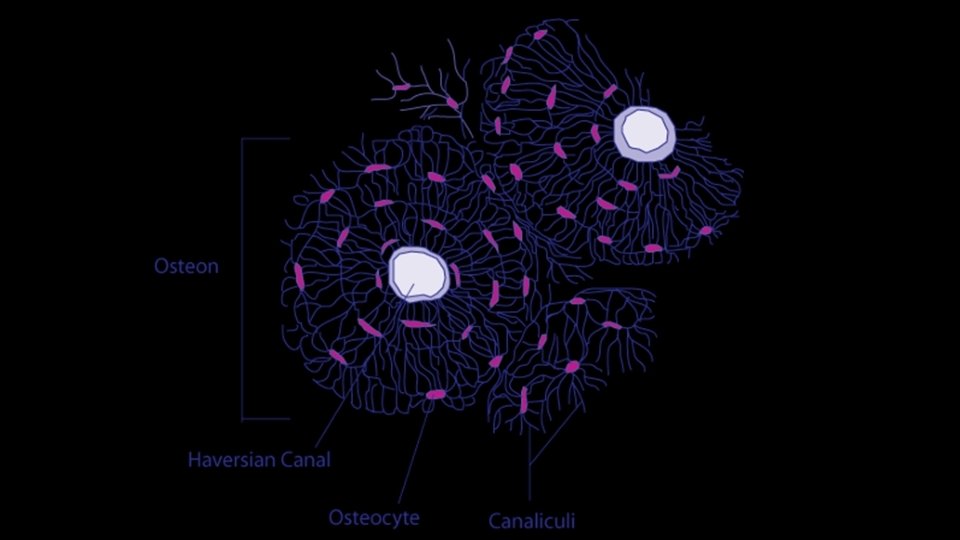

Microscopic Anatomy of Bone Figure 5. 3 Copyright © 2003 Pearson Education, Inc. publishing as Benjamin Cummings Slide 5. 10 b
 · Rings of compact bone around central canal")
Microscopic Anatomy of Bone · Lamella(e) · Rings of compact bone around central canal · Lacunae lie between rings · Lacuna(e) · Cavities containing osteocytes · Arranged in concentric rings Copyright © 2003 Pearson Education, Inc. publishing as Benjamin Cummings Figure 5. 3 Slide 5. 11 a
 · “little” canals · Contain cytoplasmic extension")
Microscopic Anatomy of Bone · Canaliculi (-us) · “little” canals · Contain cytoplasmic extension of osteocytes · Form a transport system- oxygen and nutrients from Haversian canal osteocytes via canaliculi Copyright © 2003 Pearson Education, Inc. publishing as Benjamin Cummings Figure 5. 3 Slide 5. 11 b

Microscopic Anatomy of Bone Volkmann’s canal/Perforating Canal Transverse canal- runs horizonatally in the long bone Contains larger blood vessels Connected to blood vessels in medullary cavity and in Figure 5. 3 the Haversian Canal Copyright © 2003 Pearson Education, Inc. publishing as Benjamin Cummings Slide 5. 10 b

Checkpoint 1 2 7 6 5 4 3

BONE GROWTH
 A cartilage model is produced by chondrocytes It is surrounded")
Bone Growth: Endochondral 1) A cartilage model is produced by chondrocytes It is surrounded by perichondrium, a membrane 2) A bone “collar” is produced by osteocytes The perichondrium of the diaphysis is now termed periosteum Copyright © 2003 Pearson Education, Inc. publishing as Benjamin Cummings Slide 5. 13 a

Long Bone Formation and Growth Figure 5. 4 a Copyright © 2003 Pearson Education, Inc. publishing as Benjamin Cummings Slide 5. 14 a
 Chondrocytes hypertrophy (enlarge) and cartilage is calcified 4) A primary")
Bone Growth: Endochondral 3) Chondrocytes hypertrophy (enlarge) and cartilage is calcified 4) A primary ossification center forms · Blood vessels and osteoblasts invade the calcified cartilage · Osteoblasts lay down bone matrix · Trabeculae are formed Copyright © 2003 Pearson Education, Inc. publishing as Benjamin Cummings Slide 5. 13 a

Long Bone Formation and Growth Figure 5. 4 a Copyright © 2003 Pearson Education, Inc. publishing as Benjamin Cummings Slide 5. 14 a
 Around the time of birth, secondary ossification centers form in")
Bone Growth: Endochondral 5) Around the time of birth, secondary ossification centers form in the epiphyses · Bone formation is incomplete at birth · The skeleton is not complete until the late teens (females) or early twenties (males) Copyright © 2003 Pearson Education, Inc. publishing as Benjamin Cummings Slide 5. 13 a

Long Bone Formation and Growth Figure 5. 4 a Copyright © 2003 Pearson Education, Inc. publishing as Benjamin Cummings Slide 5. 14 a

Bone Growth: Endochondral · Epiphyseal plates allow for growth of long bone during childhood · New cartilage is continuously formed · Older cartilage becomes ossified · Cartilage is broken down · Bone replaces cartilage Copyright © 2003 Pearson Education, Inc. publishing as Benjamin Cummings Slide 5. 13 a

Long Bone Formation and Growth Figure 5. 4 b Copyright © 2003 Pearson Education, Inc. publishing as Benjamin Cummings Slide 5. 14 b

Bone Growth: Endochondral · Bones are remodeled and lengthened until growth stops · Bones change shape (how? ? DNA!!!) · Bones grow in width (osteoblasts) · Bones are remodeled by osteoclasts Copyright © 2003 Pearson Education, Inc. publishing as Benjamin Cummings Slide 5. 13 b

Long Bone Formation and Growth Figure 5. 4 a Copyright © 2003 Pearson Education, Inc. publishing as Benjamin Cummings Slide 5. 14 a

Changes in the Human Skeleton · In embryos, the skeleton is primarily hyaline cartilage · During development, much of this cartilage is replaced by bone · Cartilage remains in: · Anterior portion of the nose · Parts of ribs · Joints Copyright © 2003 Pearson Education, Inc. publishing as Benjamin Cummings Slide 5. 12

Bone Growth: Intramembranous • “Between membranes” • Compact Bone • Spongy Bone • Trabeculae • Cross-section of a trabecula Copyright © 2003 Pearson Education, Inc. publishing as Benjamin Cummings Slide 5. 12

Bone Formation: Intramembranous · Membranous bones are incomplete at birth · Fontanelles: regions of the skull that remain as membranes · “Little fountains” · Bone formation complete by age ~2 Copyright © 2003 Pearson Education, Inc. publishing as Benjamin Cummings

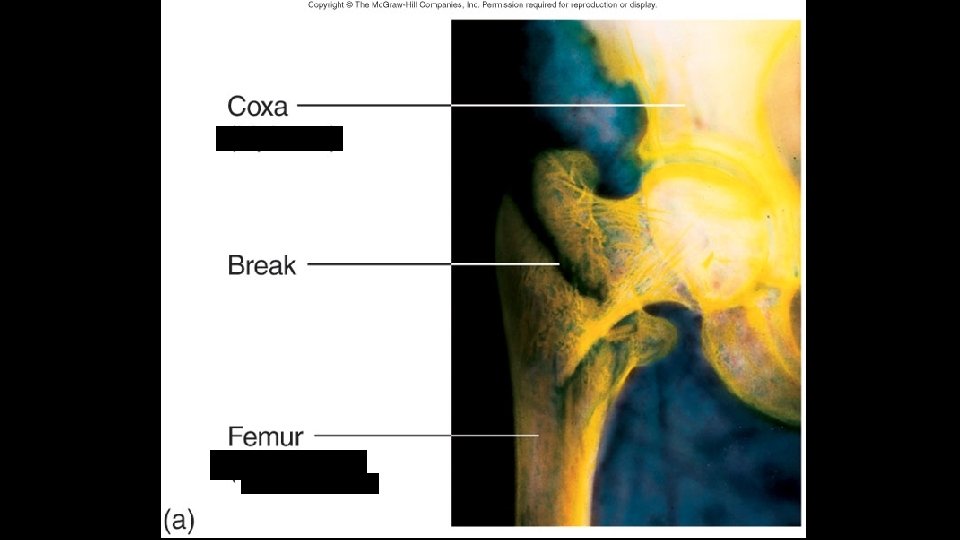
Bone Fractures · A break in a bone · Types of bone fractures · Closed (simple) fracture –does not penetrate the skin · Open (compound) fracture – broken bone penetrates through the skin · Bone fractures are treated by reduction and immobilization Copyright © 2003 Pearson Education, Inc. publishing as Benjamin Cummings Slide 5. 16

Common Types of Fractures Table 5. 2 Copyright © 2003 Pearson Education, Inc. publishing as Benjamin Cummings Slide 5. 17
 is formed · Break is splinted")
Repair of Bone Fractures · Hematoma (blood clot) is formed · Break is splinted by fibrocartilage · forms a callus (chondroblasts, other cells, and vessels) · Callus is replaced by a bone (osteoblasts) · Bony callus is remodeled (osteoclasts) Copyright © 2003 Pearson Education, Inc. publishing as Benjamin Cummings Slide 5. 18
 · Callus formation")
Repair of Bone Fractures · Fracture Repair · Hematoma formation (A) · Callus formation (B) · Bone replacement (C ) A · Bone remodeling (D) Copyright © 2003 Pearson Education, Inc. publishing as Benjamin Cummings B C D

Bone Fractures: Osteoporosis · Most common in post-menopausal women · Also occurs in males · Due to decrease in estrogen levels · Estrogen receptors on osseous tissue affect calcium deposition · Bone becomes porous, brittle · Weight-bearing exercise, calcium supplements, estrogen will help prevent osteoporosis Copyright © 2003 Pearson Education, Inc. publishing as Benjamin Cummings Slide 5. 16
- Slides: 71