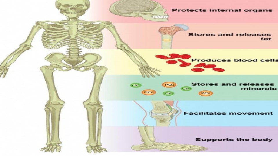
Skeletal System A Functions 1 Support 4 Mineral


 also regulates calcium. It is secreted by the thyroid when blood")


 bones b. 14 facial bones")


 cervical (7), thoracic")
There is a basic structure to a vertebrae, however, there are")


- Slides: 65
Skeletal System
A. Functions 1. Support 4. Mineral storage and homeostasis
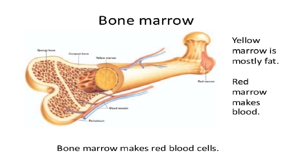
2. Protection 5. RBC production – red marrow
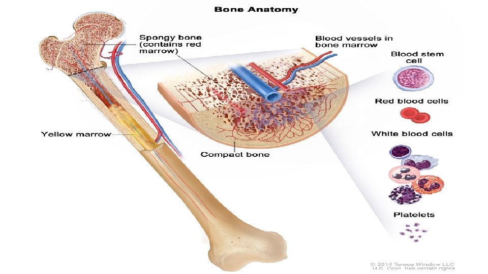
3. Movement 6. Energy storage – yellow marrow
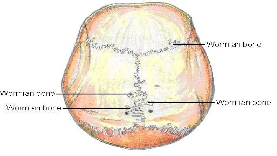
B. Bone Type 1. Long – greater length than width / one main shaft. EX – femur, toes 2. Short – equal length and width / cube shaped. EX – wrist, ankle 3. Flat – duh. EX – ribs, cranial, sternal 4. Irregular – complex shapes EX – cranial, vertebrae 5. Sutural / Wormian – bones in joints 6. Sesamoid – small bones in tendons EX – patella
C. Parts of a typical bone 1. Diaphysis – shaft 2. Epiphysis – ends 3. Metaphysis – between the diaphysis and epiphysis / growth plate exists here 4. Articular cartilage – hyaline cartilage covering the epiphysis / reduces friction & absorbs shock 5. Periosteum – membrane surrounding the bone / protection, nutrition, growth, repair, attachment 6. Medullary – marrow cavity 7. Endosteum – medullary lining
Histology A. Growth of bone 1. Bone is connective tissue with a lot of matrix. The matrix contains inorganic minerals (for hardness), collagen (for strength) and cells.
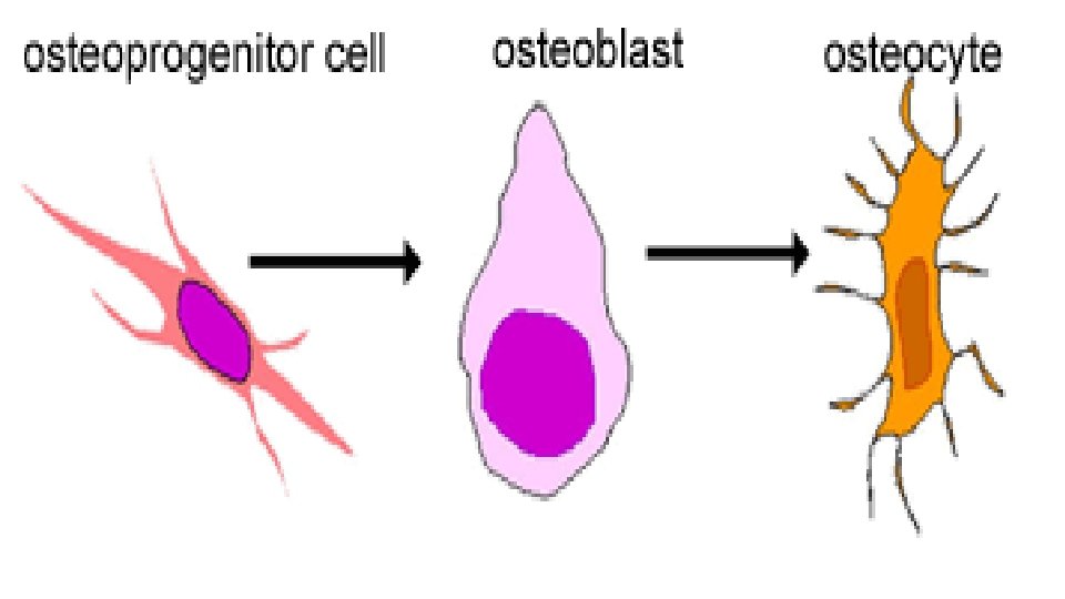
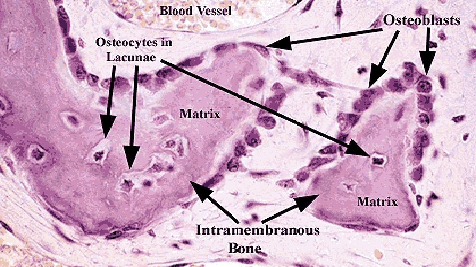
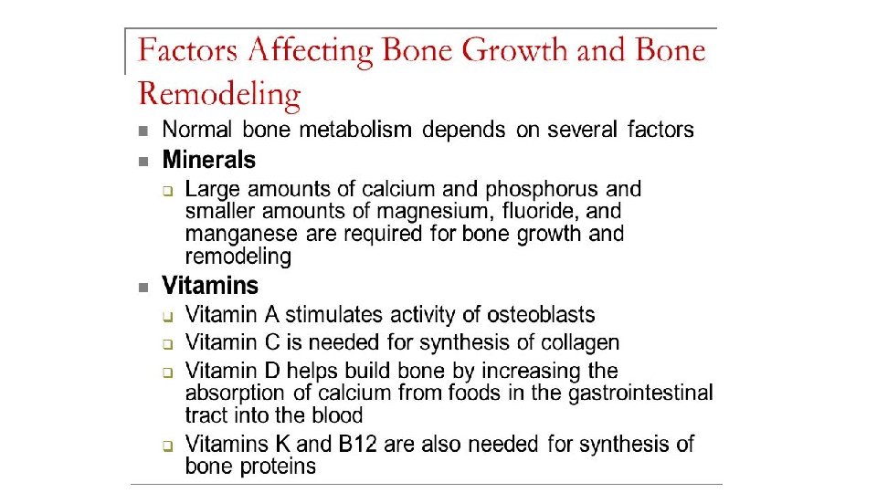
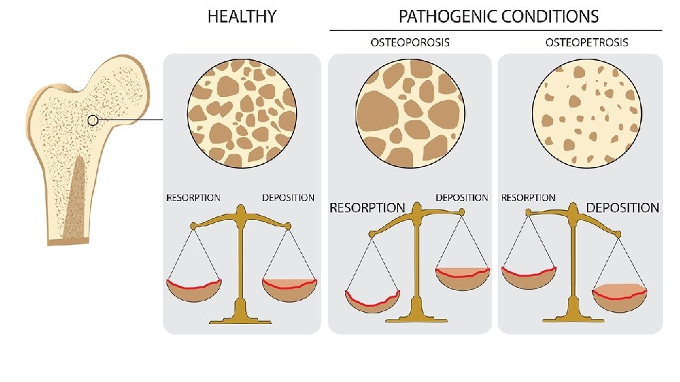
2. Bone cells a. Osteoprogenitors – these undergo mitosis to become osteoblasts b. Osteoblasts – forms bone / secrete collagen and inorganic compounds (calcium phosphate and calcium carbonate calcification) in rings around themselves. c. Osteocytes – isolated osteoblasts that have matured / perform daily cell activities d. Osteoclasts – bone resorption for growth, development, maintenance, repair
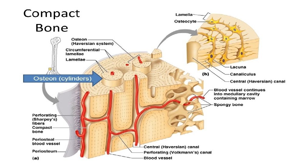
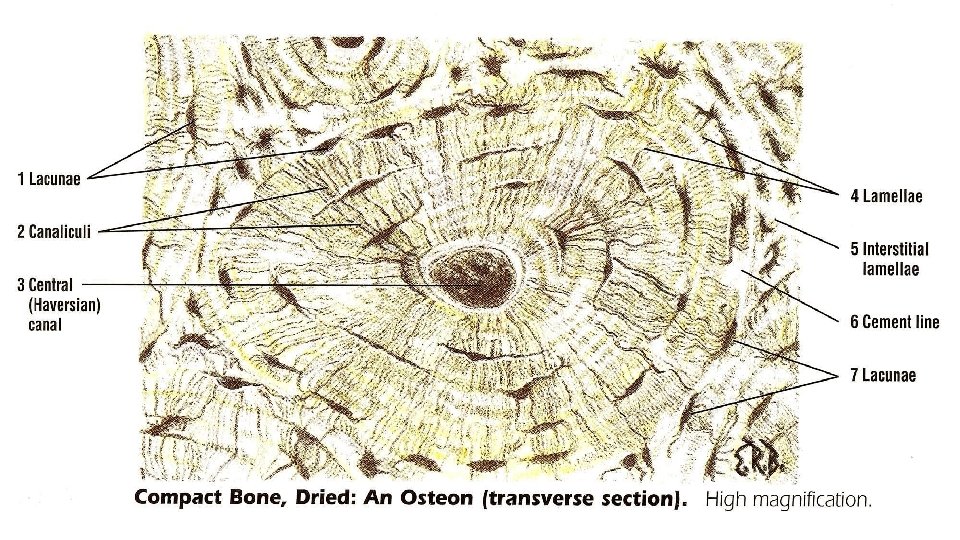
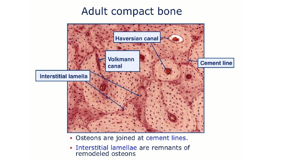
B. Compact bone 1. Osteon = Haversian system
C. Spongy bone 1. Does not contain true osteons – instead there are thin plates called trabeculae which are filled with red marrow 2. Does all RBC formation
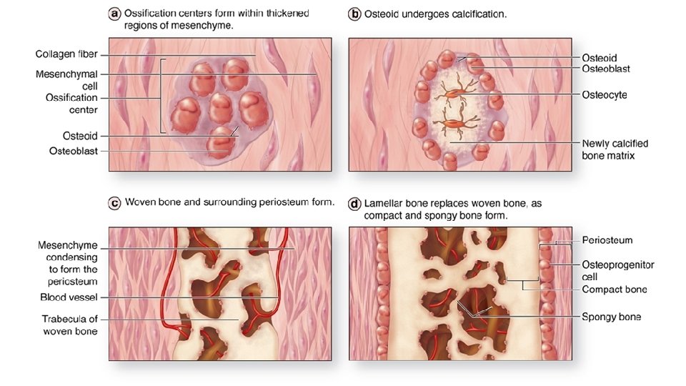
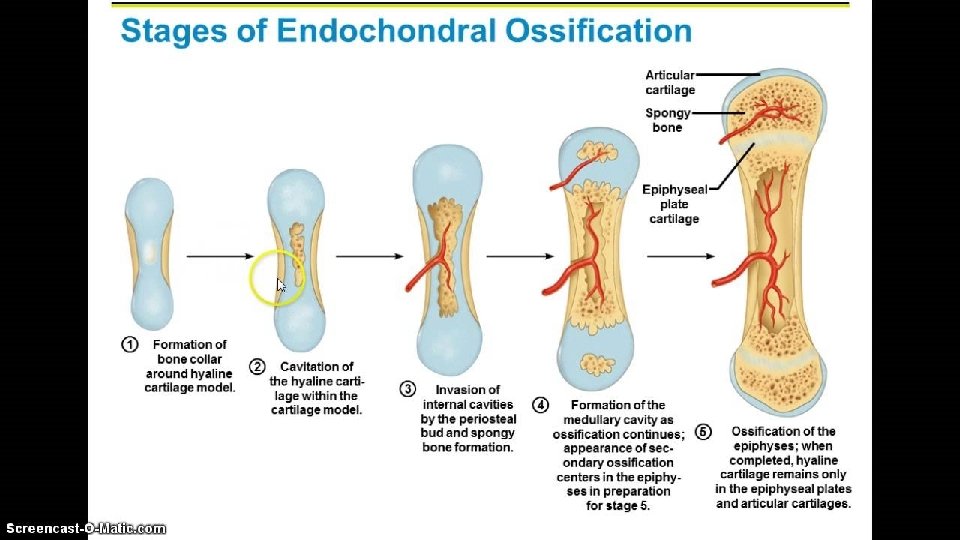
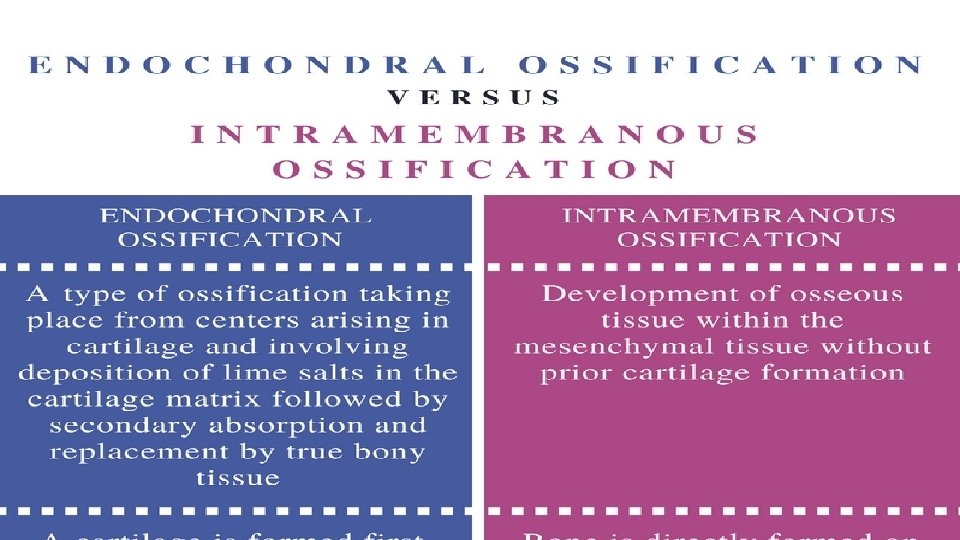
D. Bone formation 1. Ossification is the process of bone formation a. Intramembranous ossification – bone forms on or within the connective tissue b. Endochondral ossification – bone forms on a cartilage matrix * both lead to the same kind of bone
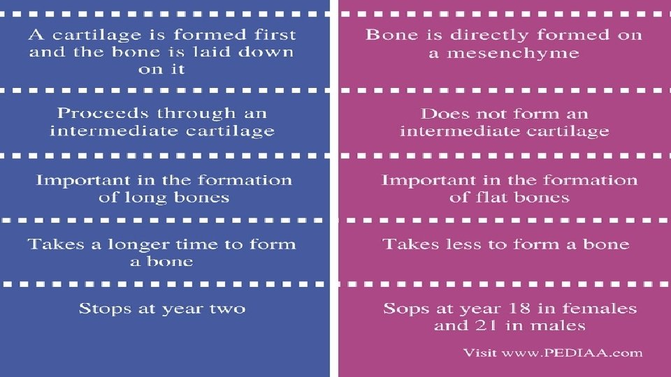
Homeostasis A. Bone growth and Maintenance 1. Remodeling – the replacing of old bone with new / constant throughout your life – you have a brand new skeleton every 2 years. 2. Depends on adequate mineral, vitamins (ACD), hormones and exercise
B. Minerals 1. Bones are the site of calcium and phosphate storage and release. 2. Parathyroid hormone (PTH) regulates blood calcium levels through a negative feedback system. If blood calcium is low, the parathyroid gland secretes PTH which causes the osteoclasts to reabsorb bone and release calcium into the blood
3. Calcitonin (CT) also regulates calcium. It is secreted by the thyroid when blood calcium is too high, causing calcium to be deposited in the bone 4. Exercise places stress upon bones causing them to deposit more minerals/collagen, making them stronger. Removal of the stress stops this process and weakens the bone (demineralization)
C. Aging 1. Aging causes a loss of calcium from the bones. In females this starts around age 30, males age 60. 2. It also leads to decreased protein formation causing a decreased collagen level in the bone. This causes brittleness.
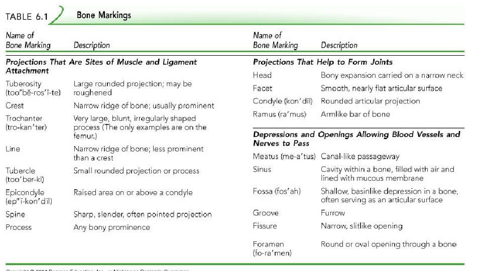
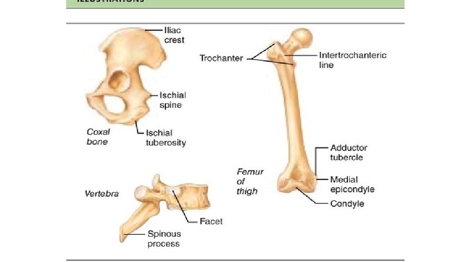
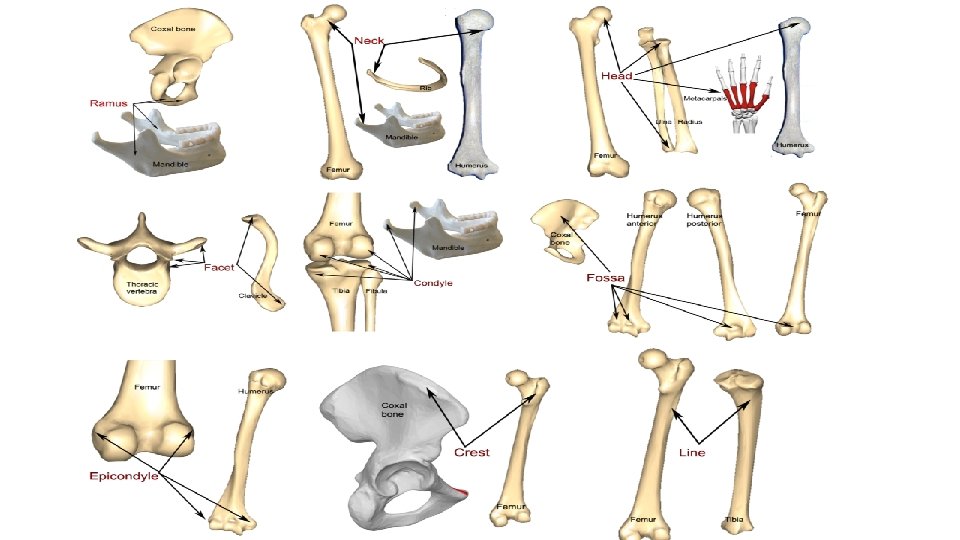
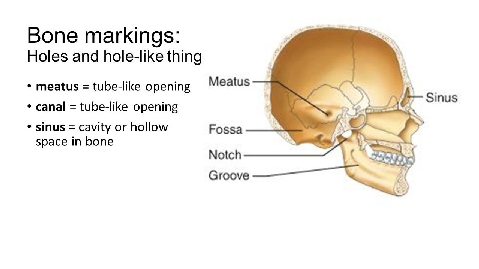
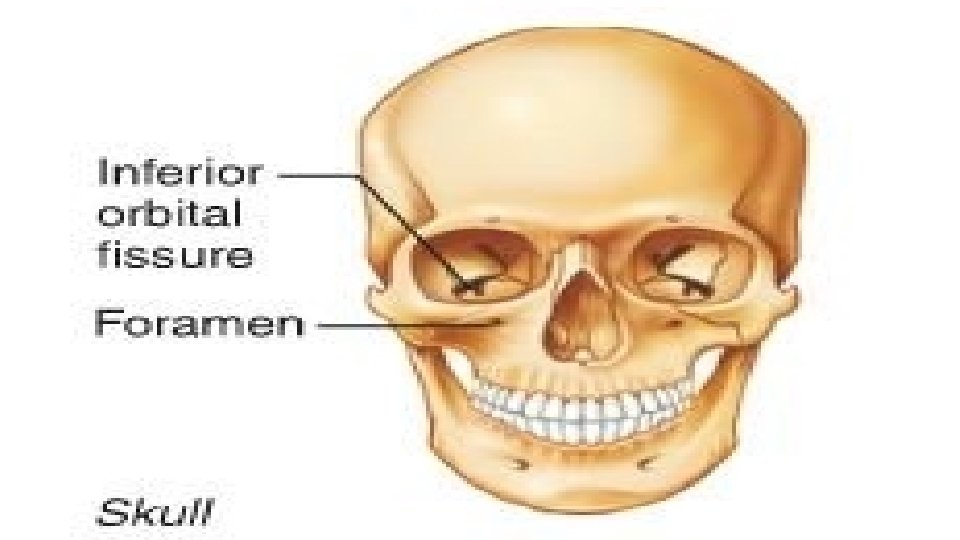
A. Bone Surface Markings The surfaces of bones have various features that adapt them to their particular function at their location in the body.
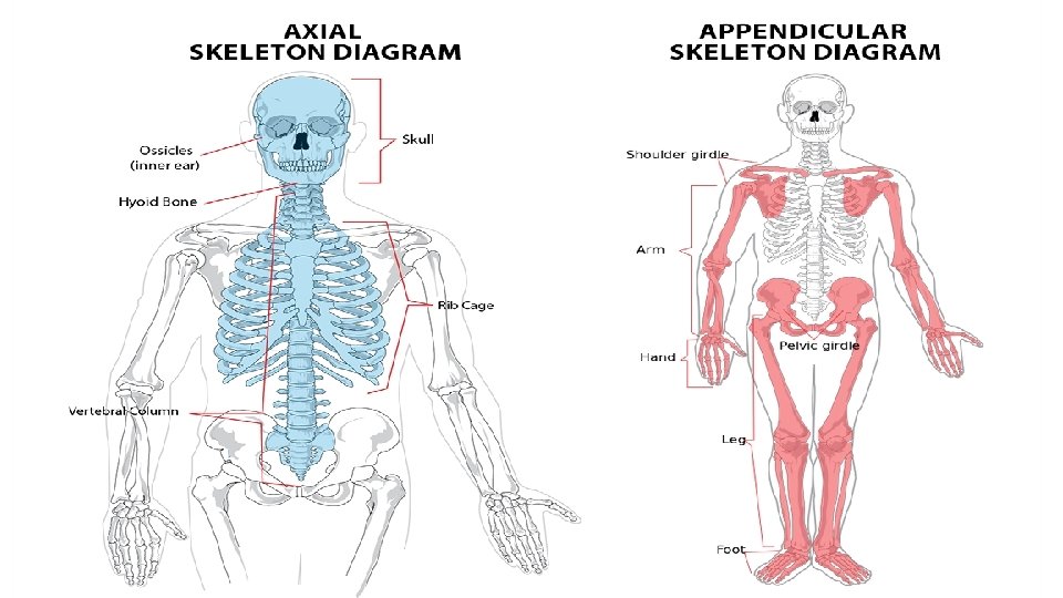
Divisions of the Skeletal System A. There a total of 206 bones in an adult skeleton. B. The entire skeleton can be divided into 2 main parts – the axial and appendicular skeletons
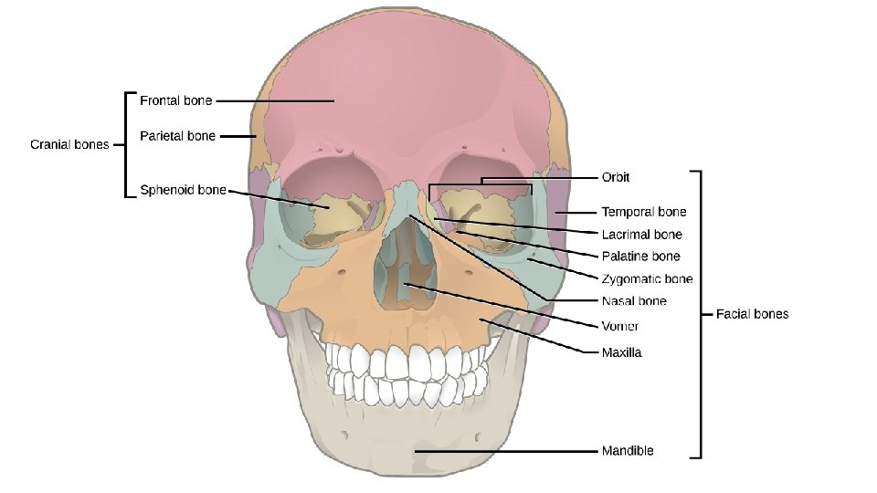
A. Axial skeleton 1. Skull a. 8 cranial (skull) bones b. 14 facial bones
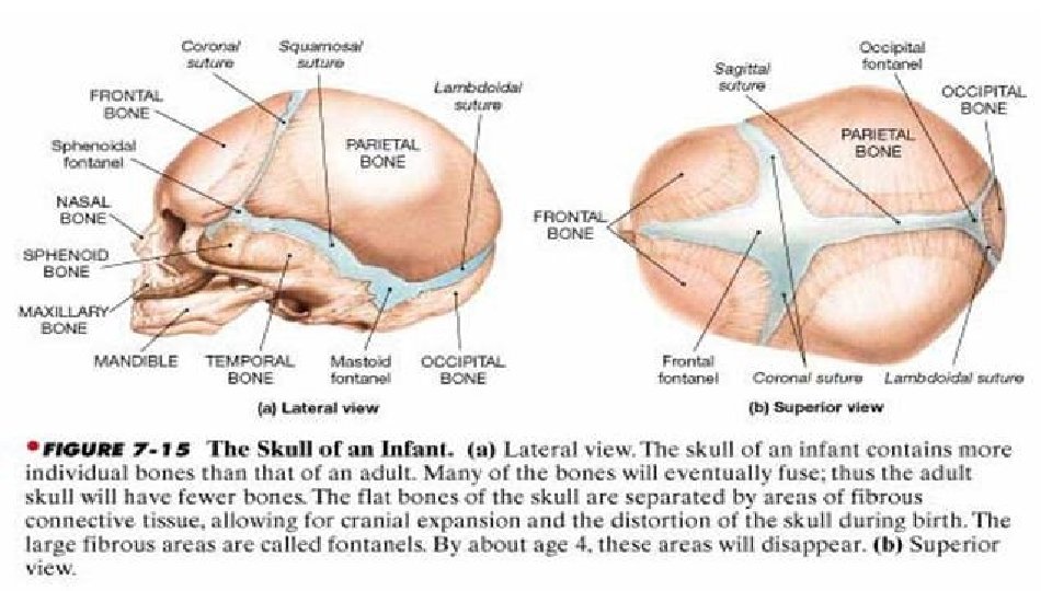
c. Sutures – the meeting place between 2 bones in the skull d. Bones to know on a picture Cranial - Frontal, parietal, temporal, occipital, sphenoid, ethmoid Facial – nasal, maxillae, paranasal sinuses, zygomatic, mandible, lacrimal bones, palantine, inferior nasal conchae, vomer e. Fontanels – these are the soft spots , connective tissue, of the skull in an infant that will become bone later
2. Hyoid bone – only bone not connected to any other bone. It is located in neck between the mandible and larynx. It functions to support the tongue.
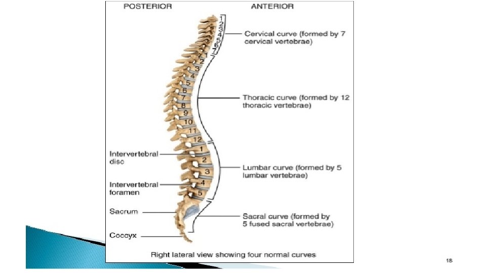
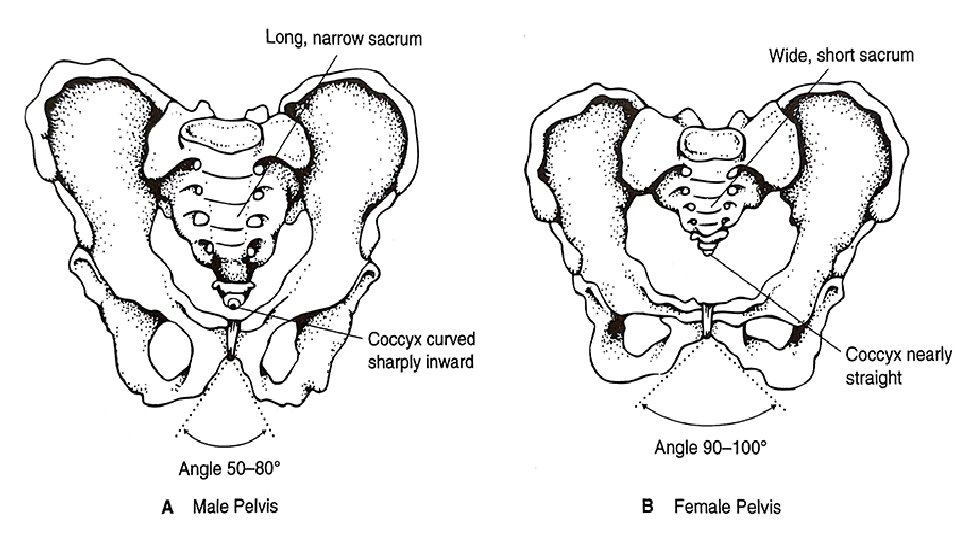
3. Vertebral column a. Divided into 5 regions (superior to inferior) cervical (7), thoracic (12), lumbar (5), sacral (5 fused = 1) and coccygeal (4 fused = 1) b. There are 4 normal curves. These help maintain balance, give strength, absorb shock and protect.
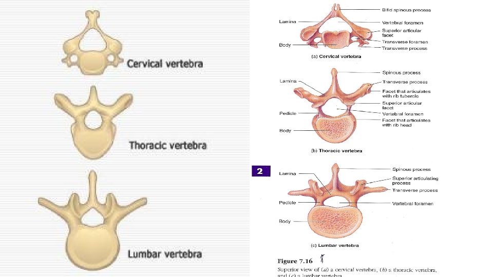
c. Vertebral structure (1)There is a basic structure to a vertebrae, however, there are structural differences amongst the 4 vertebral types. The 1 st 2 cervical vertebrae are called the atlas and axis.
4. Thorax a. This refers to the entire chest. b. It is composed of the sternum and ribs, both true (1 -7) and false (8 -12).
D. Appendicular Skeleton 1. Pectoral girdle a. This attaches an upper limb to the axial skeleton. b. It consists of the clavicle and the scapula
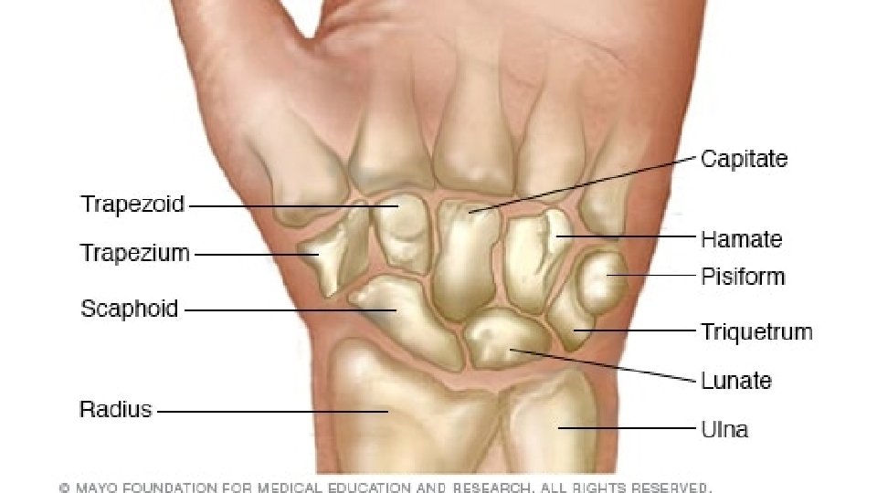
2. Upper limbs – 60 total bones a. Each upper limb contains a humerus, ulna, radius, carpals, metacarpals, and phalanges b. The carpals are each named singly.
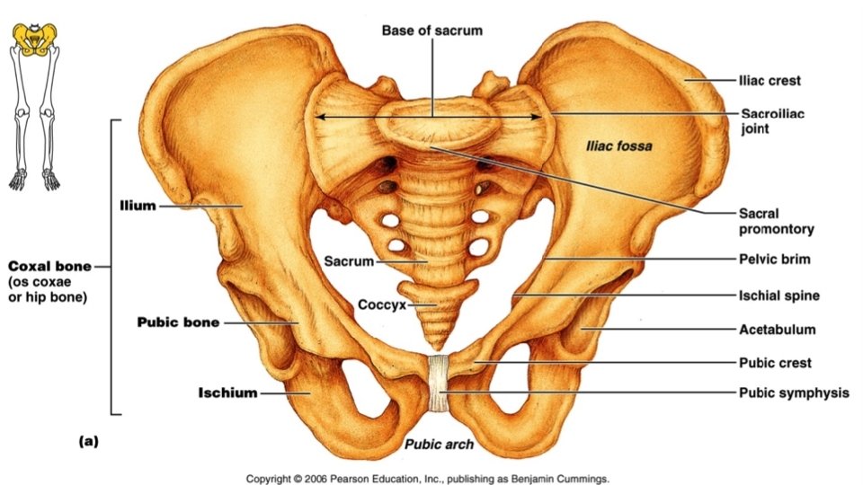
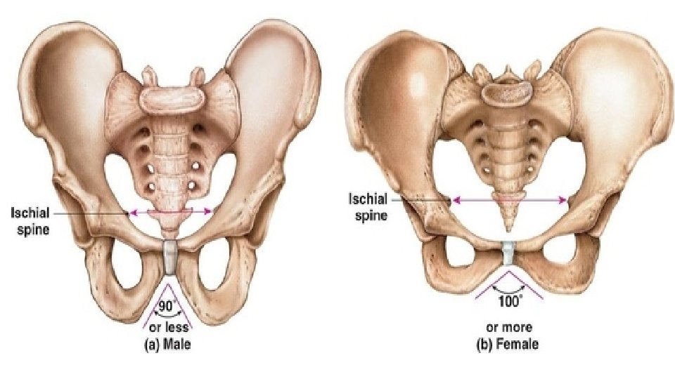
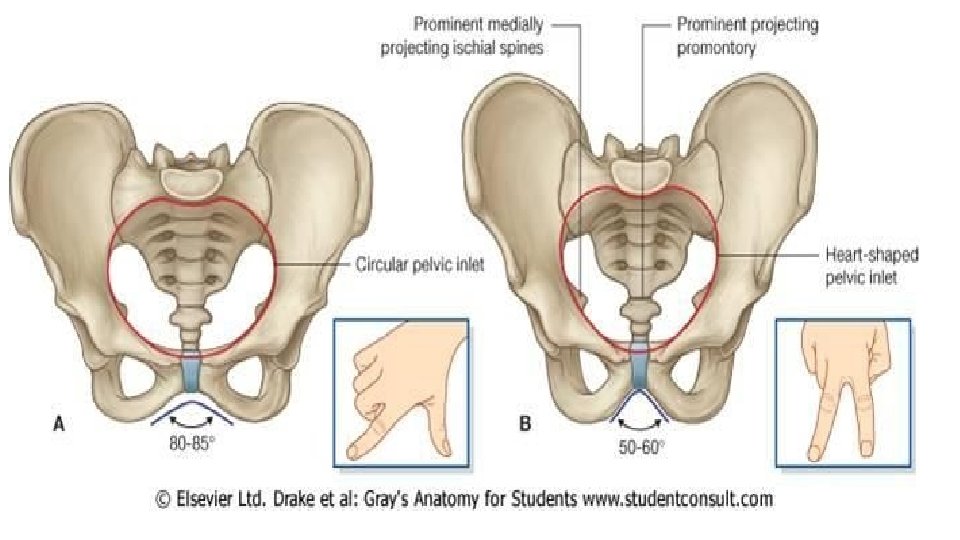
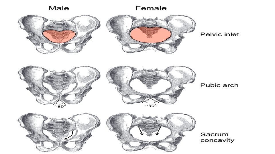
3. Pelvic girdle a. This attaches a lower limb to the axial skeleton and provides support for the vertebral column b. It consists of the 2 hipbones which are fused at the pubic symphysis.
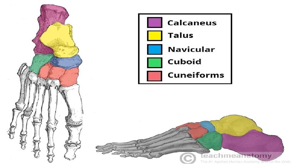
4. Lower limbs – 60 total bones a. Each lower limb has a femur, patella, fibula, tibia, tarsals, metatarsals, and phalanges. b. Shin splints are soreness / pain along the tibia caused by an inflammation of the periosteum. c. Each tarsal is named singly.
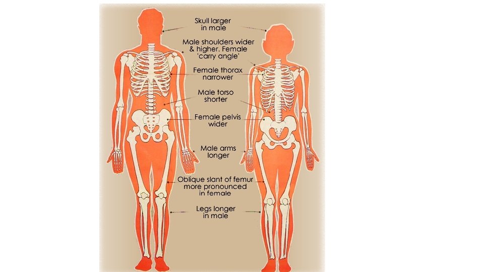
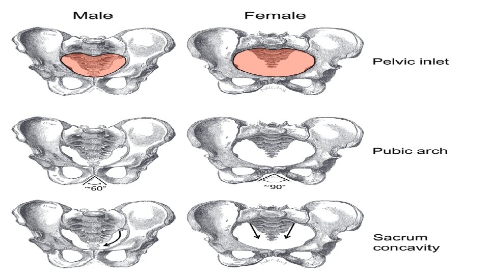
Male and Female Skeletons